A.P. and M.B. (EXAM 2)
 Call Kai
 Learn
Learn Practice Test
 Spaced Repetition
 Match
Match Flashcards
 Knowt Play
1/217

There's no tags or description
Looks like no tags are added yet.
Name | Mastery | Learn | Test | Matching | Spaced | Call with Kai | Chat |
|---|
No analytics yet
Send a link to your students to track their progress
218 Terms
Which sensory neurons do not have dendrites but receptors for physical stimuli in specialized regions
retinal photoreceptors and mechanism receptors
Anterograde definition
transport of macromolecules and mitochondria from neuron cell body to the axon
Retrograde definition
transport of macromolecules and mitochondria from axon to neuron body
what are the 4 major ionic species
Na+
k+
Cl-
Ca2+
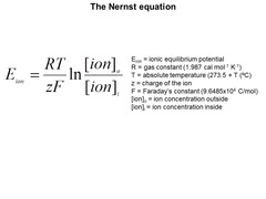
Nernst equation
used to calculate potential when the membrane is permeable to only 1 ion
Chord equation for membrane potential
membrane potential will depend on the relative conductances for the major ions and the equilibrium potential for those ions
Where
E= electromotive force (in volts)
G= conductance( in siemens) found using 1/R

electromotive force equation( V or E)
I*R
where
I= ion current
R= resistance
what happens to conductance (G) when channels open? closed?
when opened= G is high
when closed =G is low
Goldman-Hodgkin-Katz voltage equation
says that when a membrane is permeable to several different ions , the resting membrane potential depends on permeability , charge , and concentrations of all of the ions, and temperature

when the membrane is more depolarized which ion channels are open
K+ channels open
Cl- channels open
Na+ channels open
when the membrane is more hyperpolarized which ion channels are open
K+ channels open
Cl- channels open
How are graded potentials generated? describe them
By the opening of ligand-gated ion channels
small, hyper polarizing or depolarizing and decrement with distance due to ion leakage
difference between action potentials and graded potentials
action potentials are all or noting while graded potentials are incremental
in the dark , what gates are opened and what flows in
cGMP-gated channels are opened and Na+ flows in producing a dark current
How would you describe the relationship between the fraction of closed cGMP-gtaed channels and sodium current
linear
Most cells in the retina elicit what type of potentials and which cells are the exception
local; retinal ganglion
the large spikes of action potential are due to the presence of
voltage gates Na+ channels
Why can't graded potentials be propagated across long distances in neurons?
because of the leakage of plasma membrane
the velocity of the propagation depends on
the time constant( τ) of the membrane which is the time to charge and to discharge
what is the significance of the Hodgkin and Huxley model
- measured current-voltage relations
- found currents are carried by Na+ and K+ ions through axon membranes
- components of membrane conductance
resting membrane potential is
-70mV
threshold potential is
-60 mV
Depolarization potential is
+30 mV
hyper polarization potential is
-90 mV
which cranial nerves are apart of the CNS
Olfactory and optic
Simple receptors
neurons with free nerve endings
complex neural receptors
have nerve endings enclosed in connective tissue capsules
special senses receptors
cells that release neurotransmitter onto sensory neurons, initiating an action potential
Where is conduction velocity the fastest?
large diameter myelinated fibers
A and B fibers are myelinated by
Schwann cells
Are C fibers myelinated?
no
retino-hypothalamic tract
non image forming visual system
from RGC to suprachiasmatic nucleus then to pineal gland and contains body clock
retinotectal tract
to the olivary pretectal nucleus , controls pupillary light reflex
vesicular release of neurotransmitter steps
1. action potential is transmitted and depolarizes the axon terminal
2. depolarization opens voltage gated Ca channels and releases neurotransmitter
3. neurotransmitte binds to post-synaptic receptors and change the conductance in the post synaptic cell and elicit an electrical signal
Acetylcholinerase
enzyme responsible for the immediate breakdown of acetylcholine when release from the nerve ending; prevents overstimulation of cholinergic receptor sites
in the CNS what is the most common EXCITATORY neurotransmitter
glutamate
in the CNS what is the most common INHIBITORY neurotransmitter
GABA
can glutamate be inhibitory
yes ; glutamate is inhibitory for the bipolar cells of the retina
How is glutamate cleared from the synaptic cleft?
by reuptake via transporter
and
by diffusion and uptake by local glial cells
How is GABA cleared from the synapse?
reuptake using transporters on glia and
diffusion and uptake
can GABA be excitatory
yes
GABA-A receptor activation triggers membrane depolarization when there is Cl- efflux
ionotropic receptor definition and examples
receptors that are coupled to ion channels and affect the neuron by causing those channels to open
examples:
GABAR-A/C,
GLUR AMPA/KA, Nicotinic AChR(NMJ)
metabotropic receptors
receptors that are associated with signal proteins and G proteins
Examples :
GABA-B, mGLUR, MusAChR (brain only)
the ionotropic muscle type receptors are also called__________receptors and are composed of how many subunits
nicotinic receptors; composed of 5 subunits
the metabotropic muscle type receptors are also called ____________receptors and are composed of how many subunits
muscarinic receptors; composed of 1 subunit
all receptors are classified by
their agonist
receptor agonist definition
substances that mimic the action of the neurotransmitter
Exp:
Nicotine and muscarine that Mimic Ach
receptor antagonists definition
substances that block the action of the neurotransmitter
Exp:
Atropine and Curare
Receptor inverse agonists
binds to the same receptor- binding site as an agonist
, antagonizes the effects of an agonist and exerts the opposite effect by suppressing spontaneous receptor signaling (when present).
NMDA, AMPA, and Kainate are what type of glutamate receptors and are classified as?
ionotropic receptors and are fast excitatory
mGluR6 is what type of receptor and what is it responsible for
Metabotropic Group III receptor that is slow inhibitory
responsible for the ON pathway in the retina
how is glutamate release sustained in photoreceptors
a ribbon synapse that acts as a conveyor belt for the synaptic vesicles
what neurotransmitter is released by photoreceptors when put in the dark
glutamate
what are all the factors that influence post-synaptic response
1.strength, number and dendritic location
2.temporal and spatial summation between EPSP and IPSP
3.interactions between excitatory and inhibitory synapses
4. location and density of post synaptic ion channels
neuromodulator definition
substance other than a neurotransmitter, released by a neuron at a synapse and conveys information to adjacent or distant neurons, either enhancing or damping their activities.
neurohormones
produced and released by neurons in the brain, rather than by the endocrine glands, and delivered to organs and tissues through the bloodstream
Exp: pituitary gland hormones
G- Proteins
bind GTP in the active conformation and GDP in the inactive conformation
-catalyzes the hydrolysis of GTP to GDP +P
what is the difference between heterotrimeric G proteins and Monomeric G proteins
heterotrimeric G proteins have 3 subunits while monomeric have 1
what are the four families of heterotrimeric G proteins
Gαs
Gαi/Gαo/(GT*)
Gαq/Gα1
Gα12/Gα13
Gα12/Gα13 function
interacts with small (monomeric G proteins) to regulate cytoskeleton
Gαq/Gα11 function
activates beta-type phospholipase C (PLC-β) enzymes.
which hydrolyzes PIP2 => DAG and IP3 which activates protein kinase C and release stored calcium into cytoplasm respectively
Gαi/Gαo/(GT*) function
inhibits adenylate cyclase (AC)
Gαs function
stimulates adenylate cyclase (AC) and thereby decreases the production of cAMP
what is the main superfamily of the Small (monomeric) G proteins
Ras family
Activation cycle of G-protein by a G-protein- coupled receptor receiving ligand steps
1.Ligand binding to GPCRs (2) induces a conformation change
2.conformation change facilitates the exchange of GDP for GTP on the α subunit of the heterotrimeric complex
3.Both GTP-bound Gα in the active form and the released Gβγ dimer can then go on to stimulate a number of downstream effectors
4.When the GTP on Gα is hydrolyzed to GDP the original receptor is restored .
What is the significant advantage of g-protein signaling
amplification of the desired molecules
second messengers
components of molecular "circuits" inside cells that regulate metabolism, synthetic activity, ion channels, and other behavior of the cell
Main second messengers in the body are
Cyclic adenosine monophosphate (cAMP)
Cyclic guanosine triphosphate (cGMP)
Calcium (Ca2+)
Diacylglycerol (DAG)
cAMP/PKA signaling pathway allows for what and used what type of G proteins
mRNA transcription
uses Gs + Gi
PIP2/DAG/IP3 signaling pathway
ligand binds to G-protein receptor=> PIP2 is hydrolyzed => Ca2+ is released => Ca 2+ binding proteins are activates => produces DAG which activates protein kinase C
Catecholamines
dopamine, norepinephrine, epinephrine
Pupil constriction via sphincter pupil smooth muscle constriction is governed by which receptors and what pathway is the receptor a part of
governed by Cholinergic muscarinic M3 receptors
(M3 are GPCR to PIP2/DAG/IP3 pathway)
Pupil dilation via dilator muscle constriction is governed by which receptors and what pathway is the receptor a part of
governed by Adrenergic alpha1 receptors
(Alpha 1 are GPCR to PIP2/DAG/IP3 pathway)
Activation of rhodopsin by absorption of a photon leads to what and what does it cause
closure of cation channels in the plasma membrane of the rod outer segment.
This interrupts the dark current and hyperpolarizes the rod photoreceptor.
Activation of cone opsins leads to
lose cation channels in cone outer segments and hyperpolarize the cones.
Termination of the phototransduction cascade steps
(1) Inactivation of rhodopsin
(2) Inactivation of transducin
(3) Inactivation of phosphodiesterase (PDE)
(4) Activation of guanylate cyclase
spectral sensitivity
the degree to which a photopigment molecule absorbs light of different wavelengths
photoreceptors are color blind
First line of immune defense
skin and mucous membranes
second line of immune defense
phagocytic WBC
antimicrobial proteins
inflammatory response
third line of immune defense
lymphocytes and antibodies
the physical barriers of the innate immune response refers to
tightly packed epithelial cells
waterproof in skin
epithelial shedding
the physiological barriers of the innate immune response refers to
fevers
low pH
coughing/sneezing
antimicrobial molecules
interferons
interferon general mechanism
1. released from infected cell
2. after being released, it binds to surface receptors on neighbor cells
3. neighboring uninfected cell synthesized antiviral proteins that interfere with viral replication
cytokines
protein secreted by a cell that influences the cell itself or neighboring cells for the immune/inflammatory response
lysozyme
enzyme that degrades peptidoglycan cell wall of bacteria
most abundant protein found in tearfilm
Opsonins description
receptors that recognize molecules that are found on pathogens and bind to them allowing for immune cells to phagocytose the pathogen
examples are C3B and antibodies
Pathogen-associated molecular patterns (PAMPs)
Molecules (like LPS or Flagellin) associated with groups of pathogens that are recognized by cells of the innate immune system.
toll-like receptors
recognize molecular patterns unique to pathogens then stimulates the production of cytokines, chemokine, and antimicrobial peptides
when does the acquired immune response improve
after each subsequent exposure
epitope
3D molecular arrangement on antigen recognized by immune system
Cells of the acquired immune system
memory cells , B lymphocytes, T lymphocytes
B lymphocytes
produce antibody and humoral immunity
T lymphocytes
help B lymphocytes produce antibody and help destroy organism
cell mediated immunity
all cells of the immune response derive from where and as what originally
derive from the bone marrow and differentiate into CLP and CMP
most to least common WBCs
"Never Let Monkeys Eat Bananas"
Neutrophils [most]
Lymphocytes
Monocytes
Eosinophils
Basophils [least]
common myeloid progenitor differentiates into
granulocytes
mast cells
monocytes
megakaryocytes
common lymphoid progenitor (CLP) differentiates into
TC
TH
B cell
Natural killer cell
Haematopoietic stem cells differentiate into
Common myeloid progenitor and common lymphoid progenitor
granulocytes consists of
neutrophils, eosinophils, basophils
neutrophils description
most commonn
first to arrive when infection is induced
phagocytize and destroy foreign organisms