EYE MODEL
 Call Kai
 Learn
 Practice Test
 Spaced Repetition
 Match
 Flashcards
 Knowt Play
1/49

There's no tags or description
Looks like no tags are added yet.
50 Terms

Anterior cavity is anterior to the:
Lens

Anterior cavity is filled with a watery fluid called:
Aqueous humor

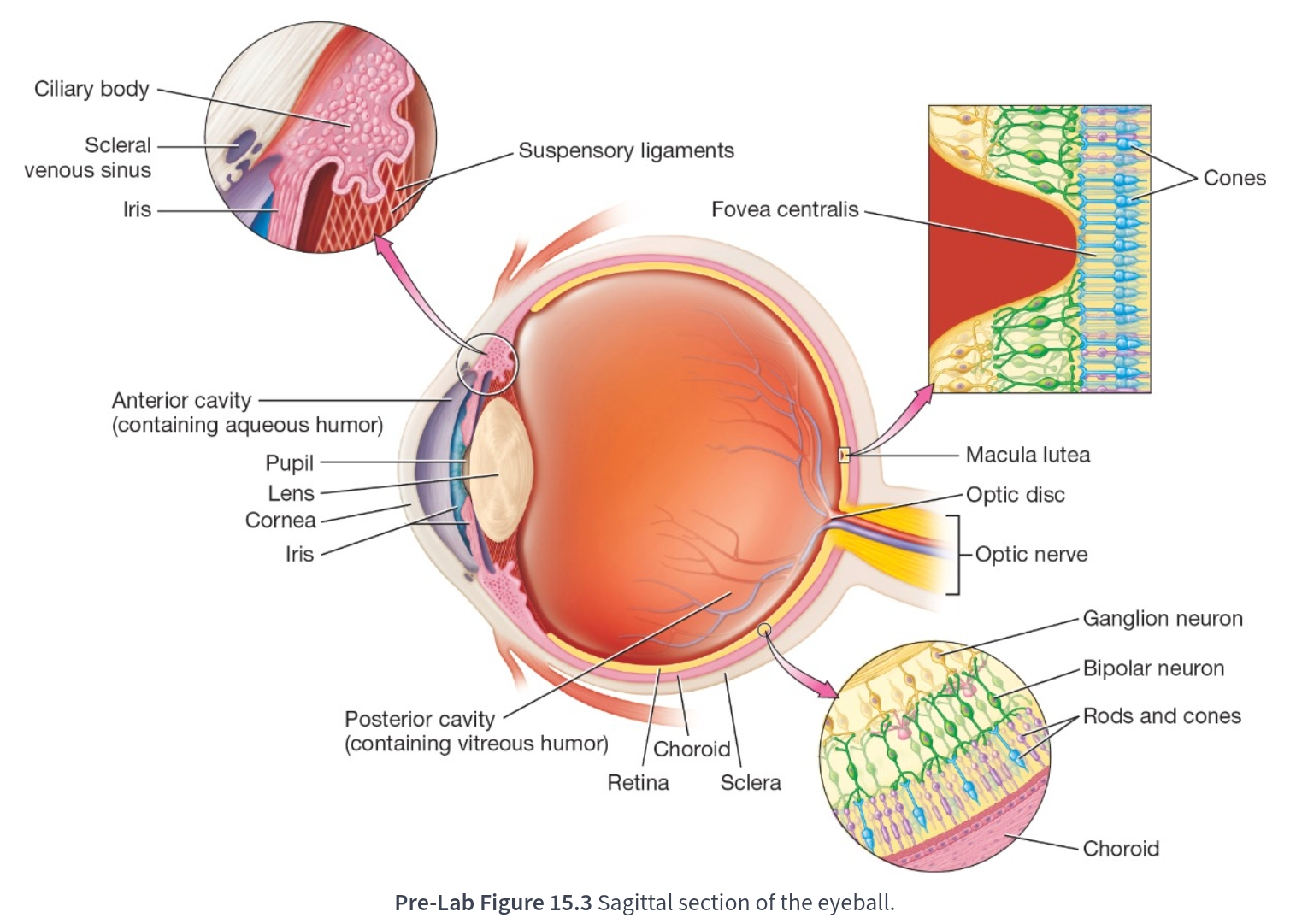
Carries most of the blood supply to the tissues of the eye:
Vascular tunic

The vascular tunic is composed of three main parts:
Choroid, Ciliary body, Iris

Makes up the posterior part of the vascular tunic:
Choroid

The choroid is brown in color to prevent:
Light scattering in the eye
Outer most layer of the eyeball:
Fibrous tunic

It is avascular and consists of two parts:
Fibrous tunic
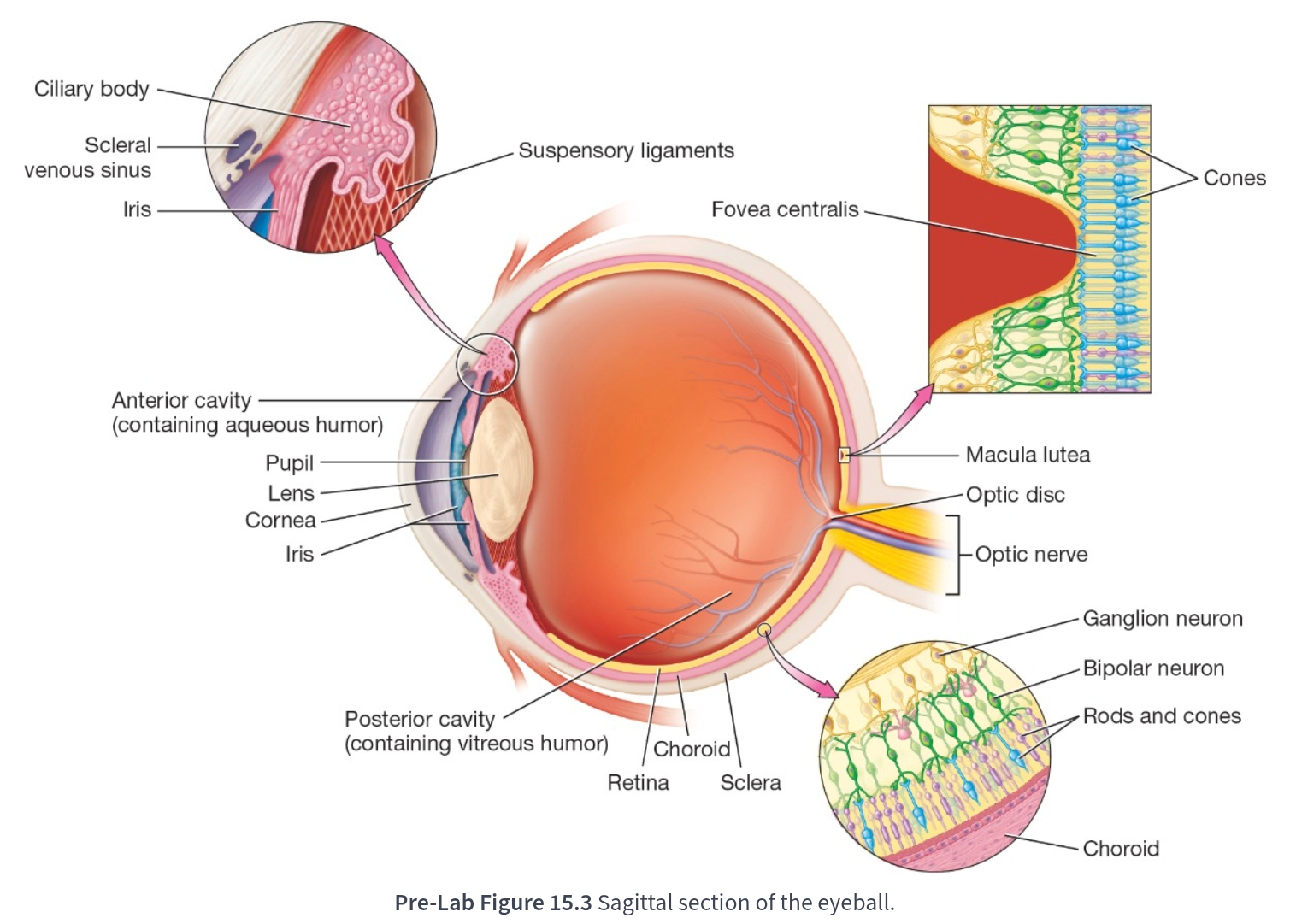
Makes up the anterior one-sixth of the fibrous tunic:
Cornea

It is the fourth refractory medium of the eyeball:
Cornea

Accounts for about two-thirds of the eye’s refractive power:
Cornea
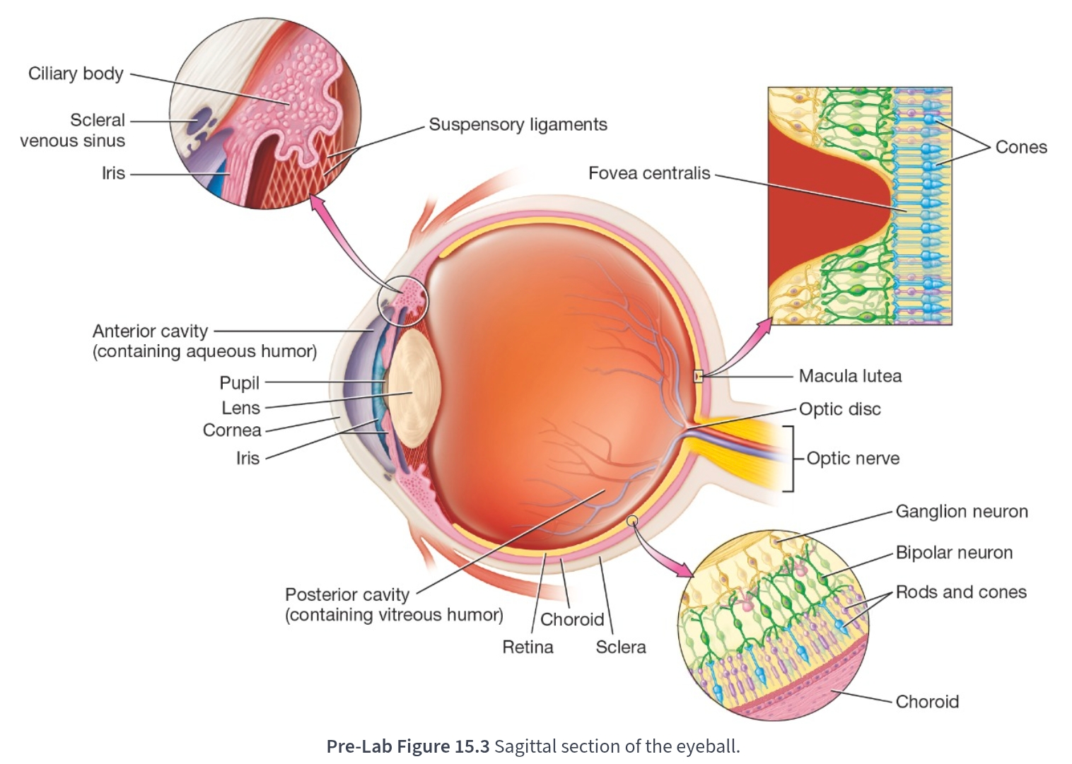
Located on the anterior aspect of the vascular tunic:
Ciliary body

Made primarily of the ciliary muscle—smooth muscle fibers that control the shape of the lens:
Ciliary muscle

Attaches to the lens via small suspensory ligaments:
Ciliary muscle
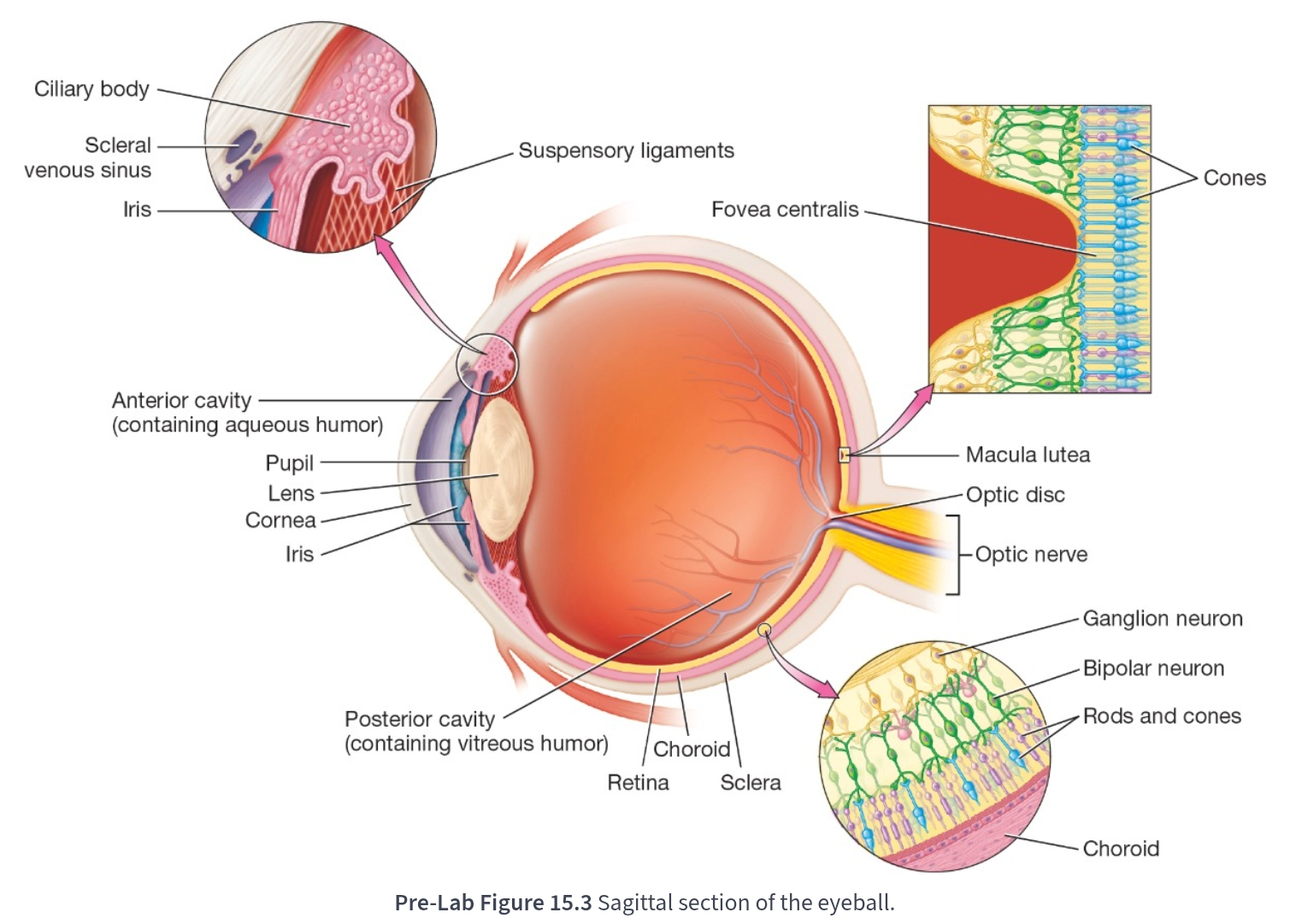
At the center of the macula lutea is the:
Fovea centralis

Contains only cones:
Fovea centralis

Responsible for color and high-acuity vision in bright light:
Cones

Most anterior portion of the vascular tunic:
Iris

Consists of smooth muscle fibers arranged around an opening called the pupil:
Iris

Refracts light coming into the eye to focus it:
Lens
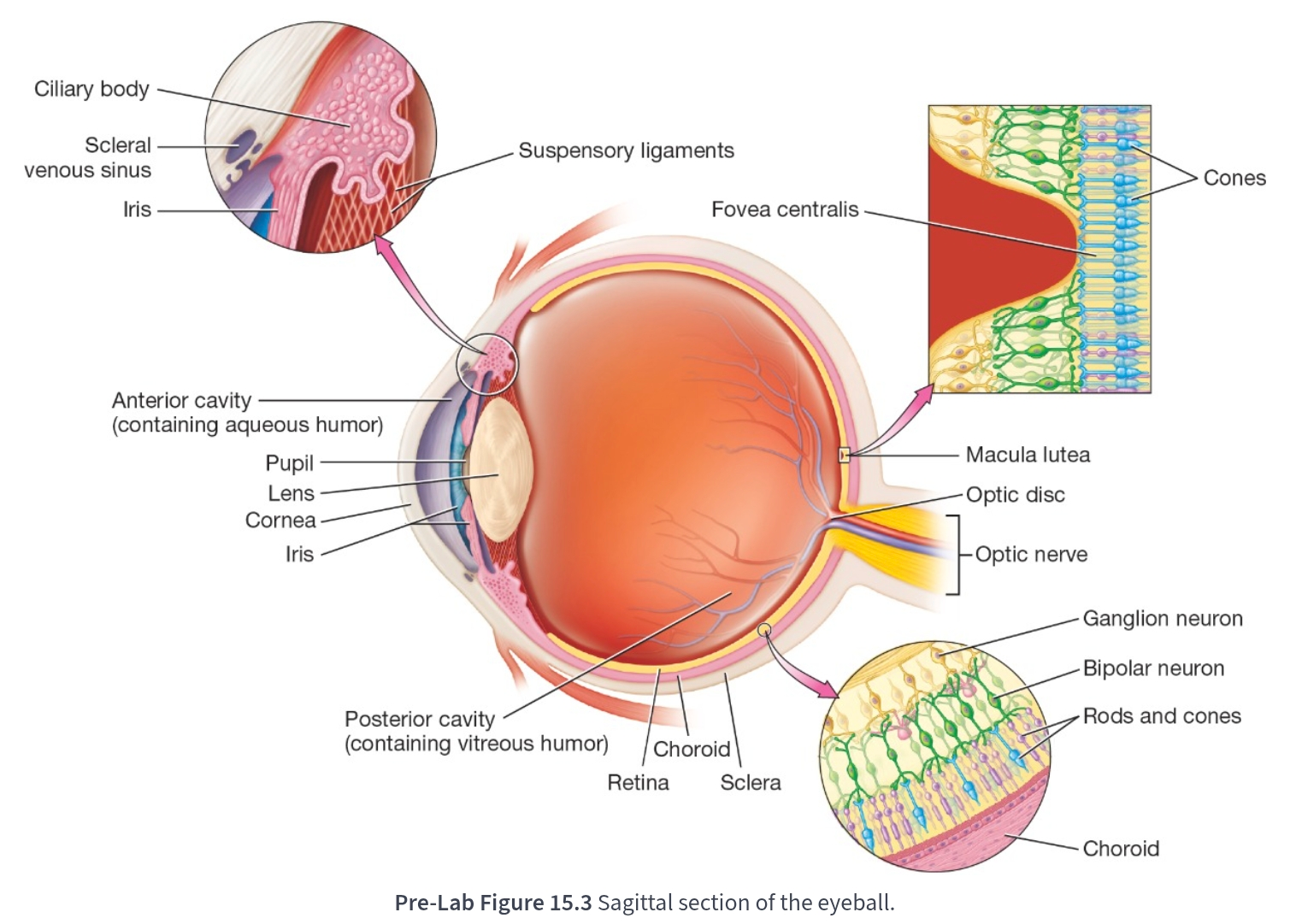
Area where cones are found in highest numbers:
Macula lutea

Also known as the blind spot because it lacks of photoreceptors means that this region can produce no images:
Optic disc
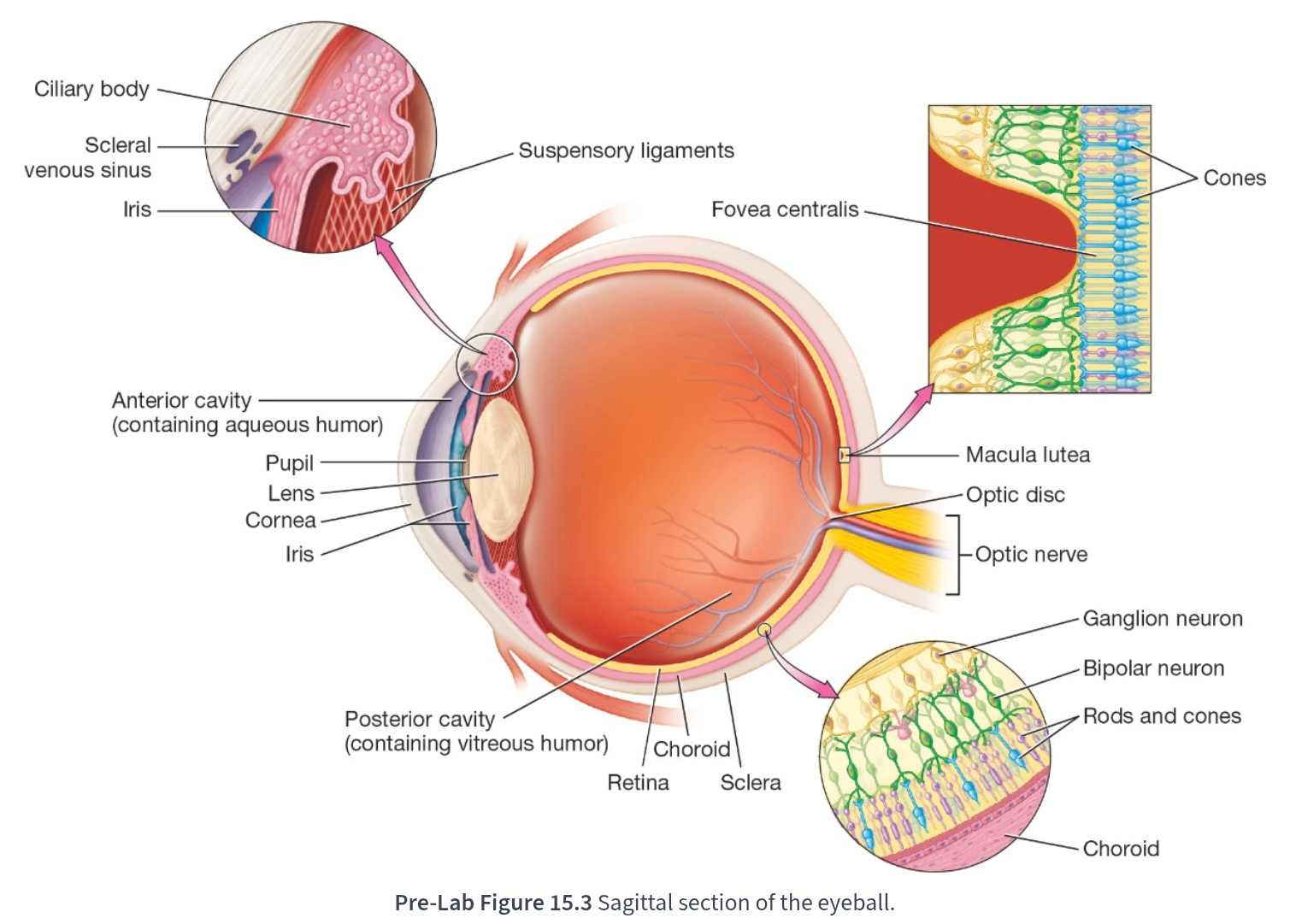
The sensory tunic consists of the:
Retina and the optic nerve

A thin, delicate structure that contains three layers of neurons:
Retina

Three layers of neurons in the retina:
Photoreceptors(rods and cones), bipolar neurons, ganglion cells

Axons of the ganglion cells converge to form the:
Optic nerve
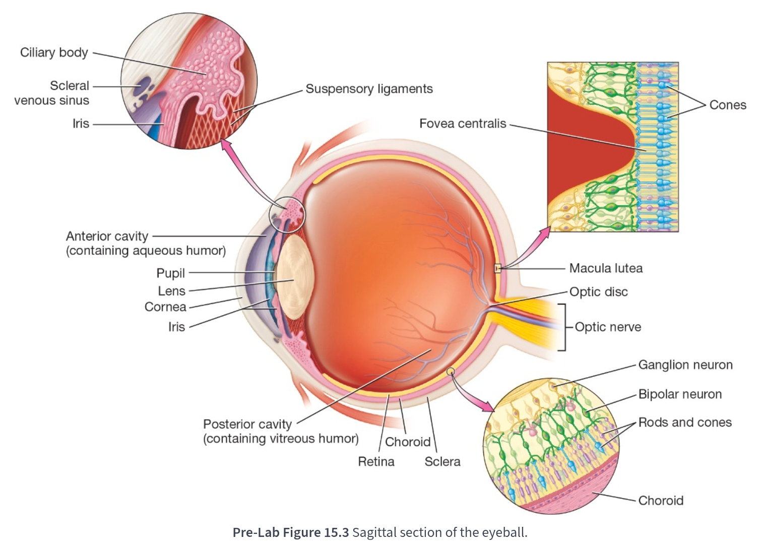
the black circular opening located in the center of the iris:
Pupil

Contains specialized photoreceptor cells—rods and cones—that convert light into electrical signals through a process called sensory transduction:
Retina

White part of the eyeball that makes up the posterior five-sixths of the fibrous tunic:
Sclera

The sclera is white because of:
Numerous collagen fibers that contribute to its thickness and toughness

Structure that drains the aqueous humor:
Scleral venous sinus

Posterior to the lens and contains a thicker fluid called vitreous humor:
Posterior cavity

Present at birth and remains relatively unchanged throughout life:
Vitreous humor
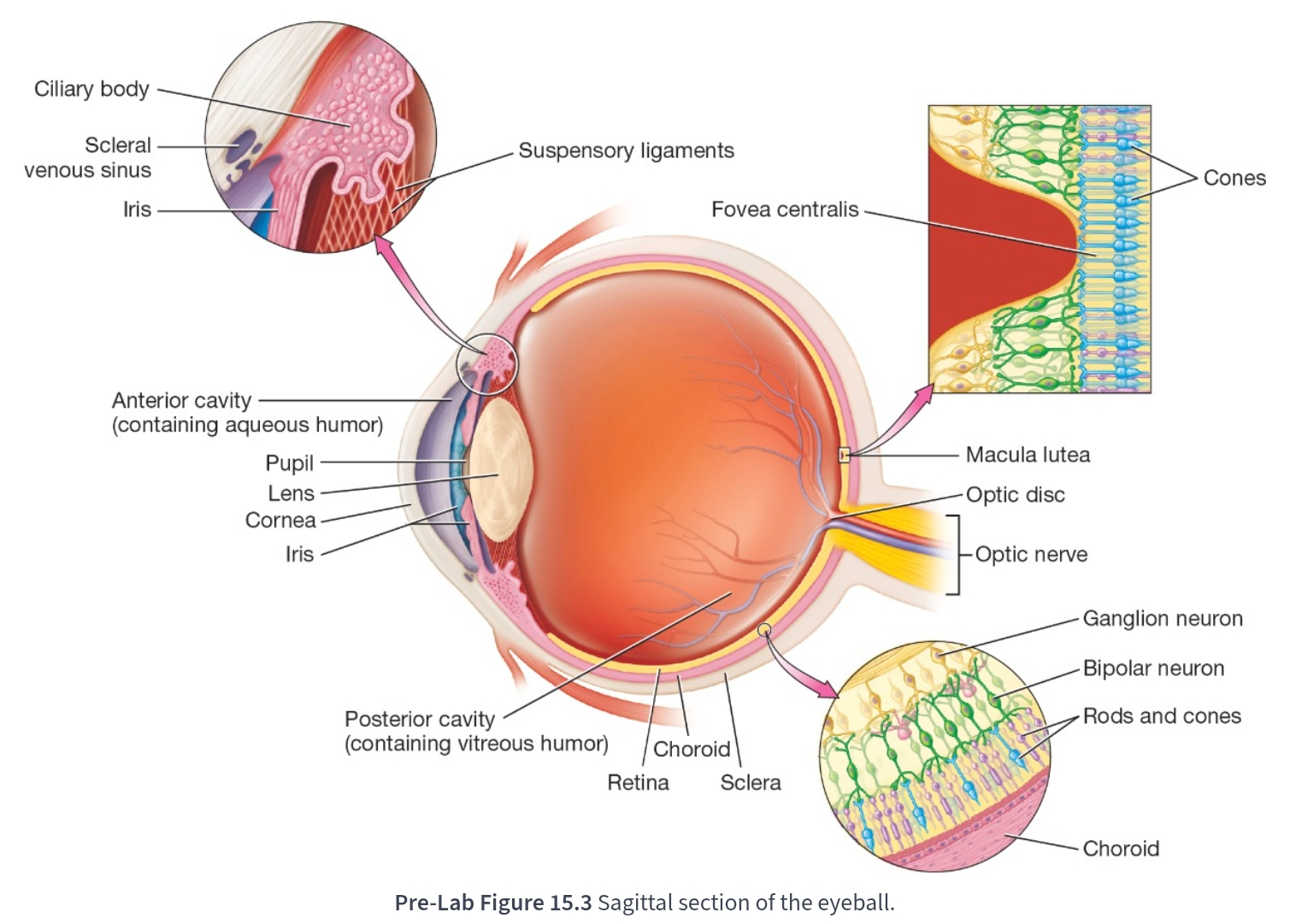
Both fluids help to refract light coming into the eye:
Aqueous humor and vitreous humor

Internal surfaces of the palpebrae are lined with a thin mucous membrane called the:
Palpebral conjuctiva
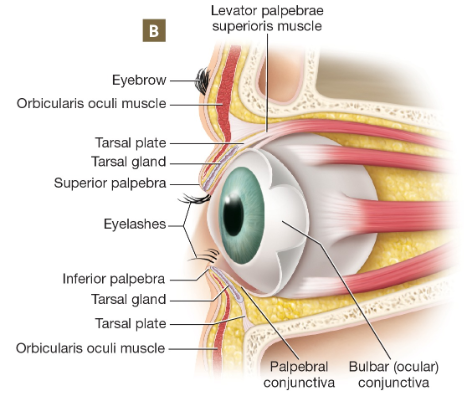
Lines much of the eyeball’s superficial surface:
Bulbar conjuctiva

Move the eyeball:
Extrinsic eye muscles

The six extrinsic eye muscles:
Lateral rectus, medial rectus, superior rectus, inferior rectus, superior oblique, inferior oblique… muscle
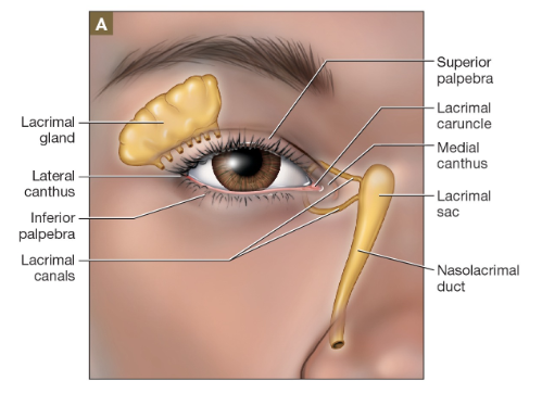
One of the most prominent external structures of the eye is the:
Lacrimal apparatus

Produces and drains tears:
Lacrimal apparatus

Tears drain into:
Small lacrimal canals near the medial canthus

Tears drain into the lacrimal canals, then drain into the:
Lacrimal sac

Found in a depression in the lacrimal bone:
Lacrimal sac

Produces the aqueous component of tears called lacrimal fluid:
Lacrimal gland
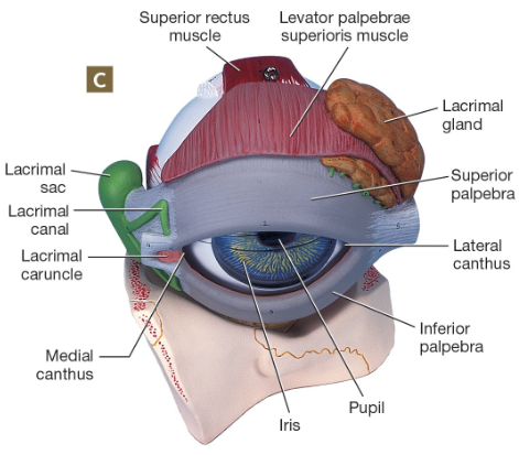
The palpebrae meet medially and laterally at the:
Medial and lateral canthi
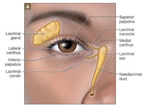
Tears travel through the ___, and finally empty into the nasal cavity, cause runny nose when you cry:
Nasolacrimal duct

Anteriorly, the eye is covered by the eyelids, or:
Palpebrae

Composed of dense connective tissue, which is associated with a sebaceous gland known as a tarsal gland:
Tarsal plate

Produce the lipid component of tears, which coats the aqueous portion and facilitates tear drainage into the lacrimal canals:
Tarsal gland
Tiny, central depression in the retina of the eye that provides our sharpest, most detailed central vision:
Fovea centralis