3- Neurology
 Call Kai
 Learn
Learn Practice Test
 Spaced Repetition
 Match
Match Flashcards
 Knowt Play
1/91

There's no tags or description
Looks like no tags are added yet.
Name | Mastery | Learn | Test | Matching | Spaced | Call with Kai | Chat |
|---|
No analytics yet
Send a link to your students to track their progress
92 Terms
approach to neuro pt’s…
.
1: list the 4 questions we want to answer when presented w/ a neuro pt
.
2: list the basic steps we go thru for these cases
.
3: why is signalment an important consideration?
1:
is the prob neurologic? (or orthopedic)?
where’s the lesion
what caused the prob
what can be done about it
.
2: hx & signalment → PE → neuro exam → min database (CBC, biochem, UA) → neurodiagnostic tests (common choices are: CSF analysis, rads, MRI)
.
3: some dz’s are more common in specific species, breeds, & ages (e.g. IVDD in chondrodystrophics, idiopathic epilepsy in dogs 1-6 y/o)
approach to neuro pt’s…
.
1: list some important pieces of info to gather from the pt’s hx
.
2: DAMNIT-V is a useful tool to use when picking your ddx.
what does it stand for
.
3: which DAMNIT-V categories should you consider if a pt presents w/…
acute problem
progressively getting worse over time
has been the same since birth
got worse then better
1: have O DESCRIBE (not interpret) what happened, part of body?, duration of signs?, prior tx?
note: can be common for O to falsely think their pet had a seizure so description is key
.
2: degenerative, anomalous, metabolic, neoplastic/ nutritional, inflam/infec, trauma/toxin, vascular
e.g. dog is acutely down → could be vascular if BV is blocked, can’t be anomalous bc its acute
.
3:
vascular or trauma
degen or neoplasia
anomaly
inflam or metabolic

1: whats the ultimate goal of a neuro exam
.
2: list the 6 components of the neuro exam
.
3: which of these ^ can we do in EVERY pt (no matter species, $ constraints, etc.)
1: localize the lesion to a specific part of the NS
.
2:
assess mental status (consciousness + behavior)
gait & posture
postural rxns
CN exam
spinal reflexes
pain sensation (spinal palpation, ± nociception)
note: CNS doesn’t have pain receptors, meninges do, so SC is NEVER a source of pain
.
3: mental status + gait/posture
neuro exam: assessing mental status…
.
we assess a pt’s level of consciousness & behavior.
1: what NS struc do each of these evaluate the fn of
.
2: list the ways we can describe consciousness
.
3: how do we assess behavior
1: cons = reticular formation (inside the brainstem; activates cortex thur visual & auditory stimuli); behavior = forebrain (AKA thalamocortical)
e.g. if pt falls aleep within 10 sec of being in exam room → indicates brainstem prob
.
2: normal, delirium, somnolence (depression), stupor, coma
.
3: combo of hx from O & observing the pt in exam room

neuro exam: assessing gait & posture…
.
1: define the following types of abnorm posture…
lordosis
kyphosis
scoliosis
.
2: whats the diff between a head tilt & head turn & what type of dz do they each indicate
1:
back curves inward
arched back (indicates pain; most common)
spine deviates laterally to left or rt
.
2:
tilt = draw line at eye level → head tilt if this isn’t horizontal → indicates vestibular lesion (in brainstem or ear) (tip: focus on the eyes NOT the ears! or you can be tricked)
turn = pt physically turns head → indicates forebrain lesion
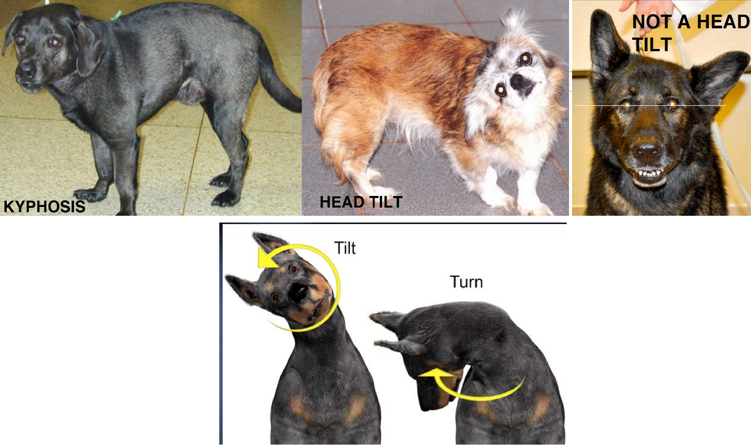
neuro exam: assessing gait & posture…
.
1: list the anatomical structures the gait exam assesses the fn of
.
2: how do we do a gait exam?
.
3: list the 3 questions we want to answer from the gait exam
.
4: define the following neuro terms…
tetrap- (e.g. tetraplegia)
parap-
hemip-
monop-
1: forebrain, CBM (cerebellum), brainstem, SC (spinal cord), LMN → EVERYTHING!
.
2: walk pt in straight line away & towards you, watch all 4 limbs, posture, & coordination
.
3:
is it norm or abnorm?
if abnorm, which limbs (e.g. ipsilateral limbs)?
what’s the prob: ataxia (neuro specific), paresis (± neuro specific), paralysis (neuro specific; is pt ambulatory?), or lameness (usually ortho prob)
e.g. of how to describe a gait: “non-ambulatory paraplegia”
.
4:
all 4 limbs
pelvic limbs only
one sided (e.g. both limbs on left side of body)
1 limb only
neuro exam: assessing gait & posture…
.
define the following neuro terms & list one clinical sign that it can cause…
paresis
paralysis (-plegia)
ataxia
weakness (indicates partial loss of motor fn) → falling while walking
complete loss of motor fn → dragging pelvic limbs when walking
incoordination → swaying side to side
neuro exam…
.
there’s 3 types of ataxia: vestibular, cerebellar, & proprioceptive.
1: list the most common clin signs assoc w/ each type
.
2: walk thru the process of determining which type a pt has
1:
vestib: head tilt, loses balance when looking up, ± circling
cereb: dysmetria/hypermetria (AKA high stepping), tremors
prop (most common): paresis
.
2:
is pt’s head abnorm?
no = proprioceptive ataxia
yes = …
does pt have head tremors?
yes = cerebellar ataxia
no = …
pt has head tilt + somnolence + prop deficits = central vestibular ataxia
pt has head tilt + norm postural rxns & mental status = peripheral vestibular ataxia
.
note: can have >1 but rare; can’t say pt has ataxia if have paralysis bc must be able to walk to appreciate it

give the common name of each cranial nerve and its basic function in the body
.
which CN do we NOT routinely assess during a neuro exam
I: olfactory → smell (sensory)
II: optic → vision (sensory)
III: oculomotor → eye muscle mvmt (motor), parasymp to pupil
IV: trochlear → eye muscle mvmt
V: trigeminal → sensory to face/teeth/eye (oph & maxillary branches), motor to mastication m. (mandib branch)
VI: abducens → eye muscle mvmt
VII: facial → motor to facial exp, eye, ear, lip, nostril mm.; parasymp to lacrimal gland
VIII: vestibulocochlear → sensory for auditory & balance
IX: glossopharyngeal → sensory & motor to tongue
X: vagus → autonomic & homeostasis function
XI: accessory → motor to neck & shoulder mm.
XII: hypoglossal → motor to tongue
.
CN I
neuro exam: CN exam…
.
1:
how do you test the menace response & what’s the normal response
what NS structures &/or CN’s does this test evaluate the function of
.
2:
how do you test pupil symmetry & PLRs & what’s the normal response
what NS structures &/or CN’s does this test evaluate the function of
1:
cover one eye → move closed fist toward P’s medial and lateral sides of the eye → P blinks & backs away
CN II/2 (for vision), thalamocortex, CBM, CN VII/7 (moving head/eye) → signal travels thru all of these structures
note: pt won’t have menace until ~4 m/o
.
2:
symmetry = shine light at face & pupils should be same size; PLR = shine light at 1 eye & it constricts (direct PLR), then check other eye & it should be constricted too (consensual PLR)
CN II/2 & III/3

neuro exam: CN exam…
.
1: define nystagmus
.
2: there’s 3 ways to classify nystagmus: physiologic, pathologic, & direction.
what NS structures &/or CN’s must be intact if a pt has physiologic nystagmus (AKA oculocephalic reflex) & how do we determine this is intact
.
3: how can the direction of nystagmus be helpful info
1: rapid, uncontrollable, & repetitive eye mvmt’s
.
2: CN III, IV, VI, VIII (3, 4, 6 move the eye; 8 picks up stimulus & sends to brain); medial longitudinal fasciculus (MLF)
move head side to side → eyes move in opposite direction of head turn, staying fixed on a point in space
.
3: direction can be horizontal, vertical, or rotational & vert is a strong indicator of central vestibular dz
neuro exam: CN exam…
.
1:
how do you test the palpebral reflex & what’s the normal response
what NS structures &/or CN’s does this test evaluate the function of
.
2:
how do you test nasal sensation & what’s the normal response
what NS structures &/or CN’s does this test evaluate the function of
1:
tap sides of eyelid → pt blinks
oph & max branches of CN V/5 (sensory) & CN VII/7 (motor)
.
2:
gently touch both nostrils → pt reacts by pulling away
thalamocortex & maxillary branch of CN V/5 (sensory)
neuro exam: CN exam…
.
1:
how do you assess the fn of the mastication mm. (e.g. temporalis & masseter m)
what NS structures &/or CN’s does this test evaluate the function of
.
2:
how do you assess the fn of the facial expression mm.
what NS structures &/or CN’s does this test evaluate the function of
.
3: how do you determine that CN VIII/8, IX/9, & X/10 are intact
1:
look & palpate for atrophy → unilateral indicates trigem tumor or myositis; bilateral indicates MMM (masticatory m. myositis), corticosteroids, or cachexia
CN V/5 (motor)
.
2:
look for facial symmetry → e.g. one side of lip is droopy
CN VII/7 (motor)
.
3: pt doesn’t have head tilt, ataxia, or dysphagia/dysphonia

1: list the 4 signs that, in combination, indicate a pt has Horner’s syndrome
.
2: this dz occurs from a dysfunction in the tectotegmentospinal tract
walk thru the flow of this pathway/tract
1:
miosis (excessive pupil constriction)
ptosis (drooping upper eyelid)
enophtalmus (eyeball sunken in)
protrusion of 3rd eyelid
.
2: hypothalamus → brainstem → SC → exits CNS at T1-3 as sympathetic neuron → travels back up to ear to reach target struc in orbit & face → causes pupil dilation, eyelid elevation, normal globe position

neuro exam: postural reactions (PRs)…
.
1: these tests don’t help us w/ lesion localization, instead they simply tell us if the pt’s problem is neuro in origin.
why?
.
2: why isnt this a replacement for the gait exam?
.
3: describe how to perform the following PR tests & describe a normal result…
conscious proprioception
hopping
tactile positioning
1: bc PRs stimulate the NS all the way from the toes → SC → brain stem → cortex → muslces
so it literally uses the entire CNS (except CBM) & PNS
so… any neuro defect = this will be absent
.
2: this assess the conscious proprioception pathway, gait exam assess uncons prop
.
3:
one hand supports the pt’s weight, the other gently flips over the paw → pt corrects it
pull shoulder/tibia laterally → pt hops
hold pt, cover their eyes, & walk toward flat surface → pt brings out leg in anticipation of standing on it (great test for cats bc its quick)

neuro exam: spinal reflexes…
.
1: whats the diff between a reflex & response/reaction
.
2: whats the purpose of testing spinal reflexes
.
3: what does it mean if spinal reflexes are…
norm or increased/spastic
decreased/flaccid or absent
1: both relay sensory info but…
reflex stays local (receptor → SC → muscle contraction)
rxn includes the cortex in its pathway (e.g. menace, postural, proprioception)
.
2: evaluates the fn of specific nerves & segments of the SC
.
3:
lesion is cranial to the SC segment being tested → AKA lesion is in the UMN
lesion is in the sensory or motor part of the reflex arc being tested → AKA lesion is in the LMN
.
note: many reflexes can be tested but only a few are reliable

neuro exam: spinal reflexes…
.
how do perform the following spinal reflexes & describe a normal result
patellar reflex
extensor tone of the limb
flexor tone of the limb
perineal reflex
cutaneous trunci (AKA panniculus) reflex
.
describe how to use an absent cutaneous trunci reflex for lesion localization
semi-flex the limb & gently hit patellar tendon w/ hammer → pt kicks its leg
apply pressure in palmar surf of paw → pt extends its leg
gently pinch pt’s toes → pt pulls leg toward body, fexing all joints
stroke perineal region → anal sphincter contracts
pinch just lateral to the spine working cd to cranial → bilateral muscle contraction
note: extensor tone & pat reflex are most reliable
.
the point where you transition from absent (abnorm) to present (normal) reflex → count 2 vertebral bodies cranially → this is where the lesion is in the SC → indicates theres a disruption somewhere along sensory &/or motor pathway connecting skin, SC, muscle, lateral thoracic n.
this is bc the sensory nerve you’re stimulating enters the SC cd to the vert body that corresponds to that SC segment (AKA verteb location of lesion & SC segment don’t line up exactly)

neuro exam: spinal reflexes…
.
name the nerve(s) & segments of the SC that are normal if a pt has a normal ___:
flexor tone in pelvic limb (lateral digit specifically)
extensor tone in pelvic limb
patellar reflex
sciatic n. | L6-S2 (note: in chronic pt w/ sciatic dysfunction, may see super flexed hocks bc of weak gastroc m.)
femoral n. | L4-L6
same ^
.
trick:
“Femoral” “Four” “Front” (bc it makes leg move toward front/cranial of dog
“S”econd, “S”ciatic, “S”ix

neuro exam: spinal reflexes…
.
name the nerve(s) & segments of the SC that are normal if a pt has a normal ___:
extensor tone in thoracic limb
flexor tone in thoracic limb
perineal reflex
cutaneous trunci reflex
radial n. | C7-T1
all nerves in brachial plexus | C6-T2
pudendal n. | S1-S3
lateral thoracic n. | C8-T1

neuro exam: pain sensation (AKA sensory evaluation)
.
1: whats the purpose of spinal palpation
.
we can assess for pain in 2 ways: spinal palpation & nociception
2: how do we perform a spinal palpation & describe a normal result
.
3: how do we do cervical palpation
.
4: how do we assess nociception & describe a normal result
which pt’s do we assess this in?
1: differentiates between painful & nonpainful dz
.
2: start at T1/2 and move caudal → use one hand to support underside of body, use the other to push fingers just lateral to spinous processes → no pain response
tip: don’t start cd to cran bc many dogs have lower back probs; skip abd area bc can’t differentiate between neuro & abd problems
.
3: push fingers in between transverse processes on lateral aspects of neck (cervical spine anatomy is diff than thoracolumbar)
.
4: pinch digits w/ hemostats → behavioral/conscious response like turning head, vocalization, etc. indicates they still have some type of sensation (VERY poor prognosis if absent)
paralyzed pt’s ONLY bc this assess for deep pain & regular pain is intact in pt’s that can walk
.
note: unlike cutaneous trunci reflex, the location at which we palpate pain is the exact location of the lesion

lesion localization…
we localize lesions in a hierarchal fashion.
.
list the locations in the ___ we can localize a lesion to:
CNS
PNS
.
SC
.
brain
.
5: brainstem
brain or SC
nerves (neuropathy), NM junction (junctinopathy), muscle (myopathy)
.
(cran to cd) C1-5, C6-T2, T3-L3, L4-S3
note: T3-L3 is most common bc its a massive section
.
thalamocortex (includes forebrain + cerebrum + prosencephalon), brainstem, or CBM
.
5: midbrain (mesencephalon), pons, rostral medulla, cd medulla

lesion localization…
.
to localize lesions we must understand the diff between UMN & LMN.
1: describe the location & fn of UMN’s
.
2: list clinical signs suggesting a UMN lesion/dysfunction
.
3/4: answer the same for LMN’s
1:
L: brain & SC (stays in the CNS)
F: they’re like the “boss” that regulates LMNs (usually by inhibiting their activity)… can initiate voluntary mvmt, provide postural adjustment (prevents pt from falling to the side), & maintain muscle tone by facilitating flexor mm & inhibiting extensor mm
.
2: paresis/paralysis, norm to inc musc tone & reflexes, mild (disuse) musc atrophy
trick: LMN is like a car engine, it can fire on its own (w/o stim from UMN, via reflex arcs) & UMN is the driver & brakes. UMN let us voluntarily control mvmt & inhibit LMN activity; no UMN = engine runs but control is lost
.
3:
L: starts in SC grey matter → nerve root → spinal n. → peripheral n → NM junction → muscle
F: manifests voluntary & reflex motor activity
.
4: paresis/paralysis, dec to absent musc tone & reflexes, fast/severe (neurogenic) musc atrophy
.
trick: when determining UMN vs LMN, use “RAT” → reflexes, atrophy, tone


lesion localization in the SC…
.
which region(s) of the SC has a lesion if a pt has ____
abnormalities in all 4 limbs
abnormalities in pelvic limbs only
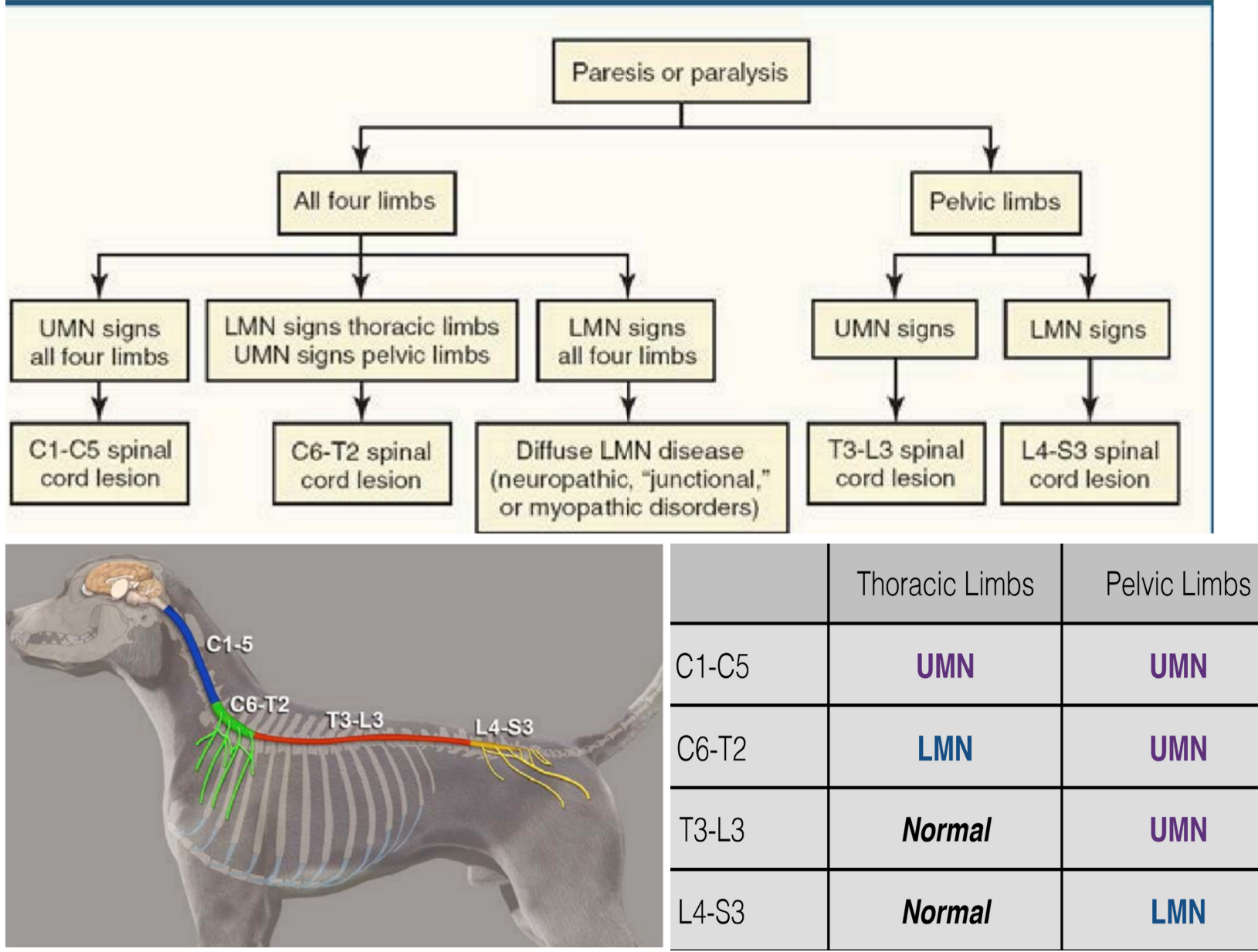
paresis/paralysis + LMN signs in pelvic limbs
paresis/paralysis in all limbs + LMN signs in thoracic limbs + UMN signs in pelvic limbs
C1-5 or C6-T2
T3-L3 or L4-S3
L4-S3: bc this is location of lumbosacral plexus (CB’s innervating pelvic limb)
C6-T2: bc this is location of brachial plexus
.
tip: 1st try to ID affected limbs w/ gait exam, then confirm w/ postural rxns


lesion localization in the SC…
.
which region(s) of the SC has a lesion if a pt has ____
paresis/paralysis + UMN signs in pelvic limbs (most common presentation!)
paresis/paralysis + UMN signs in all limbs
paresis/paralysis + LMN signs in all limbs
extensor tone inc + flexor reflex dec in thoracic limbs, + pelvic limb ataxia, + UMN signs in pelvic limbs
T3-L3: bc there’s no LMN signs (= not L4) & thoracic limbs norm (= not C1 or C6)
C1-5: can’t be C6 bc no LMN signs
lesion ISN’T in SC → its in nerve roots, peripheral n., NM junction, or muscle (WON’T cause proprioceptive ataxia bc this affects white, not grey matter of SC)
C6-8: this is a more specific scenario bc this region innervates muculocutaneous n. which normally flexes thor limb, so radial n. is normal (VERY common lesion localization in CSM pt’s)
.
note: 3 is a common situation in pt’s w/ wobbler syndrome (cervical spondylomyelopathy)
tip: 1st try to ID affected limbs w/ gait exam, then confirm w/ postural rxns
lesion localization in the SC…
.
1: at which vertebral body does the SC end in the dog? how does this affect the organization of the SC in the vertebral column, specifically in the lumbar region?
.
2: describe the presentation of a dog w/ a lesion in vertebral body L6-S1 (AKA lumbosacral dz)
1: L5 (SC ends before vert column) → sections L4-S3 of the SC live inside vertebrae L4-5
trick: “5” looks like “S” & S1/2/3 live in L5 which is the end of the SC
.
2: pain but NO paraplegia & they recover much easier bc there’s no CNS located here, just PNS

exceptions to lesion localization in the SC…
.
1: describe the clinical presentation of “spinal shock” & name the SC section that the lesion will be in
.
2: answer the same for schiff sherrington syndrome
1: pt has acute paraplegia/paresis w/ dec extensor tone & flexor reflexes, but patellar reflex is norm → only lasts a few hours to days then pt is normal
lesion between T3-L3, specifically lesion is in femoral n., but weird bc pat reflex is norm & extensor tone isn’t
e.g. hit by car
.
2: acute paraplegic dog that when placed in lateral recumbency, will lay w/ super stiff neck & thoracic limbs (see pic) & flaccid pelvic limbs → bc propriospinal tract is damaged & this NORMALLY inhibits extensor tone in thor limbs + neck
lesion between T2-L5

lesion localization in the brainstem…
.
1: list 3 major clinical signs indicative of a lesion in the brainstem
.
2: name the anatomic region that has a lesion if a pt has a CN ___ deficit & list common clinical signs assoc w/ this
III/3 & IV/4
V/5
IX/9, X/10, XI/11, XII/12
1: changes in mental status (usually somnolence), CN 3-12 deficits (bc their nuclei are in stem), tetraparesis + ataxia (bc their pathway runs thru stem)
note: paresis/ataxia caused by SC lesions wouldn’t have mentation changes + CN deficits!
.
2:
mesencephalon → ocular paralysis, mydriasis (persistent pupil dilation)
pons &/or rostral medulla → atrophy of mastication mm.
cd medulla → dysphonia, dysphagia, sterterous breathing, tongue paralysis
.
note: rostral medulla is very common & cd medulla is least common region of brainstem to have a lesion
vestibular disease…
.
1: list typical clinical signs
.
2: name the anatomic region that has a lesion if a pt has ____ vestibular dz & list UNIQUE clinical signs assoc w/ it
central
peripheral
1: head tilt; ± vestibular ataxia, strabismus, nystagmus, & (narrow) circling
.
2:
rostral medulla of brainstem → proprioceptive deficit, somnolence (mentation change), vertical nystagmus, ± CN VI/6, VII/7, VIII/8 deficits (bc this is where their nuclei live)
ear (bc this is where the receptors live) → no prop or mentation changes
lesion localization in the cerebellum…
.
1: list typical neuro signs (AKA what are “CBM signs”)
.
2: list the 2 signs that make this unique from lesions in other locations of the NS
1: head &/or whole body tremors, head & limb ataxia (wide based stance/gait, truncal sway, dys/hypermetria), NO menace response ipsilaterally (bc it goes thru CBM)
± vestib signs (if lesion is in flocciulonodular lobe of CBM)
.
2: NO prop deficits or weakness (won’t be falling down)!!!
lesion localization in the thalamocortex…
.
1: list typical clinical signs
.
2: name 4 neuro tests we do to assess thalamocortical fn & describe the result in abnorm pt’s
.
3: a pt presents w/ circling to the left + absent menace, nasal sensation, & prop on the right side.
where is his lesion?
1: abnorm behavior (according to O & observation) & compulsiveness, (wide) circling (usually towards side of lesion), normal gait (don’t need corticospinal tract to walk), SEIZURES (focal or generalized), central blindness (running into walls)
.
2: assess mental status, menace response, nasal sensation, postural rxns (prop & hopping)
last 3 will have CONTRALATERAL deficits (contralateral to the lesion bc pathway crosses to opposite cortex)
.
3: left thalamocortical region of brain
neuropathology…
.
1: there’s many mechanisms of edema in the CNS. describe each of the following….
vasogenic edema
cytotoxic edema
.
2: describe the gross appearance of edema in the brain
1:
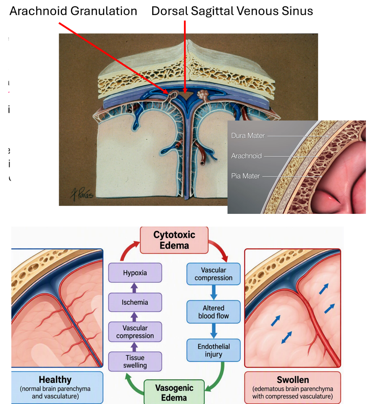
typical type of edema where smth triggers fluid to leak out of BV’s (AKA BBB is broken)
edema INSIDE of cells (AKA cell swells), esp astrocytes bc they’re super sensitive to hypoxia → Na/K ATP pump fails → Na (& water) accum inside the cell
.
2: very puffed up, sulci & gyric aren’t cleanly separated
.
reminder: normal fn of astrocytes is to serve as the extracellular space in CNS & regulate water balance via ion pumps on their surface

neuropathology…
the edema cycle can occur anywhere in body but its esp important in the CNS.
.
1: describe what the edema cycle is
.
2: walk thru the entire cycle, make sure to emphasize the most ciritcal initiating steps
1: a positive feedback loop that occurs when one form of edema initiates another, this loop drives itself & results in inc intracranial pressure & possibly death
.
2: smth causes physical injury to endothelial cells → fluid leaves BV’s which initiates….
vasogenic edema → tissue swells → BV’s are compressed → ischemia → hypoxia, which damages astrocytes & initiates…
cytotoxic edema → even more BV compression → more altered blood flow → more endo injury → repeat!
.
red = most critical steps

neuropathology…
.
1: describe how fluid leaking out of CNS BV’s is reabsorbed in NORMAL animals
.
2: use this ^ knowledge to explain why edema can QUICKLY induce ischemia, specifically in the CNS
1: fluid that leaks out combines w/ CSF & gets reabsorbed via 2 ways…
glymphatics system: glial cells (mainly astrocytes) push fluid thru lymphatics when you sleep
natural flow of CSF: fluid leaves ventricles & drains into arachnoid granulations
.
2: this system has a volume capacity (remember: CSF must be resorbed at constant rate bc its made at constant rate) so when capacity is exceeded → fluid stays in tissue → edema… + the CNS naturally has little extra space so BV’s are easily compressed → ischemia
neuropathology…
.
1: use normal physiology to describe where you expect to see the worst damage when the brain faces ischemia
.
2: describe how severe CNS edema can result in CBM & cerebrum herniation
1: remember… CNS cells are VERY energy hungry + BV’s start at surf & dive into cerebral GM… so the deeper in GM you go, the worse ischemia is…
this results in a gradient of damage: deepest tissue will have pannecrosis (everything is dead), superficial will have selective necrosis (cells w/ higher energy needs, like neurons die first)
note: WM isn’t affected as much bc its just a bunch of axons, so doesn’t need as much energy
.
2: edema → intracranial pressure will inc & push the brain backwards/caudal bc there’s no more space → CBM can be pushed out thru foramen magnum → herniation (+ can result in seizures, head pressing, etc.)

neuropathology…
.
walk thru the pathophysiology of rodenticide (AKA bromethalin) toxicity, include the end result & what clues we can use for dx
bromethalin uncouples/blocks oxidative phosphorylation (this is how ATP is made) → abaility to make ATP dec → Na/K ATP pump on the surface of astropcytes fail → Na (& water) accum inside astro → cytoxic edema (+ edema cycle!) → intracranial pressure inc (bc astro swell & there’s no room in CNS for them to expand)…
ani dies acutely or…
edema spreads into myelin sheaths in WM & we’ll see normal axon surrounded by swollen sheath → can see on histopath to dx cause of death

neuropathology…
.
1: walk thru the pathophysiology of epsilon toxin (made by c. perfringens)
.
2: describe the gross lesion we’ll see if we catch this dz EARLY in its progression
1: toxin which enters blood → crosses BBB → damages CNS endo cells → vasogenic edema → edema cycle continues until the brain is complete jelly/necrotic
note: this causes edema in MANY organ systems, including CNS
.
2: BV’s leak in a specific pattern causing focal symmetrical encephlomalacia ± hemorrhage (its symmetrical bc this location is extra susceptible/sensitive to hypoxia)
reminder… malacia: softening of CNS tissue caused by liquefactive pannecrosis & we can see this grossly

neuropathology…
.
1: whats the diff between encephalomalacia & polioencephalomalacia
.
2: what does polioencephalomalacia look like grossly
.
3: name your 4 ddx if you see polioencephalomalacia
1: enceph is softening/necrosis of brain tissue (“encephalo” = brain, “malacia” = softening), can include GM, WM, or both
polio is SPECIFICALLY softening of GM in the brain (“polio” = GM)
.
2: GM is pale & swollen + there’s a loss of distinction between the GM & WM
.
3: all of the following cause this lesion…
rapid dec in plamsa osmolality (e.g. pt is super dehydrated & you fluid overload them)
bac toxin exposure
thiamine deficiency
lead toixicity

neuropathology…
.
walk thru the pathophysiology of thiamine (AKA vit B1) deficiency in ruminants
ruminants normally get most of their thiamine from rumen microbes, so…
poor diet → dec in rumen pH → rumen microbes can’t thrive & thiamine prod dec → vasogenic edema → edema cycle → polioencephalomalacia
note: normal fn of thiamine is to support neuron function & endo cell health

neuropathology…
.
1: explain why hemorrhage in the CNS is more concerning than the simple fact that its taking up space & compressing tissue macroscopically
.
2: does a concussion (head trauma) always lead to hemorrhage?
.
3: infarcts can occur foacally, MF, or widespread.
whats the diff between grossly seeing a red vs pale infarct
.
4: list the 4 causes of infarction in the CNS
1: seeing macro compression (hemorrhage), indicates there’s microscopic compression too which indicates the edema cycle (ischemia) is going on!
the combo of physical compression + ischemia can lead to infarction!
.
2: no!
.
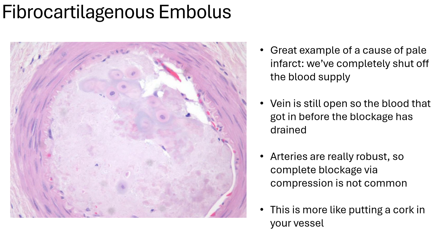
3: red indicates the infarct started as hemorrhage or congestion of blood (usually bc of venous blockage)
pale indicates infarct was caused by arterial blockage = no blood could enter the tissue (e.g. fibrocartilagenous emboli)
.
4: arteriospasm, vasculitis, embolism, vascular compression
neuropathology…
.
1: describe the signals that maintain BV dilation/patency in a normal animal
.
2: name the adaptive response BV’s have when there’s hemorrhage in the subarachnoice space of the CNS
.
3: describe how this can progress to delayed-onset arteriospasm & what the result is
.
4: why is hemorrhage in the SC (caused by spinal trauma) esp problematic & what does this lesion look like grossly
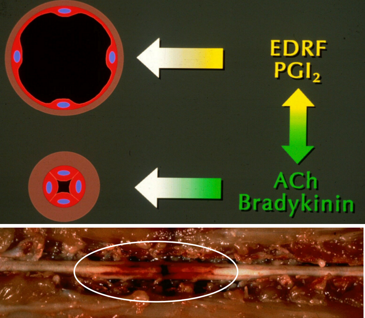
1: this is mediated by factors released by endo cells like endo relaxation factor (EDRF) & prostacyclin → AKA this is an active process, BV’s aren’t just naturally open, they need signals to keep them from collapsing
.
2: constrict to stop the bleeding
.
3: during hemorrhage, oxyhemoglobin & acute phase proteins like bradykinin are secreted → they trigger free radicals to accum → radicals damage endo cells → less EDRF is made → arteries constric…
if this signaling is prolonged (constriction continues several days after initial injury), constriction becomes EXCESSIVE (AKA delayed onset arteriospasm) → ischemia
.
4: the myelomalacia (pannecrosis of SC) can easily ascend & descend thru the SC bc fluid easily flows thru WM (compared to GM) & in the SC, the WM is adjacent to subarachnoid space (where hemorrhage comes from)… as this progresses, pt loses neuro fn until eventually it can’t breath & dies
grossly: see pic, the circle shows necrosis & hemorrhage extending cran & cd
reminder: SC anatomy is reverse of brain, WM on outside, GM on inside
.
key point: keep an eye on these pt’s!! they may initially look fine, but can progress to this a few days later!
neuropathology…
.
1: walk thru the pathophysiology of histophilus somni (bacteria) infection
.
2: describe how this can cause more than just infarction in the CNS
.
3: what type of gross lesions does this cause & explain why the brain is the best place to look for lesions
1: this bac grows in the walls of BV’s which makes it SUPER inflammatory → vasculitis → enodthelial damage → edema cycle + hemorrhage → infarction in the CNS
.
2: eventually a clump of bac + inflam cells can break off BV wall, form an emboli, travel thru blood, & lodge somewhere else to cause infarction in heart, lungs, etc. (AKA thrombotic meningoencephalitis)
.
3: MF hemorrhagic infarcts (see pic) → look in brain GM, esp near the meninges, bc BV’s dive into GM from the meninges at a VERY high density & BV is where this dz originates
.
note: one cause of infarction in the CNS is vasculitis

neuropathology…
.
your pt has an infarct in the SC GM w/o any WM involvement. name the artery you’re most suspicious of being the problem and explain your answer
ventral spinal artery bc SC has 2 sources of blood: intercostal & ventral spinal a.
GM is ONLY supplied by ventral spinal a.
WM has a lot of redundancy in its blood flow
so lesion in ventral spinal a. = GM of SC is screwed but WM should be okay

neuropathology…
one cause of infarction in the CNS is vascular compression & the most common cause of compressive SC injury is IVDD.
.
walk thru the pathophysiology of IVDD leading to infarction
the IVD degenerates over time in all dogs but this occurs at an accelerated rate in chondrodystrophic dogs (dachsunds, etc.)
as the disc degenerates, the NP pushes dorsally on the AF (bc the AF is thin dorsally)
eventually, the AF becomes so weak it ruptures → NP escapes → disc herniation → BV’s supplying SC can become compressed (reminder: SC has limited vascular supply) → ischemia → if prolonged, infarction
also, if the rupture is traumatic/rapid, the leptomeningeal BV’s can tear → hemorrhage in subarachnoid space → ascenidng-descending myelomalacia
.
note: IVDD doesn’t ALWAYS lead to infarction, this specifically occurs when BV’s become obstructed

review of intervertebral disc (IVD) anatomy…
.
1: describe the relationship between the vertebral canal, SC, IVD, & intervertebral foramen
.
2: what parts of the vert column don’t have IVD’s
.
3: describe the 2 struc that make up a IVD:
nucleus pulposus
annulus fibrosus
.
4: function of IVD?
1:
vert canal is the tube-like space inside the vert column (AKA the spine) that holds & protects the SC
SC is a struc found inside the canal
IVD’s sit in between the vert bodies that make up the vert canal
inter for is gaps in the canal where spinal nn. exit
.
2: they’re all along spine except C1-2 & sacral region
.
3:
inner ring made of gel-like material (this is the part that usually herniates in IVDD)
annulus fibrosus: outer ring surrounding ^ thats made of collagen
.
4: shock absorber & provide cushion to spine as it moves

IVDD (intervertebral disc disease; AKA disc herniation) is a very common dz.
.
1: list the possible outcomes of herniation (AKA what structures get compressed)
.
2: which IVD’s is this unlikely to occur
.
3: IVDD is classified into 4 types. list each of them
1: NP (the part that herniates) can just be a bit displaced, compress the SC, or exit the SC & compress nerve roots
.
2: T1-2 & T8-9 bc these discs have extra ligaments from ribs keeping them in place
.
3:
hansen type I (AKA type I disc extrusion or type I disc disease) → this is ACUTE dz where AF ruptures
acute noncompressive nucleus pulposus extrusion (ANNPE)
acute hydrated nucleus pulposus extrusion (HNPE)
hansen type II (AKA type II disc protrusion) → only CHRONIC form, AF progressively gets weaker
.
tip: “protrusion” = AF remains intact, “extrusion” = NP breaks thru AF, “contusion” = bruising of the SC that can occur during severe disc extrusion if NP violently hits SC

type I IVDD / IVDE (IVD extrusion)…
.
1: typical signalment (include breed & age)?
.
2: typical hx?
.
3: clinical signs?
1: mainly small breed, chondrodystrophic dogs (bc they have chondroid/cartilage degen that starts <1 y/o; esp dachsunds & pekingese), usually >2 y/o, avg age is 6 y/o
.
2: ACUTE or chronic w/ acute worsening back pain + sudden onset of paralysis/paresis
.
3: thoracolumbar (AKA back) pain, prop ataxia, prop deficits, paraplegia, ± intact nociception, ± non-ambulatory paresis, ± urinary incontinence
.
note: commonly occurs in discs between t11/l3 (around lumbosacral junction)

type I IVDD / IVDE (IVD extrusion)…
.
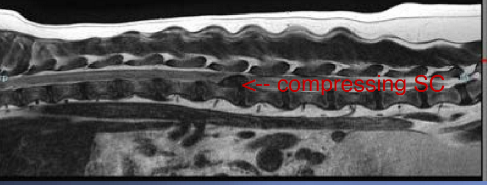
1: make & justify a diagnostic plan
.
2: describe the features in this rad that suggests IVDE
1: survey rads (use as screening test to make a presumptive dx) → this reqt sedation + analgesia for pain; will examine disc space, foramina, & articular processes
myelography for more severe pt’s (inject contrast into subrachnoice space)
but… definitive dx reqt CT or MRI bc can see disc extrusion
.
2: in a norm lateral view, intervertebral foramen looks like a horse head → if horse head is smaller or more opaque (like this pic) = abnorm & indicates disc extrusion
.
note: may see mineralized discs on rads but this doesn’t cause signs, its not a dx

acute non-compressive nucleus pulposus extrusion (ANNPE)…
.
1: typical hx?
.
2: pathogenesis & outcome?
1: hx of severe trauma like gunshot, car accident, intense exercise, etc.
.
2: small fragment of NP hits SC at a VERY high velocity → causes contusive SC injury & significant neuro deficits
this is non-compressive (unlike IVDE), instead its CONTUSIVE
hydrated nucleus pulposus extrusion (HNPE)…
.
describe what this is & how its sim/diff than IVDE
in IVDE, disc is usually dehydrated & mineralized bc its been degenerating & this is what compresses the SC
.
HNPE is acute like IVDE but… the disc is still hydrated so a fluid like material compresses the SC… the liquid can reorganize and disppear on its own
acute spinal cord injury (ACSI, AKA spinal trauma)…
.
1: the etiology can be external or internal.
list e.g. of each
.
2: typical clinical signs? include common primary & secondary lesions/problems
.
3: important considerations when performing the neuro exam?
.
4: make & justify a diagnostic plan
1: external = hit by car, falls, fights, gunshot; internal = IVDE (specifically talking about high kinetic injury that leads to SC trauma)
.
2: ACUTE onset of paraplegia; primary= contusion (more common) or compressive injury; secondary = biochemical changes, ischemia, electroylytes shifts, apoptosis, etc.
.
3: avoid postural rxns & palpation bc pt is painful + make sure to assess motor fn & nociception bc absence indicates a very poor px
.
4: combo of hx of trauma + fractures/luxations present on CT
can use rads as screening tool but CAN’T rule out ACSI if they’re norm bc minor fractures are often missed
.
notes:
-if dz is chronically proressing, must rule out ACSI!
-nociception is the most important px indicator in this dz
1: list the 3 conditions/complications that MUST be on our radar for pt’s presenting w/ acute paresis/palegic
.
.
.
2: describe how to recognize if a pt has progressive myelomalacia & why it is absolutely CRITICAL that you identify this
1: spinal shock, shiff-sherrington syndrome, progressive myelomalacia (necrosis of the SC)
.
2: check their cutaneous trunci reflex 2x/day for multiple days, if the cutoff moves cranially → indicates lesion is extending cranially & suggests prog myelomalacia! (moving cd indicates dz is improving)
MUST euthanize these pt’s bc if it progresses to phrenic n, pt will die a very painful death
.
note: prog myelomalacia is the only complication that will kill a paraplegic dog
fibrocartilaginous embolic myelopathy (FCEM; AKA spinal stroke)…
.
1: pathogenesis?
.
2: typical signalment (breed)?
.
3: typical hx?
.
4: clinical signs & neuro exam findings?
1: a fragment of fibrocartilagenous material (usually a piece of the IVD NP) enters the vasculature supplying the SC & causes an infarction (unknown how the cartilage enters BV in the first place)
AKA this is a non-traumatic, ischemic form of myelopathy that can affect ANY segment of the SC
.
2: large breed dog, schnauzer, or sheltie
.
3: acute onset of paraplegia, ~50% of cases have a hx of intense exercise (e.g. dog was running around dog park, suddenly yelps, & goes down)
.
4: NO spinal pain (bc there’s no nociceptors in SC parenchyma), VERY asymmetric neuro signs (e.g. only dragging one leg, asymmetric cutaneous trunci reflex), ± LMN signs
.
note: key way to differentiate this from IVDD & ASCI → both of these have pain on spinal palpation, FCEM doesn’t
fibrocartilaginous embolic myelopathy (FCEM; AKA spinal stroke)…
.
1: list 4 ddx, besides FCEM, for pt’s w/ acute asymmetric neuro deficits
.
2: make & justify a diagnostic plan
.
.
3: most ppl don’t have access to a MRI, so list 5 criteria that, if a pt meets, makes FCEM our #1 ddx
1: ANNPE, meningomyelitis, myelitis, IVDE (usually not this asymmetric)
note: both inflam dz’s present as neuro deficits (“myelitis”) w/o spinal pain BUT tend to worsen over time or wax & wane; best way to dx these is w/ CSF analysis
.
2: MRI for definitive dx (only modality where we can visualize SC); CBC/chem to rule out thrombocytopenia & infectious dz (since this is a vascular process); ± rads, CT, CSF to rule out other dz
.
3:
pt has an acute onset, non-progressive myelopathy
pt is a large/giant breed dog, schnauzer, or sheltie
pt has CLEAR asymmetric neuro signs (e.g. cutaneous trunci reflex)
no spinal pain
clinical improviement withi 3-7 days
list the top 3 ddx ddx for dogs presenting w/ acute paraplegia (AKA “pt was norm last night & now it can’t walk”)
acute IVDD: type I, ANNPE, or HNPE
acute SC injury/trauma (ACSI)
fibrocartilagenous embolic myelopathy (FCEM)
.
notes:
-first ddx for small dogs is ALWAYS IVDD
-less common ddx: discospondylitis & spinal neoplasia
clinical approach to pt’s w/ chronic paraparesis…
.
1: right off the bat, what body systems should we consider
.
2: asking if the pt is worse or better after rest helps us determine the body system. why?
.
3: if neurologic, why is it important to determine if the pt is ataxic?
.
4: describe how to test the ____ & what a positive result means
crossed extensor reflex
babinksy reflex
1: neuro, orthopedic, or systemic dz → many older dogs have a mix but we need to identify the PRIMARY prob
hint: ortho dz will NEVER cause prop ataxia or paralysis
.
2: rule of thumb = ortho conditions are worse & SC conditions are better after rest
.
3: prop ataxia indicates a chronic MYELOPATHY (think → IVDD, DM, neoplasia)
.
4:
pinch left paw (for e.g.) for withdrawal reflex, if rt leg reflexively extends = abnorm & indicates a chronic UMN lesion
stimulate bottom of foot, if toes stretch out = abnorm & indicates chonric UMN lesion
define:
myelopathy
radiculopathy
neuropathy
dz of the SC
dz of the nerve roots
dz of the nerves
1: neuromuscular dz is a specific cause of weakness.
list the classic neuro exam findings that indicates a pt has neuromusc dz
.
2: list 5 ddx for pt’s w/ chronic, progressive paraparesis
.
3: list common diagnostics used for these pt’s
1: generalized weakness but NO ataxia (bc its a dz of the motor unit, sensory pathways remain intact & ataxia is a sensory prob), norm to dec reflexes, ± prop deficits, ± muscle atrophy
3 locations to localize this to: nerve (neuropathy), muscle (myopathy), or NM junction (junctionopathy)
.
2: IVDD type II, degenerative myelopathy, spinal neoplasia, degenerative lumbosacral stenosis, discospondylitis
.
3: spinal rads, CT (± myelogram), MRI, CSF analysis
type II IVDD / IVD protrusion…
.
1: pathogenesis?
.
2: typical signalment (include breed & age)?
.
3: typical hx?
1: chronic, slow degeneration of the fibers in the IVD’s AF eventually causing disc to protrude dorsally & compress the SC; usually affects >1 disc
.
2: middle-aged to older, large breed, non-chondrodystrophic dogs
.
3: progressive weakness over weeks to months or occasionally disc can rupture causing acute on chronic presentation
.
note: this is the most common cause of parapresis in animals & most common location is T3-L3
type II IVDD / IVD protrusion…
.
1: neuro exam findings?
.
2: make & justify a diagnostic plan
1: neuro deficits will be localized to affected region(s), mild to mod thoracolumbar pain, but overall gait is norm bc SC learns to sacrifice less important axons over time (AKA the SC compensates)
.
2: survey spinal rads as a starting point to rule out other ddx (like neoplasia & disco), may see narrowing of IVD space, bony bridging under vert bodies, but not definitive dx
use MRI for defintiive dx (or CT myelogram but CT isn’t great for soft tissue like IVD): can see SC, vertebra, & IVD’s; if present, can see disc protrusion outside of dura mater, meninges being compressed, dehydrated NP, etc.
.
pic of CT myelogram: white arrow is narrow IVD space w/ contrast pushed dorsally (indicates disc protrusion & SC compression), green is spinous process of C7 which is used as a landmark

1: spinal neoplasia is a catch all term referring to any primary or secondary tumor/neoplasm affecting the ____?
.
2: spinal neoplasia is classified based on its location.
define & list common ddx of each…
extradural
intradural-extramedullary
intramedullary
1: vertebrae, meninges, SC, or nerve roots
.
2: tumor is…
outside of meninges → ddx = cancer originates in bone (sarcoma, MM, plasma cell), soft tissue (lymphoma), or metastatic
within dura but outside of SC → ddx = meninigioma, nerve-sheath tumor, lymphoma
within SC → ddx = primary NS tumor (glioma, empendymoma), lymphoma, or metastatic (hemangiosarcoma)
.
note: lymphoma is always on ddx bc it manifests in many ways


spinal neoplasia…
.
1: typical signalment (age)?
.
2: typical hx?
.
3: make & justify a diagnostic plan to determine whether or not a mass is present
.
4: describe the radiographic feature in the pic that suggests neoplasia
1: >5 y/o (but can be any age)
.
2: chronic, slow, progressive signs, ± pain (if affecting nerve root or bone)
acute worsening of signs is possible (e.g. bone tumor causes fracture, tumor suddenly bleeds)
note: neuro findings will localize lesion
.
3: start w/ CBC/chem/UA → rarely useful but may pick up signs of lymphoma
ALWAYS do thoracic rads bc lungs are easiest place to find evidence of metastatic dz
CT or MRI for definitive dx! (MRI is preferred)
± spinal rads, abdominal U/S (easy way to look for signs of lymphoma or hemagiosarc like mass in abdomen before doing $$$ MRI)
.
4: soft tissue opacity ventral to SC → suggests spinal hemangiosarcoma
spinal neoplasia…
.
give the pros & cons of the following imaging modalities used to dx this dz:
radiographs
CT
MRI
.
after determining a pt has a mass, what diagnostics should we use to identify the TYPE of mass
1:
pros: can see bony neoplasia, pathologic fractures & widening of SC (due to mass)
cons: can’t rule out neoplasia if normal, sedation reqt to get proper positioning
.
2:
pros: quick, GA isn’t needed, great bony detail, cheaper than MRI
cons: poor CNS soft tissue detail
.
3:
pros: great detail of CNS & bones, can pick up on secondary inflam in SC
cons: $$$, takes a couple hours, GA reqt, poor bony detail compared to CT
.
for definitive dx: biopsy
other options: CSF (rarely see neoplastic cells but will in lymphoma) & FNA (if tumor is extradural; can see round cell tumors & carcinomas)
degenerative myelopathy (DM)…
.
1: what is this?
.
2: etiology?
.
3: typical signalment?
.
4: typical hx?
1: degenerative dz of the SC WM that leads to diffuse axonal degernation & demyelination
.
2: caused by genetic mutation in SOD1
.
3: >5 y/o (avg is 9 y/o); common in boxer, GSD, corgi, & other large breed dogs
.
4: progressive weakness & ataxia over the past 6-12 months
degenerative myelopathy (DM)…
.
1: clinical signs progress thru 4 stages. describe each stage
.
2: make & justify a diagnostic plan
1:
dz ALWAYS start in T3-L3 (cd thoracic spine) = UMN paraparesis & prop ataxia
MANY months (6-12 months) later = progresses to LMN paraparesis/plegia
MANY months (14-24) later = LMN paraplegia & thoracic limb paresis
end stage = LMN tetraplegia + brain stem signs (difficulty swallowing, etc.)
key point: pt will have LMN paraplegia BEFORE thoracic limb weakness + NO SPINAL PAIN in DM!!
.
2: hisopath of SC is only way to get definitivie dx
so antemortem, must rule out all other causes of chronic pareparesis w/ MRI + CSF analysis
can further support dx w/ genetic test & trial of anit-inflam fose of prednisone
genetic test: pt can be normal, carrier w/o clinical signs, or 2 copies of gene (indicates they’ll prob develop DM at some point)
pred: pt won’t improve if they DM, will improve if they have smth else like neoplasia, etc.

degenerative lumbosacral stenosis (AKA cauda equina syndrome)…
.
1: what is this & etiology?
.
2: typical signalment & hx?
.
3: describe the type of posture these pt’s have & explain why
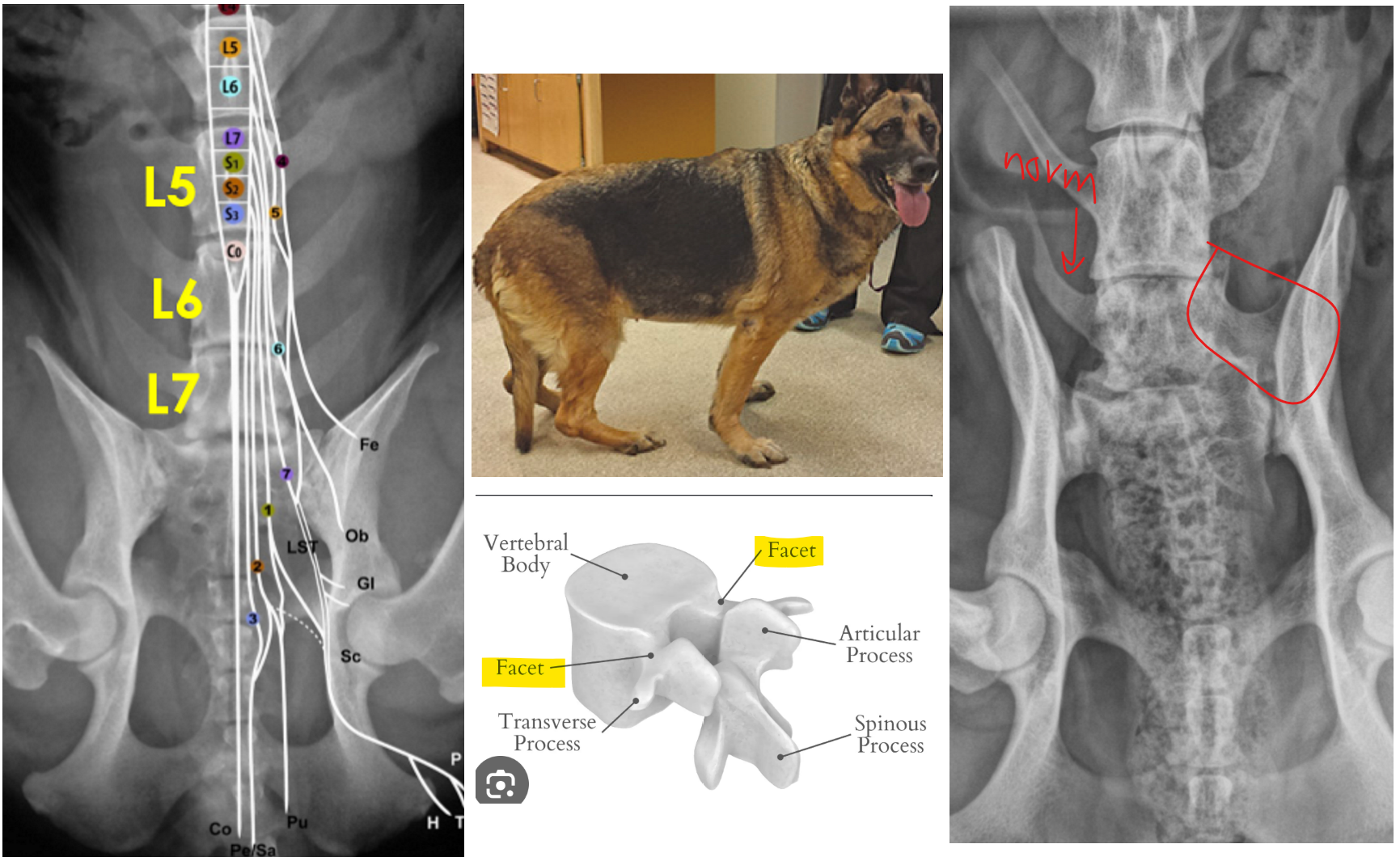
1: radiculopathy characterized by a vertebral canal stenosis (abnorm narrowing) in the L7-S1 disc space caused by type II IVDD, ligamentous proliferation, & articular facet proliferation
facet = bony surface connecting adjacent vertebral bodies
.
2: middle aged to older, large breed dogs w/ hx of progressive cauda equina signs like fecal incontinence (bc of dec anal tone)
.
3: dropped hocks (bc of sciatic weakness), tail held low to ground (dec tail tone), walk w/ hunched pelvis (weak)
bc of location (L7-S1), sciatic n. (& thus flexor mm) are affected but femoral n. (extensor mm) AREN’T so pt remains wt bearing → AKA pt is lame or paraparetic, NOT plegic or ataxic

degenerative lumbosacral stenosis (AKA cauda equina syndrome)…
.
1: neuro exam findings?
.
2: make & justify a diagnostic plan
1: mild gait changes, prop deficits in pelvic limb, dec flexor reflex in pelvlic limb, mild to mod pain in LS region
.
2: gold standard = MRI bc can see ligaments, IVD, nerve roots, etc.
BUT can rule out other ddx & dx w/ spinal rads by looking for: narrow IVD space, sclerotic endplates, sacral osteochondrosis, spondylosis deformans, & transverse process of L7 starts to fuse to ilium (AKA “transitional vertebrae”)
.
note: this dz is very sim to cervical spondylomyopathy but in LS region
discospondylitis…
.
1: what is this & SPECIFIC etiology?
.
2: typical signalment (breed & age)?
.
3: clinical signs?
.
4: most common location affected?
1: bacterial or fungal infection of the IVD’s & end plates (usually) caused by hematogenous spread
most common agents = s. aureus & s. pseudintermedius
less common but possible = brucella canis (zoonotic!!), aspergillus (fungi)
.
2: young to middle aged, large breed dogs
.
3: SEVERE PAIN!!, ± systemic signs (fever, lethargy, anorexia), ± neuro deficits from soft tissue prolifertaion or pathologic fracture pushing on SC
.
4: lumbosacral & thoracic vertebral column
.
note: this is the MOST painful spinal dz!!!

discospondylitis…
.
1: make & justify a diagnostic plan
.
2: list the 2 tests we want to run once we dx disco
.
3: define spondylosis & explain why vets commonly misdiagnose disco for spondylosis
1: very extensive… start w/ min database: CBC (inflam leukogram), chem (normal, ± inc globulins), UA & urine culture for susceptibility profile (looking for UTI)
spinal rads: usually enough to dx → look for lysis & sclerosis of IVD end plates, widening or collapse of IVD space, & fractures
note: changes lag behind signs, so appear norm if taken too early
± CT if rads are norm… pick MRI over CT if neuro deficits are profound, bc can better assess edema, inflam, etc.
.
2: blood culture w/ susceptibility profile to ID the bug & pick the right abx (but usually low yield) + brucella test
.
3: non-inflam, degen SC condition characterized by osteophytes growing along the edge of vertebrae (its essentially OA of the spine)…
bc of superimposition, the osteophytes look like they’re inside the vert canal so vets ID them as the cause of neuro signs (ataxia, etc.) BUT spondylosis will NEVER enter the vert canal so we must further investigate to find the cause of neuro probs (& it could be disco)!

list the key features to help differentiate a pt w/…
degenerative myelopathy
luumbosacral stenosis
discospondylitis
all are causes of progressive paraparesis…
no spinal pain
pt is lame or paraparetic BUT not plegic or ataxic
pt is SCREAMING in pain, mild to no neuro signs
1: list the 3 main struc assoc w/ spinal pain & list ddx for each
.
2: another cause of spinal pain is neuropathic, secondary to synringomyelia (AKA chiari-like malformation).
describe what this is & name the most common breed
1:
nerve roots → pain secondary to… IVDE, cervical spondylomyelopathy, lumbosacral stenosis
meninges → meningomyelitis, meningitis
vert body periosteum→ discospondilitis (can also progress to root & meninges), tumor, trauma
.
2: fluid filled cavities/cyst/syrinx form inside the SC which makes it hard for info to travel & pt becomes painful
cavalier king charles spaniel
.
reminder: a pure myelopathy WON’T cause pain!

cervical IVDD (C-IVDD)…
.
1: is this an extrusion or protrusion IVDD?
.
2: typical signalment (breed & age)?
.
3: most common vertebrae affected in small vs large breed dogs
1: either
.
2: small breed dog (but any breed possible), >2 y/o (avg is 6 y/o)
.
3: small = c2-3, large = c5-6 & c6-7
cervical IVDD (C-IVDD)…
.
1: clinical signs?
.
2: specific signs when cranial vs caudal cervical discs are affected?
.
3: make & justify a diagnostic plan
1: neck pain (AKA cervical hyperesthesia; MOST important sign), guarded neck posture (neck tucked in & held low), ± kyphosis secondary to pain, prop ataxia, ± lameness (if nerve root is compressed), tetraparesis, rarely tetraplegia
note: paralysis more common in thoracolumbar IVDD bc vert canal is larger in cervical region so there’s more open space which makes disc more likely to displace laterally & compress n. roots
.
2: cranial = hypereshtesia; cd = neuro deficits (bc this is location of cervical intumescence so SC occupies larger portion of vert canal)
.
3: sim to IVDD in TL region.. rads possible but CT/MRI best to visualize compression
.
reminder of normal anatomy: C2 has a unique look & there’s no IVD between C1-2

atlantoaxial subluxation (AAS)…
.
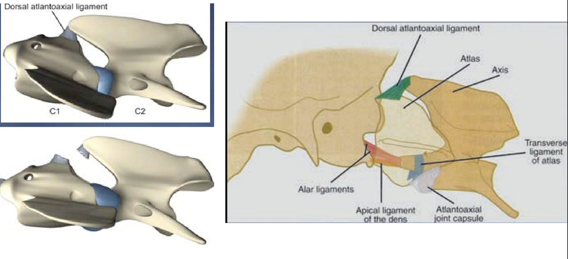
1: describe the normal ligament anatomy of the atlantoaxial joint
.
2: define AAS
.
3: AAS can be congenital (most common) or acquired. describe each
1: there’s no IVD between C1-2 so jt is supported by 4 ligaments → 3 of them (apical, alar, transverse) are diretly attached to the dens (AKA odonotoid process)
so, if smth happens to the dens, the jt loses 75% of its support…
.
2: failure of the struc support ligs or processes in the C1-C2 region leading to subluxation
.
3: congenital = pt born w/ absent dens (aplasia), small dens (hypoplasia), or absent ligs
acq = trauma causes dens fracture or lig rupture
atlantoaxial subluxation (AAS)…
.
1: typical signalment (breed & age)?
.
2: clinical signs?
.
3: make & justify a diagnotic plan
1: toy breeds (esp yorkie & poodle), <2 y/o
.
2: cervical hyerpesthesia, prop ataxia, tetraparesis
signs can be acute, chronic, or wax & wane
.
3: rads is enough! → will see big gap between axis & atlas (normal = they overlap)
note: analgesia & sedation reqt bc pt in pain

steroid responsive meningitis-arteritis (SRMA)…
.
1: what is this?
.
2: typical signalment (breed & age)?
.
3: clinical signs?
.
4: make & justify a diagnostic plan
1: immune-mediated response against meninges & arteries in CNS thats related to excessive prod of IgA in the CSF & serum
etiology unkown
.
2: VERY clear breed disposition… Beagles,Bernese mountain dogs, boxers, english Pointers, Goldens, other medium-large sized dogs (trick: BBB-PG)
6 m/o - 2 y/o (RARELY in dogs > 2 y/o)
signalement is HUGE in dx this dz!!
.
3: acute onset of diffuse, severe spinal pain; reluctant to walk; stiff gait; rigid neck; FEVER (systemic signs bc its along the whole spine); ataxia; rarely tetraparesis/plegia (usually no neuro deficits)
.
4: most sensitive way to dx = CSF analysis close the neck → fluid is cloudy, hemorrhagic, or xantochromic; SEVERE neutrophilic pleocytosis (AKA very high count & most of them are neutrophils)
can do CBC → neutrophilic leukocytosis
.
note: dogs dont get bacterial meningitis like ppl

cervical spondylomyelopathy (AKA CSM, wobbler syndrome, cervical instability, etc.)…
.
1: what is this?
.
2: this dz has two forms. describe the typical signalment (breed & age) & lesions of each
osseous associated (OA-CSM)
disc associated (DA-CSM)
.
3: typical hx (acute or chronic)?
1: neck spine dz causing static or dynamic compression of SC & nerve roots
.
2:
giant breed dogs <3 y/o (esp great dane); pt has bony proliferation causing dorsal compression
large breed dogs >6 y/o (esp doberman); pt has ventral SC compression
note: pt’s can have both
.
3: hx of chronic progressive signs, some pt’s are acute on chronic
.
this is a VERY common dz of the cervical spine

cervical spondylomyelopathy (AKA CSM, wobbler syndrome, cervical instability, etc.)…
.
1: clinical signs?
.
2: what location is this localized to on neuro exam?
.
3: make & justify a diagnostic plan
1: cervical hyperesthesia (usually only present on palpation), mild to severe prop ataxia (worse in pelvic limbs making them wobbly), thoracic limbs vary (mild to severe ataxia, spasticity, short strides), “2 engine gait”
occasionally non-amb tetraparesis
note: these pt’s can have compression w/o any signs
.
2: C1-C7
.
3: best option = kinematic/dynamic MRI (this is MRI w/ neck in natural standing position, makes lesion more obvious) → look for compression
can use rads as screening tool → irregular, arthrtitic bony margins (if OA-CSM)

list the top 4 ddx for a pt w/ cervical spine dz
cervical IVDD
atlantoaxial subluxation (AAS)
steroid respnsive meningitis-arteritis (SRMA)
cervical spondylomyelopathy (CSM)
.
other ddx:
acute SC trauma → hx of trauma
spinal neoplasia → chronic or subacute, ± pain, ± large dog
FCEM → acute, asymmetric, no spinal pain
discospondylitis, meningomyelitis, syringohydromyelia
define the following terms invlved in movement disorders…
tremor
myoclonus
tetanus
tetany
1: involuntary, rhythmic oscillatory mvmt of any body part → categorized as high/low frequency & high/low amplitude; can occur at rest, action, etc.
.
2: repeated, often arrhythmic, shock-like jerks (AKA sudden contraction of a musc group followed by immediate relaxation) → e.g. chomping the jaw
ANY meningoencepalitis can cause this but commonly assoc w/ CDV
.
3: continuous contraction of extensor mm. → e.g. pt is super stiff/spastic in all 4 limbs
.
4: intermittent & variable contraction of extensor mm.
define the following terms involved in movement disorders…
myotonia
fasciculation
dyskinesia
dystonia
1: sustained m. contraction w/ delayed relaxation (think of fainting goats)
.
2: contraction of m. fibers assoc w/ the motor unit, usually under the skin only w/o limb/body mvmt
.
3: general term for any abnorm, sudden, involuntary mm. mvmt; can be hyper or hypokinetic; e.g. twisting, jerking
dogs are commonly hyperkinetic (e.g. hyperflexion of limbs)
.
4: specific type of ^ characterized by prolonged mm. contraction w/ a fixed twisting posture (e.g. pt pulls head to left & extends arm to rt)
paroxysmal dyskinesia…
.
1: whats the diff between this ^ & dyskinesia
.
2: name the neuro dz that this commonly gets misdiagnosed as & explain why
1: dyskin is general term for involuntary abnorm mvmt; paroxy is a subtype characterized by episodic, sudden attacks of these mvmt’s interspersed w/ completely normal periods
.
2: seizures! they present similarly but these pt’s won’t get better w/ seizure meds so we MUST have paroxy in our ddx for epileptic pt’s
.
note: studies show these pt’s have a gluten intolerance & signs improve w/ gluten free diet
compare & contrast paroxysmal dyskinesia vs epileptic seizure…
locatio of primary lesion?
presence of facial involvement?
does pt remain mentally conscious during episode?
presence of pre-ictal signs?
duration?
presence of autonomic signs?
will frequency of episodes dec or inc over time?
will pt respond to antiseizure meds?
p = basal nuclei; s = cerebral cortex
p = no (can have head involvement); s = common (facial mvmt pathway goes thru cortex)
p = yes; s = consciousness altered
p = no; s = yes
p = sec to hrs; s = sec to 2 min (pt would die from hr long seizure)
p = less common; s = more common
p = dec; s = inc
p = no; s = usually

1: walk thru the diagnostic approach when presented w/ a pt w/ tremors (BEFORE doing any type of diag tests, this is just from hx & PE)
.
2: there’s MANY etiologies of generalized tremors in dogs.
list the 2 most common
1: consider…
signalment: some dz’s are VERY breed or age specific (e.g. young → think CBM or hypomyelination)
onset: aucte → think toxin or encephalitis, chronic + non-progressive → think congenital
is it episodic or constant?
mm involved? frequency & amplitude of tremor?
systemic signs (fever, etc.)?
all ^ will help narrow ddx
.
2: intoxication & steroid responsive tremor syndrome
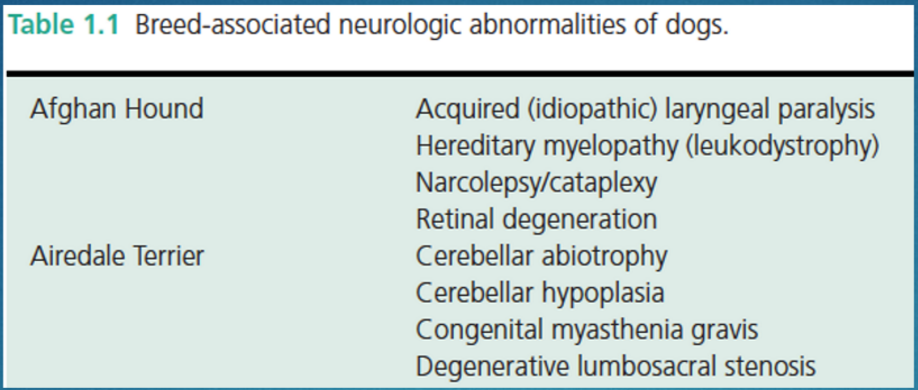
cerebellar hypoplasia (CH)…
.
1: etiology (2, be specific)?
.
2: what age do CBM signs begin & describe their progression
.
3: make & justify a diagnostic plan
.
4: ddx if the signs become progressive (2)?
1:
pt gets via intrauterine infection: panleukopenia (cats) attacks granular layer, herpesvirus (dogs) attacks gran & purkinje layer
or genetic: chow chow, irish setter, fox terrier
.
2: as soon as pt starts to walk & remain static thru life (don’t get better or worse)
.
3: MRI + histopath (will see dec # of purkinje cells) but most won’t do this so…
use combo of signalment, hx, signs, & no progression
note: purk cells are the only motor cells in CBM
.
4: cerebellar abiotrophy & feline niemann-pick dz (AKA sphingomyelinosis; this is a genetic storage dz)
cerebellar abiotrophy…
.
1: define abiotrophy
.
2: etiology?
.
3: signalment (breed & age)?
.
4: clinical signs?
.
5: make & justify a diagnostic plan
1: spontaneous cell death → this dz mainly affects purkinje cells
.
2: genetic or immune mediated
.
3: breeds = setter, collie, bull mastiff, coton de tulear, staffordshire terrier, etc.
onset varies by breed (so signalment is key)
beagle, poodle, samoyed: 2 w/o
brittany spaniel: 7-13 y/o
.
4: CBM signs tend to be progressive (can also spread to SC, cerebrum & change behavior, etc.)
.
5: signalment, signs, MRI (WM & GM won’t be well defined like norm), histopath

1: describe the relationship between meningoencephalitis & CBM signs
.
2: you’re presented w/ a pt w/ CBM signs. whats a clue indicating that the pt has meningoencephalitis, not CBM dz
.
3: list 2 causes of meningoencephalitis
1: its usually causes seizures & vestib signs BUT any inflam CNS dz can cause CBM signs!!!
.
2: pt has MF lesions (e.g. vestib signs = brainstem, CBM signs = CBM) → this is hallmark sign of inflam/infectious CNS dz
.
3: granulomatous meningoencephalitis (GME; imm mediated dz) or infectious (CDV, FIP, herpesvirus, neospora)
neosporosis…
.
1: what is this/pathogenesis?
.
2: clinical signs & age of onset?
.
3: make & justify a diagnostic plan, include results
.
4: why should this ALWAYS be on your ddx for pt’s w/ CBM signs (esp dogs living in rural areas)
1: infectious dz causing a progressive necrotizing cerebellitis
note: this is chronic but pt can go down acutely
.
2: progressive CBM signs + mild vestib signs in ADULTS
.
3: bloodwork → CK is VERY high
note: CSF may be norm in late stage bc inflam process is over
.
4: you WILL kill the pt if you give them steroids
cerebellar neoplasia…
.
1: clinical signs?
.
2: list 2 types of tumors this is usually caused by
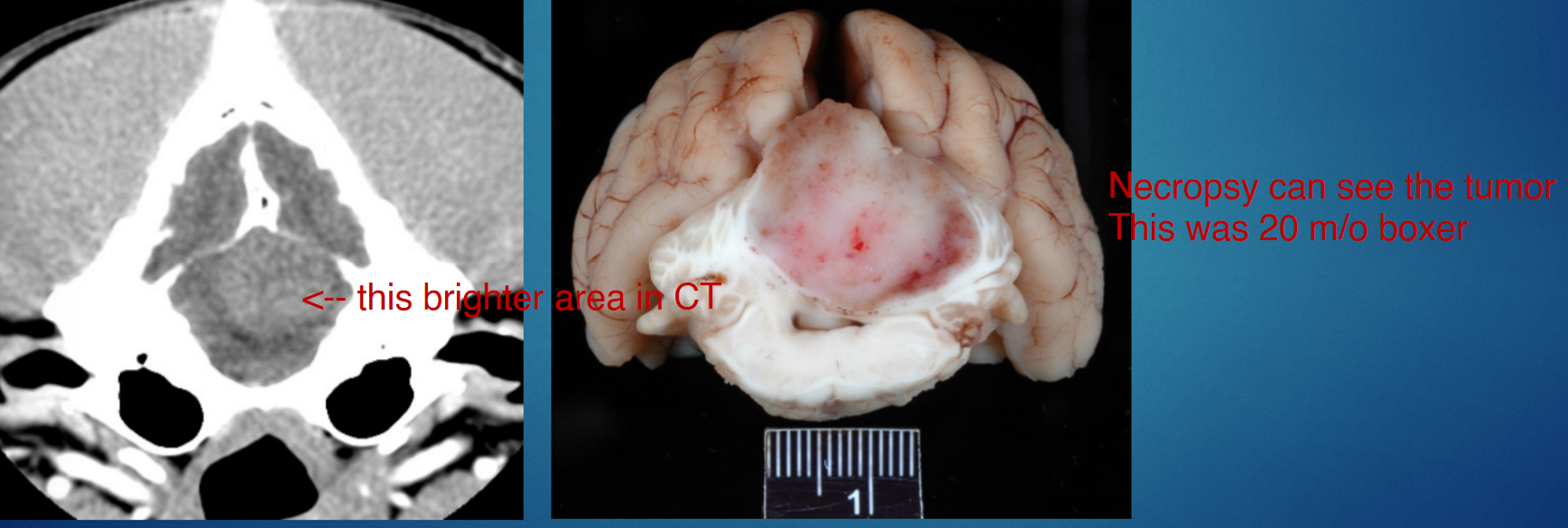
1: asymmetric signs, rarely CBM signs
.
2: medulloblastomas (esp young dogs), oligodendrogliomas
.
note: this is VERY rare, most CNS tumors affect cerebral cortex
steroid responsive tremor syndrome (SRTS; AKA immune mediated cerebellitis / white shakers)…
.
1: what is this / clinical signs
.
2: typical signalment (age & breed)
.
3: make & justify a diagnostic plan, include results
1: form of encephalitis that’s likely imm mediated (etiology unknown) characterized by an acute onset of high frequency, low amplitude, whole body tremors
less common signs: head tilt, dec menace, opsoclonus (eye moves all over the place, diff than nystagmus)
.
2: white small breed dogs < 4 y/o (e.g. maletese, west highland white terrier) → BUT ANY AGE/BREED CAN GET THIS!!
.
3: most sensitive test is CSF → VERY mild mononuclear pleocytosis, but can be norm :(
CBC/chem & imaging are useless bc will be norm
.
note: if you give steroids, pt gets better
besides CBM dz, SRTS, & a myelin disorder…
list & give e.g. of 3 ddx for a pt w/ acute onset of tremors)
#1 = toxicity, esp from mycotoxins/penitrem
others: theobromine (chocolate), organophosphates, carbamates, lead, etc.
.
medication: metoclopramide (#1), metronidazole, diphenhydramine, imipramine, ivermectin, etc.
metabolic dz: hypocalcemia (#1), hypoglycemia (esp if rapid onset)
canine leukodystrophies are dz’s affecting WM
these are lumped into myelin disorders
uncommon in dogs
Canine Leukodystrophies are a specific group of inherited, genetic disorders that fall under the umbrella of dysmyelination
dysmyelination is not the same thing as "myelin disorders," but rather a specific type of myelin disorder. "Myelin disorders" is a broad umbrella term that encompasses any condition where the protective myelin sheath around a dog's nerves is abnormal, deficient, or damaged.
ddx for pt’s w/ tremors
cerebellar dz (congenital or acquired)
steroid responsive tremor syndrome (SRTS)
metabolic dz (hypocalcemia, hypoglycemia)
toxin/medication exposure
myelin disorder (hypo or dysmyelination)
(episodic head tremors?)