Small Animal Wounds - Initial Approach
 Call Kai
 Learn
Learn Practice Test
 Spaced Repetition
 Match
Match Flashcards
 Knowt Play
1/20

There's no tags or description
Looks like no tags are added yet.
Name | Mastery | Learn | Test | Matching | Spaced | Call with Kai | Chat |
|---|
No analytics yet
Send a link to your students to track their progress
21 Terms
Again, describe the inflammation phase of wound healing…
In the first 48hrs -
Platelet plug formation
Cytokines release from damaged tissue cells → inflammatory response
Initial vasoconstriction → vasodilation
Increased blood flow and vascular permeability at the wound
WBCs (mainly neutrophils and macrophages) migrate into the wound
Attack bacteria, remove damaged tissue and foreign material
Describe the proliferative phase of wound healing…
2-5 days -
Fibroblasts and capillaries migrate into the wound
Creates framework for other cells to grow on
For a surgical wound:
Epithelial cells cover wound within 48hrs
For open wound:
Granulation tissue fills wound
Acts as scaffolding for migrating epithelial cells
Describe the maturation and remodelling phase of wound healing?
Takes MONTHS to a year
New collagen fibers organise along lines of tension
Scar then CONTRACTS, closing in the wound on the edges
** scar is 20% weaker than normal tissue
What are the two main factors that can affect wound healing? Think of some examples of each…
Local
Perfusion, infection (causes prolonged inflammatory phase), tension and movement at the wound
Foreign material and fluid accumulation can also prolong inflammatory phase, acting as sites for infection
Systemic
Stuff like age, current medications, and nutrition can slow wound healing
Steroids → inhibit inflamm. response, prolonging it
Neoplasia and metabolic diseases can do the same
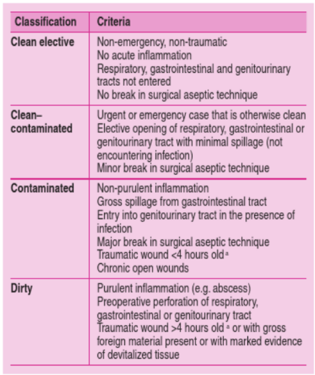
What are the different types of wounds you could have categorized by cleanliness? How does this affect your approach to treatment?
Categorized by:
Clean - treated via primary closure
Clean contaminated - can be treated via primary closure in some cases; other cases, delayed primary closure is best
Contaminated - best treated via secondary closure; provide open wound management first day, then closure 5-10 days later
Dirty - SECOND INTENTION healing best for these kinds of wounds
Describe the different methods for wound closure… (hint: 4)
Primary Closure:
Immediate closure of healthy viable tissue without tension
Good for surgical wounds, some traumatic wounds
Delayed Primary Closure:
Used for wounds with minimal contamination, questionable viability, or tension
Close defore granulatoin tissue starts but after bacteria and foreign material has been removed
Manage as an open wound initially, then close by days 2-5
Secondary Closure:
Closure once GRANULATION TISSUE has been formed
Good for contaminated wounds
Open wound management then closure at days 5-10 when healthy granulation tissue is present
Second Intention Healing:
Essentially, wound is left to heal on its own
Reserved for dirty and infected wounds, or large skin deficits that cannot be closed
What are the Halstead Principles for closure of a simple, surgical wound?
1) Gentle tissue handling
2) Accurate hemostasis
3) Preservation of local blood supply
4) Aseptic technique
5) Close tissues without tension
6) Careful approximation of tissues
7) Ensure no dead space
How do you perform open wound management?
Aim to provide optimal conditions for wound healing or prepare the wound for delayed closure… may require anesthetics, multiple dressings, and may take long. Prepare the owner for the timeline and the price
Once the patient is stable, you will want to:
Identify and control any infection
Remove debris and necrotic tissue
Prevent further contamination and damage to the wound
First:
Cover wound with sterile dressing or pack with sterile K-Y jelly, then clip a wide margin of fur around the wound
Then lavage
Then debride
Then dress
How would you lavage a wound and why?
Reduces bacterial contamination, removes foreign material, and rehydrates necrotic tissue
Use saline or LRS
Use a high volume; attach a giving set w/ a 3 way tap, attached to a 20-30mL syringe and 19g needle
Will generate enough pressure to remove debris, but not enough to drive bacteria into deeper tissues
How would you go about debriding a wound?
Surgically or non-surgically
Surgical:
Remove devitalized or contaminated tissue
Use sharp dissection with a scalpel OR
Scrape with blade
Rub with dry swab
Non-surgical:
Used to remove debris that cannot be removed by lavage or surgical means
Uses DRESSINGS to debride the wound
Moisture dilutes exudate and draws it out, hydrating necrotic tissue. Material loosens and dries into the dressing, which is then removes
Can also cause injury to normal tissue
What are the three different layers of bandaging?
Primary or contact layer - can be adherent or non-adherent
This controls the wound environment
Debrides infected or necrotic tissue
Absorbs exudate
Protects the wound and promotes healing
Can provide analgesia
Secondary layer
Absorbs excess fluid, holds primary layer in place and provides structure to bandage
Tertiary layer
Outer layer secures dressing in place and protects the secondary layer
What are the different kinds of adherent contact layers of bandaging, and what are they most commonly used for?
USED IN THE INITIAL STAGES OF WOUND MANAGEMENT ONLY
Used to DEBRIDE the wound
Should only be used initially and the dressing be changed every 24 hours
Includes “wet to dry” dressings and “dry to dry” dressings
Dry to dry self explanatory -
“Wet to Dry” dressings -
Sterile woven swabs, soaked in hartmann’s / sterile saline and squeezed dry, are placed over the wound
Followed by dry sterile swabs
Fluid, exudate, and material is wicked from the contact layer as the swabs dry
Swab then adheres to the wound, and when removed for dressing change, layer of nastiness is stripped
Can also damage healthy tissue
IS PAINFUL!!! Should be done under sedation
Wound is then lavaged and redressed
Change to non-adherent dressings when all necrotic tissue is removed and granulation tissue begins to form
What is the goal of non-adherent dressings? What are the two main different kinds you could use?
AIM: To promote granulation and epithelialization of the wound
Provites a moist wound environment
Doesn’t cause trauma to the surface of the wound
Types of non-adherent dressings:
Foam dressing
Absorbent
Provides a semi-permeable membrane to allow O2 exchange and controlled evaporation
Keeps wound moist but not wet
Hydrogels
Maintain moisture; donate and traps water to absorb wound exudate and rehydrate wound
Encourages natural debridement and sloughing
Good for dry wounds
** Hydrogels can be used WITH foam dressings for debridement; painless to remove, so may be useful for an animal where repeated sedation isn’t the best
Change every 24-72hrs
Lavage out at each dressing change
What does healthy granulation tissue look like?
Red
Uniform and flat
Progressing with each bandage change
Minimal exudate
What does NON-healthy granulation tissue look like?
Pale
Not-progressing
Usually due to necrotic debris or infection
What is the purpose of the secondary layer of the dressing?
Holds the contact layer in place
Absorbs exudate that passes through contact layer
Supports and protects the wound; provides padding
Examples: Soffban, cotton wool, conforming gauze bandage
What is the purpose of the tertiary layer of the dressing?
Holds the intermediate layer in place
Protects the other layers from trauma and contamination
Care with elastic self-adherent dressings; may become too tight
When should you use antibiotics for wound healing?
Is indicated in most traumatic wounds; should be used in conjunction with lavage and debridement of open wounds
If its a non-healing wound, culture it AFTER lavage
Do not give an open ended course, give a therapeutic course then reassess
What are two different topical wound treatments you could use, and what are their properties?
Silver dressings
Antiseptic and antibacterial
In the presence of exudate, silver ionizes to release active silver ions
Manuka honey
Antibacterial and anti-inflammatory
High osmotic potential DEBRIDES the wound and draws out exudate
Promotes rapid formulation of granulation tissue
What is one major thing that could delay wound healing if it gets contaminated? What would you do to combat this?
If the wound gets contaminated at ALL:
BACTERIAL BIOFILM CAN BE PRODUCED
THIS IS WHY YOU LAVAGE SO OFTEN
Products are available that disrupt biofilm when using would gels
PHMB / betaine (prontosan)
Betaine disrupts biofilm
PHMB kills bacteria
How do cats differ in their wound healing from dogs?
Have a different cutaneous vascular supply to dogs, therefore, heal more slowly
Granulation tissue is slower to appear and takes longer to totally cover the wound
Loss of the subcutis retards wound healing to a greater degree in the cat than in the dog