Heart
 Call Kai
 Learn
Learn Practice Test
 Spaced Repetition
 Match
Match Flashcards
 Knowt Play
1/82

There's no tags or description
Looks like no tags are added yet.
Name | Mastery | Learn | Test | Matching | Spaced | Call with Kai | Chat |
|---|
No analytics yet
Send a link to your students to track their progress
83 Terms
Most common type of birth defect
structural heart issues present at birth-congenital heart defects
Acyanotic defects
left to right shunting of blood
Ventricular septal defect(VSD)
Atrial septal defect(ASD)
patent ductus arteriosis(PDA)
coarctation
Cyanotic defects
right to left
tetralogy
transposition
truncus arteriosus
total anomalous pulmonary venous return
hypoplastic left heart syndrome
What are risk factors for CHD?
heredity, genetic disorders (Down’s syndrome), fetal substance exposure(tobacco or meds), maternal health status (diabetes, obesity, etc.)
Manifestations of CHD
heart murmurs, dyspnea, tachypnea, cyanosis, fatigue, chest pain/discomfort, difficulty gaining weight
HEART FAILURE MAY DEVELOP
Alterations Resulting in Decreased Cardiac Output
NERVES AND HORMONES—> HR(BPM)
BV AND VASCULAR RESISTANCE—>STROKE VOLUME(ML/BEAT)
Pericarditis
causes
Inflammation of pericardium by viral infection, thoracic trauma, myocardial infarction, tuberculosis, malignancy, and autoimmune conditions■

Pericarditis may cause…
pericardial effusion: Fluid accumulatesIn space between pericardial sac and heart; swollen tissue creates friction

What is a complication of pericarditis?
manifestations of this
Cardiac tamponade: life-threatening cardiac compression from fluid accumulation (pleural effusion)
falling arterial pressures, rising venous pressures, narrowing pulse pressure, and muffled heart sounds

Constrictive pericarditis
manifestations
loss of elasticity from chronic inflammation
pericardial friction rub (grating sound); sharp, sudden, severe chest pain that increases with deep inspiration and decreases when sitting up/leaning forward; dyspnea; tachycardia; palpitations; edema; flu-like symptoms
Pericarditis acronym
FRICTION
F- friction rub
R- relieved pain when sitting up/ leaning forward
I- increased pain with inspiration
C- chest pain (sharp, stabbing)
T- tachycardia
I- increased pain when lying flat
O- overall malaise and fever
N- not relieved by medication
Infective Endocarditis
formerly called
caused by
Infection of endocardium and heart valves
formerly called bacterial endocarditis
(commonly caused by streptococcus and staphylococcus)
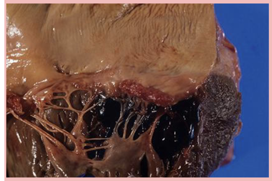
What can form and travel due to Infective Endocarditis?
Thrombi form that travel(emboli), causing microemboli and microhemorrhage
Infective Endocarditis signs and symptoms
FROM JANE
Fever(flu-like symptoms)
Roth spots(retinal hemorrhages)
Osler nodes(small, painful, raised, red or purple bumps on finger and toe pads)
Murmer(new onset)
Janeway lesions(small, painless, red or purple flat spots on palms or soles of feet)
Anemia
Nail-bed hemorrhages(splinter)
Emboli
also petechiae
Risk factors for Infective endocarditis
IV drug use, valvular disorder, prosthetic heart valves/implanted devices, rheumatic heart disease, aortic coarctation, congenital heart defect, Marfan Syndrome
Life-threatening complications for IE
myocardial infarction, stroke, or pulmonary embolism
Infective Myocarditis
complications
uncommon and poorly understood inflammation of myocardium
heart failure, cardiomyopathy, dysrhythmia, thrombus formation
Infective Myocarditis caused by
Manifestations
infections(viruses, bacteria, parasites)
drugs and vaccines
Autoimmune diseases
palpitations, cardiomegaly, pale and cool extremities, syncope, decreased urine output, leg swelling(fluid stays in legs)
Infective Myocarditis acronym
HEARTFAIL
H- Heart murmur
E- Edema (peripheral or pulmonary)
A- Arrhythmias
R- Rapid pulse (tachycardia)
T- Tiredness and weakness
F- Fever(flu-like symptoms)
A- Aching chest (angina-like pain)
I- Increased respiratory rate (dyspnea)
L- low cardiac output
Valve stenosis
narrowing; less blood can flow through the valve
decreased cardiac output, increases workload—>hypertrophy
atresia: failure to open valve
Valve regurgitation
insufficient closure; bidirectional blood flow
decreases cardiac output, increases workload on the heart—>hypertrophy and dilation of ventricles
Valvular disorders are caused by
congenital defect, infective endocarditis, rheumatic fever, myocardial infarction, cardiomyopathy, heart failure
manifestations vary and reflect depending on valve and changed cardiac flow
Cardiomyopathy
acquired or inherited conditions
weaken and enlarge the myocardium(heart muscle)
makes it harder for the heart to pump blood to body
can lead to heart failure
3 types…
dilated (too big), hypertrophic (too thick) and restrictive (too stiff)cardiomyopathy
Dilated Cardiomyopathy
who is at high risk
caused by
most common type: primary myocardial dysfunction —> ventricular dilation and cardiomegaly —>decreased cardiac output and blood stagnation
high risk: african american men and O.A.
Causes: ■ chemotherapy, alcoholism, cocaine abuse, pregnancy, infection, thyrotoxicosis, diabetes mellitus, neuromuscular disease, hypertension, coronary artery disease, med hypersensitivity
Dilated Cardiomyopathy manifestations: appear as compensatory mechanisms fail
breathing changes and nonproductivecough ⚬Fatigue⚬ dysrhythmia(s), cardiac pain and abnormalities⚬ dizziness, activity intolerance⚬abnormal lung sounds⚬peripheral edema⚬Ascites, hepatomegaly⚬weak pedal pulse⚬cool/pale extremities⚬poor capillary refill⚬jugular vein distension
Hypertrophic Cardiomyopathy mainly affects who and what?
what happens to ventricle
systolic function in men and sedentary individuals; ventricle wall becomes thick and unable to relax
Hypertrophic Cardiomyopathy risk factors
hypertension, obstructive valvular disease, thyroid disease, dominant genes
Hypertrophic Cardiomyopathy manifestations
dyspnea/intolerance on exertion, syncope(fainting), orthopnea(dyspnea when lying down), angina, dysrhythmia, left ventricular failure, myocardial infarction, fatigue
Restrictive Cardiomyopathy
rigid ventricles affects diastolic function
poor prognosis
common in south and central America, India, Asia, and Africa
caused by amyloidosis, hemochromatosis, sarcoidosis, radiation, connective tissue diseases, myocardial infarction, cardiac neoplasms
Manifestations: if present, fatigue, breathing and lung changes, angina, hepatomegaly, jugular vein distension, ascites, murmurs, peripheral cyanosis, pallor
Cardiomyopathy Acronym
WEAK HEART
W- weakness and fatigue
E- edema (peripheral and pulmonary)
A- Arrythmias
K- keep cough in mind (left-sided HF)
H- Heart murmur
E- Enlarged heart (cardiomegaly)
A- Angina or chest pain
R- respiratory distress (dyspnea, othropnea)
T- tachycardia
DCM
HCM
RCM
big and stretched ventricle-weak pump
decreased contractility
systolic dysfunction
“cant pump”
thick ventricle(septum) - crowded chamber
decreased ventricular filling
diastolic dysfunction
“too thick to fill”
stiff ventricle - won’t relax
normal size, but rigid
diastolic dysfunction
“too stiff to fill”
EKG - 1 small square
.04 seconds
1 large box
.2 sec (5 small squares)
QRS complex
.04 - .12 sec
1-3 small boxes
ventricular depolarization
PR Interval
.12 - .20 sec
3-5 small boxes
pause before the pump; allows time for ventricles to fill
Electrical Alterations
Dysrhythmias - abnormal or irregular heartbeat(rate and rhythm)
classified by origin and affect on Cardiac output and BP
caused by: acid-base imbalance, electrolyte imbalance(potassium), CHD, anything creating issues with walls of heart can affect beat
manifestations vary based on dysrhythmia: sometimes asymptomatic, palpitations, fluttering, skipped beats, etc
catch dysrhythmias before they cause sudden cardiac death
Preload
volume of blood in ventricles at end of diastole(end diastolic pressure)
increased in: hypervolemia, regurgitation of cardiac valves, heart failure
Afterload
pressure needed to eject blood/resistance left ventricle must overcome to circulate blood
increased in: HTN and vasoconstriction
Congestive heart failure(HF)
inadequate pumping that leads to decreased cardiac output (CO) and increased preload/afterload
Causes: congenital defect, myocardial infarction, valvular disease, dysrhythmia, thyroid disease
Compensatory mechanisms (sympathetic nervous system, renin-angiotensin-aldosterone system, and hypertrophy) help at first, but perpetuate heart failure
Systolic dysfunction: decreased contractility(ventricles can’t pump hard enough during systole) ■ Diastolic dysfunction: decreased filling (not enough blood fills ventricles during diastole)
Mixed dysfunction: decreased contractility and filling (both systolic and diastolic)
Definition of heart failure
Ejection fraction: measurement of how much blood is pumped by left ventricle every time the heart pumps
50-70% is normal ejection fraction
HF with reduced EF < 40%
HF with preserved EF >50%
may be acute or chronic depending on underlying cause
manifestations depend on type and severity (graded I-IV) and appear as compensatory mechanisms fail
Left-sided heart failure
think left→lungs
cardiac output falls; blood backs up to pulmonary circulation—> pulmonary congestion, dyspnea, activity intolerance
causes: left ventricular infarction, hypertension, aortic or mitral valve stenosis, CAD
manifestations: pulmonary manifestations
pulmonary congestions: cough, crackles, pink-tinged sputum, tachypnea
tachycardia, cyanosis, exertional dyspnea, fatigue
Right-sided Failure
Blood backs up to peripheral circulation, causing edema/weight gain
causes: pulmonary disease, left-sided failure, right MI, pulmonic or tricuspid valve stenosis
Manifestations: systemic
peripheral edema
ascites
enlarged liver and spleen
JVD
weight gain
increased peripheral venous pressure
Alterations resulting in Ineffective Tissue Perfusion
Aneurysms
caused by weakening of an artery
commonly: abdominal or thoracic aorta; cerebral, femoral, or popliteal arteries
exsanguination(bleeding out) is a possible consequence of rupture
caused by HTN, build up of plaques(atheroscelrosis), dyslipidemia, DM, tobacco, older age, trauma, infection, congenital defects
True aneurysms affect
all three layers
Saccular aneurysm
bulge on the side (asymmetrical)⚬
Fusiform aneurysm
affects entire circumference(symmetrical)
Dissecting aneurysm
occurs in inner layers, not a true aneurysm
Manifestations of aneurysm
if present, depend on location/size; pulsating mass(esp. with abdominal aneurysm), pain, respiratory difficulty, neurologic
Dyslipidemia
high levels of lipids in the blood, which increases risk for chronic disease
lipids come from dietary sources and are produced by liver
classified based on density; triglycerides(low density lipoproteins(LDLs): “bad” cholesterol, makes up most serum cholesterol and is more invasive in nature) and protein(high density lipoproteins(HDLs); “good)” cholesterol, helps remove LDL cholesterol from blood)
manifestations: asymptomatic until it causes other diseases and complications
Atherosclerosis
chronic inflammatory disease TRIGGERED BY A VESSEL WALL INJURY(have to have this occur to develop this disease)
vessel wall injury can happen from HTN, smoking, high cholesterol(dyslipidemia), diabetes
Characterized by thickening/hardening lesions calcifying on the arterial wall—>obstructs vessel, leads to platelet aggregation, and vasoconstriction
complications: PVD(symptoms start when vessels become 70% occluded; clients may report pain in distal tissues, or chest pain if coronary artery issue) CAD, thrombi, HTN, stroke
manifestations: asymptomatic until complications develop
Peripheral Vascular Disease(PVD)
narrowing of the peripheral vessels - (arteries and/or veins)
commonly atherosclerosis in the arteries
may also be caused by a thrombus, inflammation, or vasospasm
Examples:
Thromboangitis Obliterans
Raynaud Phenomenon
Thromboangitis Obliterans
aka buerger disease
chronic inflammatory condition of the arteries
may lead to thrombosis and eventually complete occlusion of the small/medium arteries in extremities
bv are occluded so tissue becomes ischemic at first and eventually necrotic
pale, cyanotic, necrotic
MOST COMMONLY AFFECTS MALES 20-40, WHO SMOKE
exact cause unknown

Raynaud Phenomenon
vasospams of the arteries related to sympathetic stimulation—> tissues can change color; white, blue, red
associated with cold temperatures, autoimmune conditions(lupus and scleroderma), and stress
most commonly affects females between 18-30 years old
increased vessel occlusion may lead to ischemia of affected tissue(but less likely than with buerger disease)

Coronary Artery Disease
Narrowing or blockage of the arteries that supply blood to the myocardium(heart) - most commonly caused by atherosclerosis
also caused by vasospasm, cardiomyopathy, and thrombi occlusion
most common type of heart disease in the U.S. and leading cause of myocardial infarction
Nonmodifiable risk factors:
■ age: males > 45 years; females > 55 years or premature menopause■ family history: premature cad in first-degree male relatives
Modifiable risk factors:■ tobacco use, stress, obesity, physical inactivity, diabetes mellitus, hyperlipidemia, hypertension
3 main types: obstructive, nonobstructive, and coronary microvascular disease
Obstructive CAD
Plaque accumulates in the large arteries causing narrowing and decreased blood supply to the myocardium
⚬Coronary artery is occluded by more than 50%
⚬Blood flow may become completely occluded—> MI
Nonobstructive CAD:
large arteries occluded less than 50%
⚬ also caused by damage or injury to the lining of the coronary arteries that impact the ability to vasodilate in response to increased myocardial oxygen demand
Coronary Microvascular Disease
Affects the smallest arteries of the myocardium(arteries that branch off of the coronary arteries)
⚬Caused by molecular changes in small vessels aspart of normal age or damage (i.e., inflammation, diabetes, hypertension)
⚬Arteries do not respond to signals to vasodilate with increased oxygen demands on the myocardium
CAD: Angina
Stable Angina: Chest pain caused by ischemia that is initiated by increased oxygen demand(like walking up the stairs) and relieved with decreased demand (at rest)⚬ Decreased blood flow may or may not cause permanent ischemia
Unstable Angina(BAD): ⚬ unpredictable chest pain ⚬ Occurs at rest, or increases in frequency and/or intensity ⚬ Considered a “preinfarction” state (high risk for MI)
Coronary Artery Disease: Clinical Manifestations
angina that may radiate to neck, jaw, arm, or back
indigestion-like sensation
N/V
cold/clammy extremeties
diaphoresis
dyspnea, dizziness, fatigue, weakness, lightheadedness
sleep distrubances(worse when trying to sleep/lay down)
Coronary Artery Disease: acronym
H- heavy or squeezing chest pain (angina)
E- exertion triggers pain
A- anxiety or fear
R- radiating pain
T- tachycardia
P- pallor and diaphoresis
A- atypical symptoms in women/ older adults
I- indigestion or epigastric discomfort
N- numbness or tingling
S- shortness of breath
Thrombus
blood clot that forms anywhere in the circulatory system
3 conditions promote thrombus formation:
endothelial injury(physical trauma, strain, injury), sluggish blood flow,(congenital abnormalities affect venous anatomy-may thurner and paget schroetter) hypercoagulability(estrogen therapy, inflammation, dehydration)
venous thrombi more common than arterial due to lower pressure against gravity
Emboli
part or all of a thrombus breaks loose, travels through circulatory system, embeds in smaller vessel
can be air, fat, tissue, bacteria, amniotic fluid, tumor cells, foreign substance
Right side of the heart origin
venous circulation that travels first to pulmonary circulation, creating a pulmonary embolism
Left side of the heart origin:
arterial circulation and travel to other organs such as brain and heart, causing an infarction
Manifestations depend on
location of the body and whether they are venous or arterial
Indications of deep vein thrombosis(DVT)
calf swelling, erythema(swelling), and leg warmth
Pulmonary Embolism-
thrombus originates elsewhere and disrupts blood flow in the pulmonary artery or branches
Varicose veins
dilated, engorged veins r/t improper valve formation
most common in legs; may also see in esophagus, rectum, and testicles
increased venous pressure and pooling
risk factors: genetics, pregnancy, obesity, prolonged sitting or standing, alcohol use(esophagus), constipation(hemorrhoids)
manifestations: purple bulging veins, pedal edema, aching in legs, shiny, hairless pigmented skin on legs and feet, skin ulcer, possible necrosis
Lymph Edema
unusual swelling in the extremeties r/t obstruction
may be unilater or bilateral
primary caused by: rare; congenital absence or decreased lymphatics
secondary caused by: mastectomy, scarring by radiation, occlusion r/t tumors, obstructions r/t infection, injury/trauma
staging system 1-4(4 most severe)
manifests: edema and skin changes, hyperpigmentation, ulcer, thick and rough(elephant skin)
Myocardial Infarction
heart attack and acute coronary syndrome
death of the myocardium from sudden blockage of coronary blood flow
Myocardial oxygen supply cannot meet body’s oxygen demand⚬ Myocardial cells die as oxygen supply dwindles, leading to tissue necrosis
Leading cause of death in the U.S.
cardiovascular disease(usually caused by cardiac damage post MI)
Myocardial infarction risk factors
dyslipidemia, DM, HTN, stress, tobacco use
Myocardial Infarction manifestation
very similar to CAD
CRUSHING
Chest pain(intense, crushing, pressure)
Radiating pain
Unrelieved by rest or nitroglycerin(meds)
Sweating(diaphoresis)
Hard to breathe(dyspnea)
Increased hr and BP initially
N/V
Going to be anxious and fearful, dizzy
unstable angina, coughing, indigestion, elevation in cardiac biomarkers, can’t sleep
Alterations Resulting in Decreased Cardiac Output and Ineffective Tissue Perfusion
Hypertension
One of the most prevalent chronic health conditions in U.S.
Prolonged elevation in blood pressure creates excessive cardiac workload due to vasoconstriction, increasing afterload
decreased renal blood flow inappropriately activated renin-angiotensin-aldosterone system(RAAS)
risk factors: age(females increase risk after menopause), African Americans, family history, obesity, inactivity, tobacco use, high-sodium diet, low potassium/calcium/magnesium diet, high vitamin D intake(RAAS), alcohol, stress
Primary (essential) hypertension:
most common, develops gradually over time■ No identifiable cause; 95% of adult cases
Secondary hypertension:
more sudden and severe
caused by renal disease, diabetes, adrenal tumors, meds, and cocaine, and amphetamines(illicit drug use)
Pregnancy-induced hypertension:
Preeclampsia that may lead to eclampsia (increased BP may lead to seizures)
Malignant hypertension (hypertensive crisis):
intense and nonresponsive to interventions ■ BP is at least 180/120 & symptomatic
HTN can lead to
severe health complications if uncontrolled
cardiovascular disease
MI, heart failure, Left Ventricular Hypertrophy
Stroke and Brain complications
ischemic and hemorrhagic stroke
cognitive impairment and dementia
Kidney damage
chronic kidney disease
kidney failure
vision loss and retinal hemorrhages
aneurysms and Peripheral artery disease
Orthostatic (postural) Hypotension
decrease in both systolic and diastolic BP upon standing(at least 20mmHg and 10 mmHg within 3 minutes of standing up)
lack of normal BP compensation in response to gravitational changes on the circulation
acute OH or chronic OH
Symptoms of Low BP
dizzy, lightheaded, fainting or passing out, upset stomach, blurred vision, fast/shallow breathing, fatigue, weakness, tired, confusion, agitation