Lecture 8: Protein Purification and Identification: Part 2
 Call Kai
 Learn
Learn Practice Test
 Spaced Repetition
 Match
Match Flashcards
 Knowt Play
1/5

There's no tags or description
Looks like no tags are added yet.
Name | Mastery | Learn | Test | Matching | Spaced | Call with Kai | Chat |
|---|
No analytics yet
Send a link to your students to track their progress
6 Terms
Identifying polypeptides
why is it important
Three main methods to identify them
The most unique feature on a polypeptide chain is the primary structure
The sequence of amino acids in the polypeptide is derived from the genetic code, BUTmight not match the DNA sequence exactly
SDS-PAGE and immunoblotting (Western Blotting)
Used to confirm the presence of larger polypeptides
Antigen-specific antibodies bind to proteins containing specific amino acid sequences or structures
(Tandem) mass spectrometry (turning it into smaller pieces to deduce what the sequence of the amino acids are, what is present+ their sequence - more complicated)
Identify polypeptides after they are ionized and separated into small fragments
Computational analysis can be used to determine the sequence based on their mass-to-charge ratio
Immunoblotting (western blotting)
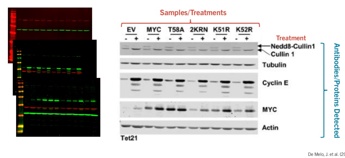
1. Proteins are separated by SDS-PAGE (so they can be removed from the gel and can bind to antibodies) and transferred to a solid-support membrane (blotting).
side note: however the polymer sheet has high affinity to all proteins (so any thing that is exposed with be bound to this sheet), including antibodies, so not interactions would happen
2. The membrane is exposed to a protein-solution (e.g. milk or BSA) to bind random proteins to any unused regions of the membrane.
like parking lot with lots of cars, no more parking spots
3. A primary antibody, specific for your POI, is added to recognize linear sequences of amino acids.
binds to the antibody
4. Secondary antibodies are specific for the Fc domain of the primary antibody and are attached to a fluorescently labeled tag or enzyme that generates a chemi-luminescent, visible product.
binds to the primary antibody already bound to the antigen, amplifying the signal and helping make it more visible
5. This may show if your POI is present in the sample and its approximate size based on where it migrated on the gel.
signals will be seem if your POI is present in the gel
Other information
tubular and actin are loading controls: has the same level fo dark band = same amount of protein eachh time
To be able to draw conclusions
Less expression in lysine (maybe some effect more studying need to be done there)
Issue with protein sequence + solution
Can get the polypeptide sequence from the DNA, however is much more complicated as sequence changes due to post-transcription and translation modifications
Also polypeptides have 20 amino acids, including different PTMs that need to be identified using just the primary DNA (more complex, labour intensive)
Solution mass spectrometry
Mass spectrometry
Peptides are bombarded by a laser to create ionized fragments
Fragments are attracted to a charged plate detector and analyzed in a mass analyzer by their time of flight
Time of flight depends on the charge and mass of the molecule
More charge = faster
Smaller = faster
Comparison to known peptides can identify the sequence of your polypeptide
Issue: some amino acids might have the exact same atomic mass

Sequencing using Mass spec
Sequences of peptides are compared to databases to identify protein present.
Each protein will be composed a certain amount of atoms that have a particular weight
If a unique fragment is identified in your sample, it is likely that the protein was present.
Fragments of many different sizes
Protein’s can be digested with a protease before MS produce smaller fragments that are easier to ionize and sequence
Can also be used to sequence a peptide that is unknown
Process
protein digestion: will break it into separate parts of the poor in, making git easier to do mass spectroscopy
