HBS 1.2 Test
 Call Kai
 Learn
 Practice Test
 Spaced Repetition
 Match
 Flashcards
 Knowt Play
1/81

There's no tags or description
Looks like no tags are added yet.
82 Terms
joints
Areas where two or more bones join together
fibrous joints
firm joints (teeth, jaw, sutures)

cartilaginous joints
slight movement bones held together by cartilage (pubic symphysis, ephihysel plate)

Synvoial Joints
most common and mobeable joint
articular cartilage
smooth covers the end of bones and cushions joints (at end of joints)

elastic cartilage
cartilage with abundant elastic fibers; more flexible than hyaline cartilage (ears, epigtous, respitory track)
fibrocartilage
dense and tough to absorb pressure and tension (under knees)
Ball + socket joint
greatest range of motion (hips and shoulders)

pivot joint
only rotates rotates around its own axis (neck, 1st and 2nd vertebre, wrist )

hinge joint
only 1 bone moves (elbow, knee)

saddle joint
type of joint found at the base of each thumb; allows grasping and rotation

condoyloid Joint
angular movements along 2 axes

planar joint
flat or slightly curved, permitting back and forth and side-to-side movements (carpals. tarsals, vertebre)

Deppression
lowers a body part (only shoulders and mandible)

elevation
raise body part (only shoulders and mandible)

rotation
The spinning motion on its axis
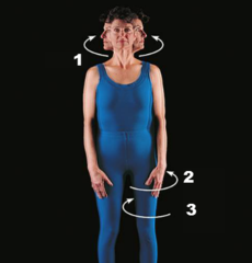
circumduction
the circular movement at the far end of a limb

flexion
Decreases the angle of a joint

extension
Increasing the angle of a joint

abduction
away from midline

adduction
back to midline

plantar flexion
pointing toes (ankle only )

Dorsiflexion
laying foot flat

elbow flexion

knee flexion

shoulder flexion

shoulder lateral rotation
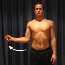
shoulder medial rotation

shoulder extension

hip extension

shoulder abduction

hip flexion

skeletel muscle
straitions, voluntary, attached to tendom, contract and convert chemical energy to mechincal force
smooth muscle
no stiations, involuntary, hollow organs walls (stomach) and blood vessel helps with digestion and absopstion
cardic muscle
striations, non- voluntary, heart, pump blood and deliver oxygen
striatons
Marked with striped, grooved, or ridged on muscle
skeletal muscle histology
striations, nuclei, 1 line is one muscle cell

smooth muscle histology
no striations, nuclei, all muscle cells are blended together

cardiac muscle histology
Branching, intercalated discs with gap junctions, striations, nucleus per cell,

tendon of diagram
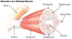
bone on diagram

Epimysium
covers the entire skeletal muscle

Perimysium
wraps around fasicles
endomysium
connective tissue wrap that surronds each muscle fiber

sarcolemma
plasma memberane around each muscle cell/fiber
muscle
organ made up of many fasicles
fasicle
bundle of muscle fibers
muscle fibers
muscle cell made up of many myofibrils
myofibril
long proteins that make up each muscle fibers/cells
myofilaments
make up myofbrils actin thin myacin thick
where calcium releases and acleyonine is put in through t tube
Sarcoplasmic Reticulum
muscle contraction 1
Signal travels down a motor neuron towards the neuromuscular junction.

muscle contraction 2
Signal reaches axon terminal causing the release of acetylcholine
muscle contraction 3
Acetylcholine binds to receptors on the surface of the skeletal muscle
mc 4
Signal spreads across the sarcolemma and travels down the T tubules.
mc 5
Sarcoplasmic reticulum releases calcium ions.
mc 6
Calcium binds to troponin.
mc 7
Tropomyosin is moved off of the myosin binding sites on the actin filament.
mc 8
Myosin grabs on (binds) to actin. (cross bridge)
mc 9
Myosin pulls the actin towards the M-line and releases ADP & phosphate. (powerstroke)
mc 10
ATP binds to myosin
mc 11
Myosin releases the thin filament
mc 12
Myosin hydrolyzes the ATP and cocks back ready to bind to actin again.
mc 13
Impulse ends.
mc 14
Calcium is pumped back into the sarcoplasmic reticulum
mc 15
Tropomyosin covers the myosin binding sites on the actin.
inter costals
orgin ribs,insertion ribs, move air out of chest

pectoralis minor
origin front of ribs 3-5 insertion part of scapula called coracial process action rotates pulls shoulder forward

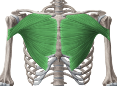
pectroralis major
origin ribs and clavicle edge, insertion humureus, action shoulder adduction medial
branchialis
origin halfwway down humeurs midway to ulna, insertion proximal ulna flexes forearm

triceps
orgin proximal half of dorsal humerus, insertion distal of the elbow on the ulna, action extends elbow

What are the requirements for a muscle's structure?
Must have at least 2 attachments and must cross at least 1 joint.
What happens to muscles when they contract?
Muscles always pull and get shorter.
What is the term for the attachment that moves in a muscle?
Insertion.
What is the term for the attachment that remains stationary in a muscle?
Origin.
What are flexors and extensors in terms of muscle function?
Flexors decrease the angle between ventral surfaces; extensors increase the angle.
How do muscles typically work in relation to each other?
Muscles always work in opposing pairs.
What do muscle striations indicate?
Muscle striations point to attachments and show direction of pull.
how muscles get names
shape, size, bones, direction of muscle fibers, function, number of origin
strain
an injury or tearing of a muscular tendon
sprain
strecthing/tearing liagment which connects bone to bone
tear
a partial or complete structural rupture of tissue fibers (muscles, tendons, liagments)