Balance Week 3
 Call Kai
 Learn
 Practice Test
 Spaced Repetition
 Match
 Flashcards
 Knowt Play
1/200

There's no tags or description
Looks like no tags are added yet.
201 Terms
B3LG1.1. Describe the developmental sequence from cardiogenic mesoderm to the four-chambered heart.
Answer: During week 3, splanchnic cardiogenic mesoderm forms paired endocardial tubes → embryonic folding brings the tubes together → fusion forms the primitive heart tube → rightward looping in week 4 establishes spatial relationships → atrial and ventricular septation occurs mainly during weeks 4–5 → neural crest contributions and conotruncal septation separate the great vessels during weeks 5–7 → aortic arch remodeling continues through approximately week 8.
Extra Information:
- Most major structural congenital heart defects originate from disturbances during weeks 3–8.
- The second heart field contributes substantially to the right ventricle and outflow tract.
- Neural crest migration is crucial for conotruncal septation and aortic arch development.
Source: B3LG1 Embryology PDF pp. 3–5 | First Aid 2025 pp. 285–286
B3LG1.1. What are the adult derivatives of the primitive heart tube regions?
Answer: Truncus arteriosus → ascending aorta and pulmonary trunk. Bulbus cordis/conus → smooth ventricular outflow tracts. Primitive ventricle → predominantly trabeculated left ventricle. Primitive atrium → trabeculated portions of both atria. Right horn of sinus venosus → smooth right atrium or sinus venarum. Left horn of sinus venosus → coronary sinus.
Extra Information:
- The primitive pulmonary vein is incorporated into the posterior left atrium, forming its smooth portion.
- The right and left common/anterior cardinal venous system contributes to the superior vena cava.
- Distinguish trabeculated atrial derivatives from the smooth atrial portions.
Source: B3LG1 Embryology PDF p. 6 | First Aid 2025 p. 286
B3LG1.1. How does normal cardiac looping establish the position of the chambers, and what happens if looping is abnormal?
Answer: The straight primitive heart tube normally undergoes rightward or D-looping. The developing right ventricle moves anteriorly and to the right, the left ventricle moves toward the left and relatively posteriorly, and the atrial region shifts superiorly and posteriorly. This establishes the basic topology needed for proper chamber alignment and great-vessel connections. Abnormal looping can produce dextrocardia, situs abnormalities, heterotaxy, or complex congenital heart disease.
Extra Information:
- Looping establishes position before final septation and valve development.
- Abnormal laterality can coexist with anomalous venous return and outflow alignment.
- The second heart field and cardiac neural crest contribute to subsequent outflow development.
Source: B3LG1 Embryology PDF pp. 5–7
B3LG1.1. Explain how the mitral and tricuspid valves, chordae tendineae, and papillary muscles develop.
Answer: Endocardial cushions develop in the primitive atrioventricular canal. Superior and inferior cushions grow and fuse to separate the common AV canal into right and left AV orifices. The central cushions contribute to septal AV valve leaflets, while lateral cushions contribute to mural leaflets. During delamination, fenestrations develop in the underlying ventricular myocardium and primitive valve tissue separates from muscle. Fibrous remodeling produces valve leaflets and chordae tendineae, while remaining myocardial attachments become papillary muscles.
Extra Information:
- Endocardial cushion abnormalities may cause AV valve dysplasia, stenosis, or atresia.
- Failure of complete fusion can produce a common AV valve in an AV septal defect.
- Abnormal delamination of the septal tricuspid leaflet contributes to Ebstein anomaly.
Source: B3S Structure ICG Answer Key pp. 17–23 | First Aid 2025 p. 285
B3LG1.1. How do the aortic and pulmonary semilunar valves develop?
Answer: The aortic and pulmonary valves develop from endocardial cushion tissue in the embryonic outflow tract. Migrating neural crest cells contribute to conotruncal cushion formation. Opposing cushions grow and spiral, then fuse to divide the outflow tract into systemic and pulmonary channels. Additional cushion tissue remodels and forms the semilunar valve leaflets. Cyclic fetal flow and mechanical loading help shape the thin mature valve cusps.
Extra Information:
- Semilunar valves develop from outflow tract cushions, while AV valves develop from AV canal cushions.
- Disruption may produce bicuspid aortic valve or pulmonary valve stenosis.
- Normal spiral septation connects the LV to the aorta and the RV to the pulmonary artery.
Source: B3S Structure ICG Answer Key pp. 15–16 | First Aid 2025 p. 285
B3LG1.1. Which embryonic developmental failures lead to common congenital cardiac structural abnormalities?
Answer: Abnormal looping or laterality → abnormal cardiac position or ventricular connections. Failed atrial or ventricular septation → ASD or VSD. Failed endocardial cushion fusion → AV septal defects and abnormal AV valves. Failed conotruncal septation → persistent truncus arteriosus. Failed outflow spiral rotation → transposition of the great arteries. Anterior malalignment of the infundibular septum → tetralogy of Fallot. Failed pulmonary venous incorporation → TAPVR.
Extra Information:
- Match the lesion to the embryologic process rather than memorizing the diagnosis alone.
- Failure of cardiac neural crest migration particularly affects conotruncal structures.
- Major defects often involve more than one developmental structure.
Source: B3LG1 Embryology PDF pp. 19–21, 25–29, 35–36 | First Aid 2025 pp. 285, 302
B3LG1.2. Explain normal atrial septation, including the ostium primum, ostium secundum, and foramen ovale.
Answer: The septum primum grows inferiorly toward the endocardial cushions, leaving the ostium primum between its lower edge and the cushions. Fusion closes the ostium primum. Before it closes completely, apoptosis creates the ostium secundum in the upper septum primum. The thicker septum secundum develops to its right but leaves the foramen ovale. The septum primum acts as a flap valve that allows fetal right-to-left atrial blood flow.
Extra Information:
- After birth, increased LA pressure presses the septum primum against the septum secundum.
- Anatomical fusion eventually forms the fossa ovalis.
- This arrangement allows fetal blood to bypass the high-resistance lungs.
Source: B3LG1 Embryology PDF p. 14 | B3S Structure ICG Answer Key pp. 3–4 | First Aid 2025 pp. 285–287
B3LG1.2. Compare the embryologic causes of secundum ASD, primum ASD, sinus venosus defect, and patent foramen ovale.
Answer: Secundum ASD results from excessive resorption of septum primum or inadequate septum secundum formation, leaving a true interatrial deficiency. Primum ASD results from failure of lower atrial septation and fusion with AV endocardial cushions. Sinus venosus defects occur near the SVC or IVC and are often associated with anomalous pulmonary venous connections. PFO results from failure of postnatal fusion of septum primum and secundum rather than loss of septal tissue.
Extra Information:
- Secundum ASD is the most common true ASD.
- Primum ASD is part of the endocardial cushion/AV septal defect spectrum.
- ASD usually produces a left-to-right shunt after birth.
Source: B3LG1 Embryology PDF pp. 14–15 | First Aid 2025 pp. 285, 303
B3LG1.2. Explain the formation of the muscular and membranous ventricular septum and the cause of the most common VSD.
Answer: The muscular interventricular septum grows upward from the primitive ventricular floor but initially leaves an interventricular foramen. Endocardial cushions and conotruncal ridges contribute tissue that aligns and fuses with the muscular septum, forming the membranous interventricular septum and closing the foramen. Failure of this fusion or membranous septal development produces a perimembranous VSD, the most common anatomic location of VSD.
Extra Information:
- Muscular VSDs arise in the trabeculated muscular septum.
- VSD hemodynamics depend on size of the defect and pulmonary vascular resistance.
- Small restrictive VSDs can cause louder murmurs than large nonrestrictive VSDs.
Source: B3LG1 Embryology PDF p. 17 | B3LG2 Acyanotic PDF pp. 3, 6 | First Aid 2025 p. 285
B3LG1.2. How do endocardial cushions form, and which intracardiac structures depend on them?
Answer: Myocardial signaling molecules, including BMP2/BMP4, Notch1, and TGF-β pathways, promote endothelial-to-mesenchymal transition of selected endocardial cells. These cells lose their epithelial organization, migrate into cardiac jelly, and produce cushion primordia. Cushion growth, fusion, and remodeling contribute to the lower atrial septum, central AV septum, membranous ventricular septum, and mitral and tricuspid valves.
Extra Information:
- Failure of cushion development or fusion can produce partial or complete AVSD.
- Complete AVSD commonly includes an atrial defect, ventricular defect, and common AV valve.
- AVSD has a strong association with trisomy 21.
Source: B3LG1 Embryology PDF p. 19 | First Aid 2025 pp. 285, 302
B3LG1.2. Explain normal conotruncal septation and the role of neural crest cells.
Answer: Cardiac neural crest cells migrate toward the embryonic outflow tract and contribute to the formation of conotruncal ridges. The ridges grow, spiral, and fuse to create the aorticopulmonary septum. The septum divides the common outflow tract into aorta and pulmonary trunk and aligns the aorta with the LV and the pulmonary artery with the RV. Its inferior connection helps complete the membranous ventricular septum.
Extra Information:
- Defective septal formation produces persistent truncus arteriosus.
- Failure to spiral appropriately produces D-transposition.
- Anterosuperior displacement of the infundibular septum produces TOF.
Source: B3LG1 Embryology PDF pp. 20–21, 25–26 | First Aid 2025 p. 285
B3LG1.2. How do abnormalities of conotruncal septation distinguish TOF, D-TGA, and persistent truncus arteriosus?
Answer: TOF results from anterior and superior malalignment of the infundibular septum, narrowing the RV outflow tract and producing a VSD with overriding aorta. D-TGA results when aorticopulmonary septation fails to spiral correctly, connecting the aorta to the RV and pulmonary artery to the LV. Persistent truncus arteriosus results when the outflow tract fails to divide, leaving one common arterial trunk supplying pulmonary and systemic circulations.
Extra Information:
- TOF cyanosis primarily depends on severity of pulmonary outflow obstruction.
- TGA produces parallel circulations that require mixing for survival.
- Truncus produces mixed blood and often pulmonary overcirculation.
Source: B3LG1 Embryology PDF pp. 25–26 | B3LG2 Cyanotic PDF pp. 4, 8, 17 | First Aid 2025 p. 302
B3LG1.3. Identify the derivatives of the first, second, and third aortic arches.
Answer: First aortic arch → much of it regresses, with remnants contributing to the maxillary arteries. Second aortic arch → largely regresses, contributing remnants such as the stapedial artery. Third aortic arch → common carotid arteries and proximal internal carotid arteries. The external carotid arteries arise as branches from this developing carotid system.
Extra Information:
- The first two arches contribute relatively small adult arterial remnants.
- The third arch is the major carotid arch.
- The distal internal carotid arteries also depend on cranial dorsal aortic segments.
Source: B3S Structure ICG Answer Key p. 9 | B3LG1 Embryology PDF p. 27
B3LG1.3. What are the right and left derivatives of the fourth and sixth aortic arches, and what happens to the fifth arch?
Answer: Left fourth arch → part of the definitive aortic arch. Right fourth arch → proximal right subclavian artery. Fifth arch → typically absent or regresses without a major adult derivative. Proximal sixth arches → proximal pulmonary arteries. Distal left sixth arch → ductus arteriosus. The distal right sixth arch ordinarily regresses.
Extra Information:
- The left subclavian artery develops mainly from the left seventh intersegmental artery.
- The right subclavian artery has contributions from right fourth arch, right dorsal aorta, and right seventh intersegmental artery.
- Remember: third = carotids, fourth = aortic/subclavian, sixth = pulmonary/ductus.
Source: B3LG1 Embryology PDF p. 27 | B3S Structure ICG Answer Key pp. 9–10
B3LG1.3. How does abnormal aortic arch remodeling produce double aortic arch and interrupted aortic arch?
Answer: Normally, selected embryonic dorsal aortic segments regress while others persist to create one unobstructed aortic arch. Persistence of the right dorsal aortic segment that should regress can produce a double aortic arch, creating a vascular ring around the trachea and esophagus. Abnormal formation or remodeling of arch segments can produce interrupted aortic arch, leaving discontinuity between the ascending and descending aortic circulation.
Extra Information:
- Double aortic arch can cause stridor, wheezing, swallowing difficulty, or feeding problems from compression.
- Interrupted arch may require ductal flow to supply the descending aorta.
- Coarctation is usually narrowing near the ductal insertion region.
Source: B3S Structure ICG Answer Key pp. 9–10 | B3LG1 Embryology PDF pp. 27–28
B3LG1.4. Trace the normal fetal flow through the ductus venosus and explain why this shunt exists.
Answer: Oxygenated blood from the placenta enters through the umbilical vein → a portion bypasses hepatic sinusoids through the ductus venosus → enters the IVC → reaches the right atrium. This allows relatively oxygen-rich placental blood to reach the heart while avoiding much of the fetal hepatic microcirculation.
Extra Information:
- The placenta, not the fetal lungs, provides oxygen during fetal life.
- The ductus venosus becomes the ligamentum venosum after birth.
- The umbilical vein becomes the ligamentum teres hepatis.
Source: B3LG1 Embryology PDF p. 30 | First Aid 2025 pp. 286–287
B3LG1.4. Trace the normal fetal foramen ovale shunt and explain its function.
Answer: Relatively oxygen-rich blood entering the right atrium from the IVC is preferentially directed through the foramen ovale → left atrium → left ventricle → ascending aorta. This preferentially supplies oxygenated blood to the coronary and cerebral circulation while bypassing the high-resistance fetal lungs.
Extra Information:
- The foramen ovale is a normal fetal right-to-left shunt.
- After birth, increased pulmonary venous return raises LA pressure and closes the flap functionally.
- Its adult remnant is the fossa ovalis.
Source: B3LG1 Embryology PDF pp. 14, 30 | First Aid 2025 pp. 286–287
B3LG1.4. Trace the normal fetal ductus arteriosus shunt and explain the effect of high fetal PVR.
Answer: Blood from the SVC enters the RA → RV → pulmonary artery. Because fetal lungs are fluid-filled and pulmonary vascular resistance is high, most RV output bypasses pulmonary capillaries through the ductus arteriosus → descending aorta → lower fetal circulation and placenta. Thus, ductal flow is normally predominantly pulmonary artery to aorta in fetal life.
Extra Information:
- The ductus arteriosus arises from the distal left sixth aortic arch.
- Its adult remnant is the ligamentum arteriosum.
- This relationship reverses in the usual postnatal PDA because pulmonary resistance falls.
Source: B3LG1 Embryology PDF pp. 27, 30 | First Aid 2025 pp. 286–287

B3LG1.4. Explain step by step why PVR falls and SVR rises at birth, and how this closes fetal shunts.
Answer: First breaths expand and oxygenate pulmonary alveoli → pulmonary arterioles dilate and pulmonary vascular resistance decreases → pulmonary blood flow and pulmonary venous return to LA increase → LA pressure exceeds RA pressure → septum primum closes against septum secundum. Simultaneously, umbilical cord clamping removes the low-resistance placenta → systemic vascular resistance increases. Increased oxygen tension and decreased circulating prostaglandins promote ductus arteriosus constriction.
Extra Information:
- The reduction in PVR is not completely instantaneous and continues over the neonatal period.
- Failure of PVR to fall adequately contributes to persistent pulmonary hypertension of the newborn.
- Ductal closure can reveal critical previously compensated congenital lesions.
Source: B3LG1 Embryology PDF p. 30 | First Aid 2025 p. 287 | B3S Structure ICG Answer Key p. 4
B3LG1.4. Match the three fetal shunts and major umbilical vessels with their adult remnants.
Answer: Foramen ovale → fossa ovalis. Ductus arteriosus → ligamentum arteriosum. Ductus venosus → ligamentum venosum. Left umbilical vein → ligamentum teres hepatis. Distal umbilical arteries → medial umbilical ligaments.
Extra Information:
- The median umbilical ligament derives from the urachus, not the umbilical arteries.
- The ligamentum arteriosum lies near the left recurrent laryngeal nerve.
- Functional closure occurs before permanent anatomical remodeling.
Source: First Aid 2025 pp. 286–287 | B3S Structure ICG Answer Key pp. 1, 4
B3LG2.1. What are the four major categories of neonatal cyanosis and the physiologic mechanisms responsible?
Answer: Congenital heart disease → right-to-left shunting, inadequate pulmonary blood flow, or mixing of oxygenated with deoxygenated blood. Sepsis → impaired oxygen delivery and potentially pulmonary dysfunction or circulatory shock. Respiratory disorders → impaired alveolar ventilation or gas exchange. Hemoglobin abnormalities → abnormal oxygen binding or transport, producing cyanosis or misleading oxygen saturation findings.
Extra Information:
- The faculty mnemonic emphasizes congenital heart disease, sepsis, respiratory disorders, and hemoglobinopathy.
- Neonatal cyanosis can be cardiac or non-cardiac.
- Identify whether the primary problem is oxygenation, circulation, mixing, or hemoglobin function.
Source: B3LG2 Cyanotic PDF pp. 30–31
B3LG2.1. How can the response to supplemental oxygen and chest X-ray findings help distinguish cardiac from pulmonary causes of neonatal cyanosis?
Answer: Severe cyanosis from a major fixed right-to-left cardiac shunt often shows limited improvement with supplemental oxygen because deoxygenated blood bypasses functional alveoli. Pulmonary disorders may show greater improvement, depending on severity. Clear or oligemic lung fields suggest reduced pulmonary blood flow. Cardiomegaly with increased pulmonary vascular markings suggests pulmonary overcirculation from significant shunting.
Extra Information:
- The hyperoxia test is a physiologic clue, not a definitive substitute for echocardiography.
- TOF may produce a boot-shaped cardiac silhouette.
- D-TGA may produce an egg-on-a-string appearance.
Source: B3LG2 Cyanotic PDF pp. 31–32 | First Aid 2025 p. 302
B3LG2.1. How should a cyanotic or shocked neonate be assessed for possible ductal-dependent congenital heart disease?
Answer: In a newborn younger than one month with unexplained cyanosis or shock, consider critical ductal-dependent congenital heart disease. Determine oxygen saturation, perfusion, pulse quality, respiratory work, and signs of metabolic acidosis. Echocardiography establishes cardiac anatomy and shunting. Severe deterioration as the ductus closes suggests a lesion relying on ductal flow for pulmonary or systemic circulation. Prostaglandin E1 may be needed to maintain patency while definitive evaluation and treatment proceed.
Extra Information:
- Pink infants with tachypnea and feeding difficulty often have pulmonary overcirculation and heart failure.
- Blue infants may have restricted pulmonary flow or severe mixing abnormalities.
- Grey, poorly perfused infants suggest circulatory shock or systemic outflow obstruction.
Source: B3LG2 Cyanotic PDF pp. 30–32 | B3S Structure ICG Answer Key pp. 3, 8
B3LG2.2. Classify ASD, VSD, AVSD, PDA, and coarctation as predominantly acyanotic lesions and explain their hemodynamics.
Answer: ASD usually shunts LA → RA. VSD usually shunts LV → RV. AVSD creates atrial and/or ventricular left-to-right communications through the central septal defect. PDA usually shunts aorta → pulmonary artery after birth. These lesions can increase pulmonary blood flow without immediately reducing systemic arterial oxygen saturation. Coarctation is principally an obstructive lesion narrowing the aorta, rather than a classic intracardiac left-to-right shunt.
Extra Information:
- Large VSD, AVSD, and PDA commonly cause early heart failure after PVR falls.
- ASD may remain asymptomatic much longer.
- Long-standing left-to-right shunts may eventually reverse.
Source: B3LG1 Embryology PDF pp. 28, 31 | B3LG2 Acyanotic PDF pp. 2–3, 11 | First Aid 2025 pp. 303–304
B3LG2.2. Identify the site and direction of shunting in tetralogy of Fallot and explain the four abnormalities.
Answer: Tetralogy of Fallot consists of infundibular RV outflow tract obstruction, VSD, overriding aorta, and RV hypertrophy. RV outflow obstruction raises resistance to pulmonary ejection → blood preferentially crosses the VSD from right to left or enters the overriding aorta → systemic desaturation. RV hypertrophy develops in response to pressure overload.
Extra Information:
- Severity of RVOT obstruction determines the degree of cyanosis.
- The faculty specifically emphasizes infundibular or subpulmonic stenosis.
- A mild obstruction can produce relatively pink TOF, while severe obstruction produces significant cyanosis.
Source: B3LG2 Cyanotic PDF pp. 3–6 | First Aid 2025 p. 302
B3LG2.2. Why does D-transposition of the great arteries require mixing for survival?
Answer: In D-TGA, the aorta arises from the RV and the pulmonary artery arises from the LV. This establishes two parallel circulations: systemic venous blood → RA → RV → aorta → body, and pulmonary venous blood → LA → LV → pulmonary artery → lungs. Without communication between the circuits, oxygenated pulmonary venous blood cannot adequately supply the systemic circulation. Mixing through a PFO/ASD, VSD, or PDA is therefore essential.
Extra Information:
- D-TGA is caused by failure of normal conotruncal spiraling.
- Prostaglandin E1 can maintain ductal communication.
- Balloon atrial septostomy improves atrial mixing when inadequate.
Source: B3LG1 Embryology PDF pp. 26, 33 | B3LG2 Cyanotic PDF pp. 17–19 | First Aid 2025 p. 302
B3LG2.2. Explain the blood mixing and shunting in persistent truncus arteriosus.
Answer: Failure of the truncus arteriosus to divide leaves a single arterial trunk receiving output from both ventricles, usually through a large associated VSD. Oxygenated and deoxygenated blood mix in the common outflow. The resulting blood supplies both systemic and pulmonary circulations. Once postnatal PVR falls, pulmonary runoff becomes substantial, increasing pulmonary blood flow and producing congestive heart failure even when cyanosis is relatively mild.
Extra Information:
- The lesion is associated with 22q11.2 deletion syndrome.
- Pulmonary overcirculation may dominate the presentation.
- Repair involves separating systemic and pulmonary outflow, including VSD closure and RV-to-pulmonary artery connection.
Source: B3LG2 Cyanotic PDF pp. 8–10 | B3LG1 Embryology PDF p. 26 | First Aid 2025 p. 302
B3LG2.2. Trace pulmonary venous blood flow in TAPVR and explain why an ASD is required.
Answer: In total anomalous pulmonary venous return, pulmonary venous blood fails to enter the LA directly and instead drains into systemic venous circulation through structures such as the SVC, innominate vein, coronary sinus, or infracardiac venous channels. Oxygenated blood returns to the right heart and mixes with systemic venous blood. An atrial communication allows some mixed blood to travel RA → LA → LV → aorta, maintaining systemic output.
Extra Information:
- Unobstructed TAPVR may produce high pulmonary flow and symptoms resembling a large ASD.
- Obstructed infracardiac TAPVR causes pulmonary venous hypertension, edema, and severe cyanosis.
- The key distinction is abnormal pulmonary venous drainage, not an isolated septal defect.
Source: B3LG2 Cyanotic PDF pp. 20–23 | B3LG1 Embryology PDF p. 29 | First Aid 2025 p. 302
B3LG2.2. How do Hypoplastic Left Heart Syndrome (HLHS) and critical neonatal coarctation produce ductal-dependent systemic blood flow?
Answer: In HLHS, severe underdevelopment of left-sided structures prevents the LV and aorta from providing adequate systemic output. Systemic venous blood enters the right heart, mixes with pulmonary venous return through an atrial communication, and RV output enters the pulmonary artery. Blood reaches the aorta through a patent ductus arteriosus. In critical coarctation, the ductus can supply descending aortic blood flow beyond the obstruction. Ductal closure therefore produces severe hypoperfusion and shock.
Extra Information:
- HLHS depends on interatrial communication for left-to-right atrial decompression and on ductal flow for systemic perfusion.
- Severe neonatal coarctation presents with poor femoral pulses and lower-body hypoperfusion.
- PGE1 can temporarily maintain systemic perfusion.
Source: B3LG2 Cyanotic PDF pp. 24–26 | B3S Structure ICG Answer Key pp. 3, 8 | First Aid 2025 p. 304
B3LG2.3. Match abnormal cardiac rotation, septation, or migration to the congenital defect produced.
Answer: Abnormal conotruncal rotation/spiraling → D-TGA. Anterior malalignment of the infundibular septum → TOF. Failure of atrial septation → ASD. Failure of ventricular septation → VSD. Failure of endocardial cushion fusion → AVSD. Failure of aorticopulmonary septal formation → persistent truncus arteriosus. Failure of pulmonary venous connection and incorporation into the LA → TAPVR.
Extra Information:
- Rotation determines the correct connections between ventricles and great arteries.
- Septation divides embryonic chambers and outflow channels.
- Migration supplies cells needed for major cardiac structural components.
Source: B3LG1 Embryology PDF pp. 25–29, 35–36 | First Aid 2025 p. 285
B3LG2.3. Which congenital cardiac anomalies are associated with trisomy 21, 22q11.2 deletion, Turner syndrome, and maternal diabetes?
Answer: Trisomy 21 → especially AVSD and also VSD/ASD. 22q11.2 deletion → conotruncal defects including truncus arteriosus and TOF. Turner syndrome → coarctation and bicuspid aortic valve. Maternal diabetes → increased risk of congenital heart defects including D-TGA and VSD.
Extra Information:
- Conotruncal anomalies reflect abnormalities involving cardiac neural crest and outflow development.
- AVSD reflects endocardial cushion development.
- Syndromic associations help connect embryology to clinical diagnosis.
Source: B3LG1 Embryology PDF pp. 19, 26, 28 | First Aid 2025 pp. 302, 304
B3LG2.3. Explain how developmental defects of pulmonary venous incorporation produce supracardiac, cardiac, and infracardiac TAPVR.
Answer: Normally, the common pulmonary vein connects the pulmonary venous plexus to the LA and becomes incorporated into its posterior wall. Failure of normal connection leaves pulmonary veins draining through alternative systemic venous pathways. Supracardiac TAPVR drains upward through a vertical vein to systemic veins. Cardiac TAPVR commonly drains to the coronary sinus. Infracardiac TAPVR drains downward toward hepatic or portal venous pathways and is especially prone to obstruction.
Extra Information:
- Obstruction produces pulmonary congestion and severe neonatal respiratory distress.
- All forms require an interatrial route to systemic circulation.
- This is a developmental venous connection abnormality.
Source: B3LG1 Embryology PDF p. 29 | B3LG2 Cyanotic PDF pp. 20–23
B3LG2.3. Explain embryologic abnormalities underlying tricuspid atresia, pulmonary atresia, and HLHS.
Answer: Tricuspid atresia results from failure of normal development of the right AV valve orifice, leaving no direct RA-to-RV inflow and usually a hypoplastic RV. Pulmonary atresia results from failure of normal pulmonary outflow formation, producing an imperforate pulmonary valve or outflow channel. HLHS is marked by severe underdevelopment of the LV and associated left-sided inflow and outflow structures. These abnormalities obstruct normal sequential cardiac blood flow and create dependence on alternative communications.
Extra Information:
- Tricuspid atresia requires an atrial communication.
- Pulmonary atresia often requires a PDA to provide pulmonary blood flow.
- HLHS requires ductal-dependent systemic circulation.
Source: B3S Structure ICG Answer Key pp. 1–3, 8 | B3LG2 Cyanotic PDF pp. 24–26 | First Aid 2025 pp. 285, 302
B3LG2.4. Why do large VSDs and complete AVSDs commonly become symptomatic weeks after birth instead of immediately?
Answer: At birth, PVR remains relatively elevated, limiting the pressure gradient and left-to-right pulmonary shunting. During subsequent weeks, pulmonary vascular resistance falls → a larger fraction of LV output passes to the right heart and lungs → pulmonary overcirculation and increased pulmonary venous return → ventricular volume overload and congestion → tachypnea, sweating with feeds, poor feeding, hepatomegaly, and failure to thrive.
Extra Information:
- Large shunts may produce substantial symptoms by 4–8 weeks.
- Higher pulmonary blood flow increases cardiac and respiratory energy demands.
- A loud murmur does not necessarily mean a large shunt.
Source: B3LG1 Embryology PDF pp. 17, 32 | B3LG2 Acyanotic PDF pp. 3, 6–7 | B3S Structure ICG Answer Key p. 7
B3LG2.4. Why may an ASD remain relatively asymptomatic into later childhood even though it produces left-to-right shunting?
Answer: The ASD connects two low-pressure atrial chambers, so the pressure difference and initial shunt load are often less dramatic than with a large ventricular defect. After birth, blood generally flows LA → RA → RV → pulmonary artery, causing gradual right-sided volume overload. Many children remain asymptomatic, but progressive right-heart enlargement can produce exercise intolerance, fatigue, palpitations, and eventual pulmonary vascular complications.
Extra Information:
- A fixed split S2 and pulmonary flow murmur are classic findings.
- The murmur is due to increased flow across the pulmonary valve rather than flow directly through the ASD.
- Large or hemodynamically significant ASDs are often closed electively.
Source: B3LG2 Acyanotic PDF pp. 11–12 | First Aid 2025 pp. 294, 303
B3LG2.4. Why can a newborn with D-TGA rapidly worsen during the first several days of life?
Answer: D-TGA creates parallel circulations requiring an intercirculatory communication for oxygenated blood to reach the systemic circuit. Early survival may depend on mixing through the PDA and PFO/ASD. As ductal constriction and changes in atrial pressure restrict mixing, oxygen delivery becomes inadequate → worsening central cyanosis, hypoxemia, metabolic acidosis, and cardiovascular deterioration.
Extra Information:
- PGE1 preserves ductal communication.
- Balloon atrial septostomy can improve atrial mixing.
- Definitive treatment is anatomical repair with an arterial switch procedure.
Source: B3LG2 Cyanotic PDF pp. 17–19 | B3S Structure ICG Answer Key p. 5 | First Aid 2025 p. 302
B3LG2.4. How does severity of pulmonary obstruction determine cyanosis in TOF from birth through infancy?
Answer: Mild RVOT obstruction may permit sufficient pulmonary blood flow, producing relatively normal oxygenation or pink TOF early in life. Increasing or dynamic obstruction raises RV outflow resistance → more blood crosses the VSD right to left → pulmonary perfusion falls and arterial oxygen saturation decreases. Crying, excitement, or exertion may worsen dynamic infundibular obstruction and trigger hypercyanotic spells.
Extra Information:
- Hypercyanotic spells are commonly associated with increased RVOT obstruction and right-to-left shunting.
- Squatting or knee-chest positioning increases SVR and favors pulmonary rather than systemic runoff.
- More severe obstruction generally produces earlier and more pronounced cyanosis.
Source: B3LG2 Cyanotic PDF pp. 4–6 | B3LG1 Embryology PDF p. 25 | First Aid 2025 p. 302
B3LG2.4. Why can persistent truncus arteriosus cause worsening heart failure after pulmonary vascular resistance falls?
Answer: Both ventricles empty into a single common arterial trunk through a large VSD, allowing oxygenated and deoxygenated blood to mix. Immediately after birth, relatively elevated PVR limits pulmonary flow. As PVR falls, blood preferentially enters the low-resistance pulmonary vascular bed → pulmonary overcirculation → increased pulmonary venous return and cardiac workload → tachypnea, failure to thrive, and congestive heart failure.
Extra Information:
- Severe pulmonary overcirculation may cause relatively mild cyanosis despite a cyanotic anatomical lesion.
- The common arterial trunk receives blood from both ventricular circulations.
- Associated 22q11.2 deletion is an important embryologic association.
Source: B3LG2 Cyanotic PDF pp. 8–12 | B3LG1 Embryology PDF p. 26 | First Aid 2025 p. 302
B3LG2.4. Why may critical neonatal coarctation cause shock several days after birth while an older child presents with hypertension?
Answer: In critical neonatal coarctation, a patent ductus may initially supply the descending aorta beyond the narrowed segment. As the ductus closes over the first days of life → descending aortic perfusion falls → poor femoral pulses, cool lower extremities, metabolic acidosis, oliguria, and shock. A less severe lesion may permit longer survival, allowing collateral vessels to enlarge, producing upper-extremity hypertension, weak leg pulses, and eventually rib notching.
Extra Information:
- Severe neonatal disease is ductal dependent.
- PGE1 can restore or preserve descending aortic perfusion.
- Older patients may have headaches, epistaxis, leg fatigue, or exertional claudication.
Source: B3S Structure ICG Answer Key pp. 3, 5–6 | B3LG1 Embryology PDF p. 28 | First Aid 2025 p. 304
B3LG2.5. Explain why left-to-right shunting usually increases as neonatal pulmonary vascular resistance falls.
Answer: In fetal life, high PVR resists pulmonary flow. With lung expansion and oxygenation after birth, pulmonary vascular resistance decreases. Meanwhile, removal of the placenta raises systemic vascular resistance. This increases the relative driving force for blood to move from the systemic high-pressure circulation toward the lower-resistance pulmonary circulation through a VSD, PDA, or other communication.
Extra Information:
- Greater pulmonary flow increases pulmonary venous return and left-sided volume load.
- Persistent excessive pulmonary flow can cause pulmonary hypertension over time.
- Defect size and chamber compliance also determine actual shunt volume.
Source: B3LG1 Embryology PDF pp. 17, 30–31 | B3S Structure ICG Answer Key pp. 1, 7
B3LG2.5. How do systemic and pulmonary vascular resistance influence right-to-left shunting in TOF?
Answer: With TOF, pulmonary blood flow is limited by RVOT obstruction. A decrease in systemic vascular resistance makes the systemic route through the overriding aorta relatively easier, increasing right-to-left flow across the VSD and worsening cyanosis. Increasing SVR raises resistance to systemic runoff and can promote more flow toward the pulmonary circulation, reducing the right-to-left shunt.
Extra Information:
- Squatting and knee-chest positioning increase systemic resistance.
- Dynamic RVOT spasm can worsen shunting independently of changes in SVR.
- The balance between RVOT obstruction and SVR strongly influences oxygen saturation.
Source: B3LG2 Cyanotic PDF pp. 4–6 | First Aid 2025 p. 302
B3LG2.5. How does ductal closure affect lesions dependent on pulmonary versus systemic circulation?
Answer: In ductal-dependent pulmonary lesions, such as pulmonary atresia, the PDA permits aorta → pulmonary artery flow. Closure reduces pulmonary perfusion and causes severe cyanosis. In ductal-dependent systemic lesions, such as critical coarctation or HLHS, the PDA permits pulmonary artery → descending aorta/systemic flow. Closure reduces systemic perfusion and can cause profound shock.
Extra Information:
- The direction of beneficial ductal blood flow depends on the underlying lesion.
- PGE1 maintains ductal patency in either form of ductal dependence.
- Ductus-dependent lesions can deteriorate after an initially reassuring neonatal examination.
Source: B3S Structure ICG Answer Key pp. 3, 8 | B3LG2 Cyanotic PDF pp. 24–26
B3LG2.5. Define pulmonary-to-systemic blood flow ratio Qp:Qs and explain how it reflects congenital shunt physiology.
Answer: Qp:Qs compares pulmonary blood flow with systemic blood flow. A ratio near 1:1 indicates approximately equal pulmonary and systemic flow. A ratio greater than 1 indicates pulmonary overcirculation, commonly due to left-to-right shunting. A ratio less than 1 suggests pulmonary blood flow is lower than systemic blood flow, as in lesions with significant right-to-left shunting and restricted pulmonary flow.
Extra Information:
- Shunt size alone does not determine flow because resistance and pressure gradients matter.
- Large left-to-right shunts can increase ventricular workload and pulmonary congestion.
- Interpret the flow ratio together with clinical findings and vascular resistance.
Source: B3LG2 Acyanotic PDF pp. 6–8 | B3LG1 Embryology PDF pp. 30–31
B3LG2.6. Explain the complete pathophysiology of Eisenmenger syndrome.
Answer: An uncorrected large left-to-right shunt → chronic elevation of pulmonary blood flow and vascular pressure → pulmonary vascular endothelial injury and remodeling → progressive increase in pulmonary vascular resistance → pulmonary arterial hypertension → increased RV pressure and hypertrophy → eventual reversal to right-to-left or bidirectional shunting → systemic desaturation, cyanosis, secondary erythrocytosis, and digital clubbing.
Extra Information:
- Classic preceding lesions include VSD, ASD, and PDA.
- Reversal occurs when pulmonary resistance and right-sided pressure become sufficiently elevated.
- The mechanism is acquired pulmonary vascular disease caused by longstanding congenital shunting.
Source: First Aid 2025 p. 303 | B3S Structure ICG Answer Key p. 3 | B3LG1 Embryology PDF p. 15
B3LG2.6. Why does Eisenmenger syndrome cause cyanosis, clubbing, and secondary polycythemia?
Answer: Pulmonary vascular disease reverses the shunt so deoxygenated venous blood enters systemic arterial circulation. Arterial hypoxemia produces central cyanosis. Chronic hypoxemia stimulates renal erythropoietin release, increasing RBC production and hematocrit. Prolonged abnormal oxygenation and vascular signaling contribute to digital clubbing.
Extra Information:
- Polycythemia increases oxygen-carrying capacity but can increase blood viscosity.
- Eisenmenger physiology generally represents advanced irreversible pulmonary arteriolar remodeling.
- Early repair of clinically significant left-to-right shunts can prevent progression.
Source: First Aid 2025 p. 303
B3LG2.6. Why can Eisenmenger syndrome from a PDA produce differential cyanosis?
Answer: A PDA connects the pulmonary artery to the descending aorta, typically beyond the branches supplying the head and upper extremities. If severe pulmonary hypertension reverses ductal flow, relatively deoxygenated blood passes from pulmonary artery → descending aorta. The lower body receives desaturated blood while the upper body may retain higher saturation, producing differential cyanosis and sometimes clubbing of the toes more than fingers.
Extra Information:
- Compare right-hand preductal saturation with lower-extremity postductal saturation.
- Differential cyanosis may also occur in other ductal shunting physiology.
- Ductal shunt reversal differs from intracardiac shunt reversal because of anatomical insertion position.
Source: First Aid 2025 p. 303 | B3S Structure ICG Answer Key p. 5
B3LG3.1. Which nonpharmacologic interventions lower BP, and what systolic reductions are reported in the faculty lecture?
Answer: DASH diet → approximately 8–14 mmHg reduction. Weight loss → approximately 5–20 mmHg per 10 kg lost. Sodium restriction → approximately 2–8 mmHg. Regular aerobic activity → approximately 4–9 mmHg. Combining interventions can produce additional blood pressure improvement.
Extra Information:
- DASH emphasizes fruits, vegetables, and reduced saturated fat.
- Activity example: approximately 30 minutes of brisk walking on most days.
- These are approximate educational estimates, not guaranteed individual effects.
Source: B3LG3 Antihypertensives PDF p. 6
B3LG3.1. Why does dietary sodium restriction lower BP, and why are salt-sensitive patients more affected?
Answer: Reduced dietary sodium decreases the amount of Na⁺ retained in the extracellular fluid → reduces osmotic water retention → lowers intravascular volume and venous return → reduces cardiac output and arterial pressure. Patients whose kidneys require higher arterial pressure to excrete a given sodium load may experience greater blood pressure changes with altered salt intake.
Extra Information:
- Renal pressure natriuresis is central to long-term BP regulation.
- Volume expansion may increase cardiac output before vascular adaptations increase peripheral resistance.
- Sodium restriction complements drug therapy.
Source: B3LG3 Antihypertensives PDF pp. 4, 6 | B3LG4 Cardiovascular Coupling PDF pp. 32–33
B3LG3.1. What additional lifestyle counseling should be included in hypertension management?
Answer: Counsel patients to maintain a healthy weight, follow a DASH-style eating pattern, restrict excess dietary sodium, perform regular aerobic exercise, limit alcohol, and avoid tobacco exposure. Dietary potassium may be increased through appropriate foods when renal function and medications permit. These measures reduce cardiovascular risk and may reduce medication requirements.
Extra Information:
- Potassium advice must account for CKD and drugs that increase serum potassium.
- Sustainable behavior changes improve long-term adherence.
- Hypertension may remain asymptomatic despite progressive end-organ injury.
Source: B3LG3 Antihypertensives PDF pp. 5–6
B3LG3.2. Which four physiologic sites can antihypertensive drugs target to reduce arterial pressure?
Answer:
Arterioles control total peripheral resistance.
Venous capacitance vessels control venous return and preload.
The heart controls cardiac output through heart rate and stroke volume.
The kidneys regulate intravascular volume through Na⁺ and water handling.
Reducing resistance, cardiac output, or circulating volume can reduce arterial pressure because MAP approximately equals CO × TPR.
Extra Information:
Cardiac output = heart rate × stroke volume.
Venous tone affects mean systemic filling pressure.
Renal volume regulation is particularly important for chronic BP control.
Source: B3LG3 Antihypertensives PDF p. 4 | First Aid 2025 pp. 289–291
B3LG3.2. Classify antihypertensive drug classes that predominantly reduce arteriolar resistance.
Answer:
Dihydropyridine calcium channel blockers such as amlodipine inhibit L-type calcium channels in vascular smooth muscle → arteriolar relaxation.
ACE inhibitors reduce angiotensin II production. ARBs block AT1-mediated vasoconstriction.
Hydralazine directly relaxes arteriolar smooth muscle.
Alpha-1 blockers reduce adrenergic vasoconstriction.
All can reduce total peripheral resistance, although their effects on volume and cardiac function differ.
Extra Information:
DHP CCBs can cause peripheral edema and reflex tachycardia.
Hydralazine may activate compensatory sympathetic tone and renin.
ACE inhibitors and ARBs also reduce aldosterone-mediated volume retention.
Source: B3LG3 Antihypertensives PDF pp. 4, 10, 14, 25
B3LG3.2. Classify antihypertensive drug classes that primarily modify cardiac output or sympathetic regulation.
Answer:
Beta-blockers reduce β1-mediated heart rate and myocardial contractility, lowering cardiac output and suppressing renin release.
Non-dihydropyridine calcium channel blockers, including verapamil and diltiazem, reduce SA/AV nodal activity and contractility.
Central alpha-2 agonists such as methyldopa and clonidine reduce sympathetic outflow, lowering vascular resistance and sometimes heart rate.
Extra Information:
Beta-blockers can be beneficial when compelling cardiac indications exist.
Non-DHP CCBs are problematic in HFrEF because of negative inotropy.
Excessive AV nodal suppression can produce bradycardia or AV block.
Source: B3LG3 Antihypertensives PDF pp. 8, 14, 37, 49 | First Aid 2025 pp. 289, 321
B3LG3.2. Classify antihypertensive agents that reduce renal sodium and water retention.
Answer: Loop diuretics inhibit NKCC2 in the thick ascending limb, producing potent natriuresis. Thiazides inhibit NCC in the distal convoluted tubule. Mineralocorticoid receptor antagonists reduce aldosterone-dependent Na⁺ reabsorption. ENaC blockers directly inhibit collecting duct Na⁺ entry. SGLT2 inhibitors reduce proximal Na⁺-glucose reabsorption. ACE inhibitors, ARBs, and renin inhibitors reduce angiotensin II/aldosterone signaling and indirectly promote sodium excretion.
Extra Information:
- Diuretics initially reduce circulating volume and cardiac output.
- Long-term thiazide BP effects also involve reduced vascular resistance.
- Potassium-sparing drugs reduce potassium excretion.
Source: B3LG3 Antihypertensives PDF pp. 16–22 | First Aid 2025 pp. 603, 625–628
B3LG3.3. Explain the RAAS activation sequence and the actions of angiotensin II and aldosterone.
Answer: Reduced renal perfusion pressure, decreased NaCl delivery to the macula densa, or increased renal sympathetic β1 stimulation promotes renin release from juxtaglomerular cells. Renin cleaves angiotensinogen → angiotensin I. ACE converts angiotensin I → angiotensin II. Angiotensin II stimulates AT1 receptors causing systemic vasoconstriction, preferential efferent arteriolar constriction, aldosterone secretion, and enhanced renal sodium retention. Aldosterone increases collecting duct Na⁺ reabsorption and K⁺/H⁺ secretion.
Extra Information:
- Angiotensin II also contributes to vascular smooth muscle growth and remodeling.
- ACE is present on vascular endothelium and is abundant in the pulmonary circulation.
- ACE also degrades bradykinin.
Source: B3LG3 Antihypertensives PDF pp. 9–10 | First Aid 2025 pp. 601, 628
B3LG3.3. Explain the antihypertensive mechanism of ACE inhibitors and their effects on renin, aldosterone, and bradykinin.
Answer: ACE inhibitors such as captopril, lisinopril, and ramipril inhibit conversion of angiotensin I into angiotensin II. Reduced angiotensin II causes less AT1-mediated arteriolar vasoconstriction and less aldosterone secretion, lowering SVR and sodium/water retention. Loss of angiotensin II negative feedback increases renin release. Inhibition of bradykinin breakdown increases bradykinin levels, contributing to vasodilation.
Extra Information:
- ACE inhibition reduces intraglomerular pressure by reducing angiotensin II-mediated efferent constriction.
- ACE inhibitors can slow adverse cardiac remodeling and progression of proteinuric kidney disease.
- The drug-name suffix is -pril.
Source: B3LG3 Antihypertensives PDF pp. 9–12 | First Aid 2025 p. 628
B3LG3.3. Why do ACE inhibitors cause dry cough and angioedema?
Answer: ACE normally breaks down bradykinin. ACE inhibition increases bradykinin and related inflammatory peptide activity. Bradykinin contributes to persistent dry cough through airway sensory and inflammatory effects. It can also increase nitric oxide and prostacyclin signaling and vascular permeability, leading to angioedema involving lips, tongue, or airway.
Extra Information:
- ACE inhibitor angioedema can occur at any point in treatment.
- Airway involvement is potentially life-threatening and ACE inhibitors should not be restarted after ACEI-associated angioedema.
- ARBs do not directly inhibit bradykinin degradation and cause far less cough.
Source: B3LG3 Antihypertensives PDF pp. 10–11 | First Aid 2025 p. 628
B3LG3.3. Why can ACE inhibitors and ARBs cause hyperkalemia and acute decline in GFR?
Answer: Reduced angiotensin II decreases aldosterone release → less ENaC-mediated sodium reabsorption and less potassium secretion in the collecting duct → potassium retention. Angiotensin II normally helps maintain glomerular capillary pressure through preferential efferent arteriolar constriction. Blocking this mechanism dilates the efferent arteriole → lowers intraglomerular hydrostatic pressure → may lower GFR and increase serum creatinine, especially when renal perfusion depends heavily on angiotensin II.
Extra Information:
- Bilateral renal artery stenosis creates particular risk of a significant GFR decline.
- Pregnancy is a contraindication because fetal renal development can be harmed.
- Monitor serum creatinine and potassium after initiation or dose escalation.
Source: B3LG3 Antihypertensives PDF pp. 10, 31 | First Aid 2025 pp. 601, 623, 628
B3LG3.3. Explain how ARBs differ mechanistically from ACE inhibitors.
Answer: ARBs such as losartan, valsartan, and telmisartan selectively block angiotensin II binding to the AT1 receptor. They prevent AT1-mediated vasoconstriction and aldosterone stimulation even when angiotensin II is formed by pathways other than ACE. Unlike ACE inhibitors, ARBs do not directly prevent bradykinin degradation, so dry cough is much less common.
Extra Information:
- Both classes can cause hyperkalemia, hypotension, and reduced GFR.
- Both are contraindicated during pregnancy.
- The drug-name suffix is -sartan.
Source: B3LG3 Antihypertensives PDF p. 10 | First Aid 2025 p. 628
B3LG3.3. What is aliskiren's mechanism, and why should direct renin inhibition not routinely be combined with ACE inhibitors or ARBs?
Answer: Aliskiren directly inhibits renin, preventing conversion of angiotensinogen into angiotensin I and reducing downstream angiotensin II production. It acts at the initiating enzymatic step of RAAS. Combining renin inhibitors with ACE inhibitors or ARBs can excessively suppress RAAS, increasing risk of hyperkalemia, hypotension, and acute kidney injury without routine additional clinical benefit.
Extra Information:
- Dual RAAS blockade is particularly problematic in diabetes or renal impairment.
- Aliskiren is contraindicated in pregnancy.
- Its clinical use is more limited than that of ACE inhibitors and ARBs.
Source: B3LG3 Antihypertensives PDF p. 10 | First Aid 2025 p. 628
B3LG3.3. What pharmacokinetic and clinical-use principles distinguish ACEIs, ARBs, and aliskiren?
Answer: ACE inhibitors include agents such as captopril, lisinopril, and ramipril, while ARBs include losartan, valsartan, and telmisartan. Their dosing and metabolism vary by individual agent, but their major shared clinical effects are RAAS suppression, vasodilation, and reduced aldosterone activity. Aliskiren acts directly on renin and is generally less frequently used. ACEIs/ARBs are preferred in hypertension with proteinuric kidney disease and are important in appropriate heart-failure regimens.
Extra Information:
- The provided lecture emphasizes mechanisms and clinical cautions more than individual drug-specific absorption or elimination parameters.
- Efferent arteriolar dilation explains renal hemodynamic changes.
- Avoid substituting an ARB for an ACE inhibitor after severe ACEI-associated angioedema without careful clinical assessment.
Source: B3LG3 Antihypertensives PDF pp. 10–12, 31, 38 | First Aid 2025 p. 628
B3LG3.3. Why are ACE inhibitors and ARBs useful in proteinuric CKD and HFrEF beyond simply lowering BP?
Answer: In proteinuric CKD, reduced angiotensin II-mediated efferent arteriolar constriction lowers intraglomerular pressure → reduces protein filtration stress and slows kidney damage. In HFrEF, reduced angiotensin II signaling decreases afterload and aldosterone-dependent fluid retention and limits maladaptive myocardial and vascular remodeling. Thus, RAAS inhibition can provide organ protection beyond the numerical BP reduction.
Extra Information:
- Monitor potassium and renal function.
- ACEIs/ARBs are particularly valuable in CKD with albuminuria.
- Reduced arterial afterload can improve forward ventricular ejection.
Source: B3LG3 Antihypertensives PDF pp. 9, 12, 38 | First Aid 2025 pp. 289, 628
B3LG3.4. Compare dihydropyridine and non-dihydropyridine calcium channel blockers by molecular target and physiologic effect.
Answer:
Both classes inhibit L-type voltage-gated calcium channels.
Dihydropyridines such as amlodipine preferentially affect vascular smooth muscle → decreased intracellular Ca²⁺-dependent contraction → arteriolar dilation and reduced SVR.
Non-dihydropyridines, verapamil and diltiazem, have greater cardiac effects → reduced SA nodal activity, AV nodal conduction, and myocardial contractility.
Extra Information:
DHP names commonly end in -dipine.
DHPs primarily lower resistance while non-DHPs also reduce cardiac output and rate.
Verapamil generally has stronger cardiac depression than diltiazem.
Source: B3LG3 Antihypertensives PDF p. 14
B3LG3.4. Why do dihydropyridine CCBs cause ankle edema, flushing, headache, and reflex tachycardia?
Answer: Dihydropyridines dilate resistance arterioles, reducing systemic vascular resistance and BP. Baroreceptor unloading can increase sympathetic activation → reflex tachycardia. Arteriolar dilation increases capillary hydrostatic pressure relative to venous outflow → more fluid filters into dependent tissues → peripheral edema. Cutaneous and cerebral vascular dilation contributes to flushing and headache.
Extra Information:
- DHP-associated edema is not necessarily due to total body sodium and water overload.
- Reflex tachycardia is more prominent with rapidly acting vasodilators.
- DHP CCBs are commonly used for chronic hypertension.
Source: B3LG3 Antihypertensives PDF p. 14 | First Aid 2025 pp. 289, 301
B3LG3.4. Why are verapamil and diltiazem avoided in HFrEF and used cautiously with beta-blockers?
Answer: Verapamil and diltiazem reduce calcium entry into cardiac cells, decreasing contractility and slowing nodal conduction. In HFrEF, negative inotropy can worsen an already weak ventricular pump. Combined use with beta-blockers can produce additive SA/AV nodal suppression and myocardial depression, increasing risk of severe bradycardia, AV block, and hypotension.
Extra Information:
- Verapamil is associated with constipation.
- Non-DHP drugs can be useful for certain supraventricular tachyarrhythmias in selected patients.
- Distinguish nodal slowing from the predominantly vascular effects of DHPs.
Source: B3LG3 Antihypertensives PDF pp. 14, 28 | First Aid 2025 p. 289
B3LG3.4. Explain why loop diuretics cause potent natriuresis and increased urinary calcium and magnesium.
Answer: Loop diuretics inhibit apical NKCC2 in the thick ascending limb of Henle → reduced Na⁺, K⁺, and Cl⁻ uptake. Reduced K⁺ recycling into the lumen diminishes the lumen-positive transepithelial potential, reducing paracellular Ca²⁺ and Mg²⁺ reabsorption. This produces potent salt excretion and increased urinary calcium and magnesium loss. Inhibition of medullary salt accumulation also reduces the kidney's ability to concentrate urine.
Extra Information:
- Agents include furosemide, bumetanide, torsemide, and ethacrynic acid.
- Loop diuretics can increase afferent vasodilatory prostaglandin signaling.
- NSAIDs may blunt loop diuretic efficacy.
Source: B3LG3 Antihypertensives PDF pp. 17–18 | First Aid 2025 pp. 625–626
B3LG3.4. Explain the clinical uses and adverse-effect mechanisms of loop diuretics.
Answer: Loop diuretics are particularly useful for volume overload, pulmonary edema, heart failure, cirrhosis, nephrotic syndrome, and selected cases of hypercalcemia or hyperkalemia. Increased distal sodium delivery and volume depletion stimulate aldosterone-dependent K⁺ and H⁺ excretion → hypokalemic metabolic alkalosis. Other adverse effects include hypomagnesemia, hypocalcemia, ototoxicity, dehydration, hyperuricemia, and sulfonamide-related reactions with most agents.
Extra Information:
- Ethacrynic acid is a nonsulfonamide loop agent but is relatively ototoxic.
- Bumetanide is more potent per milligram than furosemide, not necessarily more efficacious.
- Loop diuretics have a high ceiling because the thick ascending limb normally reabsorbs a large sodium fraction.
Source: B3LG3 Antihypertensives PDF pp. 17–18, 22–23 | First Aid 2025 p. 626
B3LG3.4. Explain why thiazides lower BP and increase calcium reabsorption.
Answer: Thiazides inhibit the apical Na⁺/Cl⁻ cotransporter NCC in the early distal convoluted tubule → reduced NaCl reabsorption and moderate natriuresis. Lower intracellular sodium increases the gradient favoring basolateral Na⁺/Ca²⁺ exchange → calcium exits the tubular cell into blood more efficiently → increased apical calcium entry and overall tubular calcium reabsorption → reduced urinary calcium excretion.
Extra Information:
- Thiazides are commonly first-line agents for uncomplicated hypertension.
- They decrease the kidney's ability to dilute tubular fluid.
- Unlike loops, thiazides decrease urinary calcium loss.
Source: B3LG3 Antihypertensives PDF pp. 19–20 | First Aid 2025 pp. 603, 627
B3LG3.4. Why do thiazides cause hyponatremia, hypokalemic alkalosis, hyperuricemia, and metabolic adverse effects?
Answer: NCC blockade reduces NaCl reabsorption in a water-impermeable diluting segment → impaired free-water excretion, which can cause hyponatremia when water intake and ADH effects are sufficient. Increased distal sodium delivery and aldosterone activity increase K⁺ and H⁺ secretion → hypokalemic metabolic alkalosis. Competition for renal organic acid transport contributes to hyperuricemia. Thiazides may also produce hyperglycemia, increased lipids, and hypercalcemia.
Extra Information:
- Agents include hydrochlorothiazide, chlorthalidone, metolazone, and indapamide.
- Thiazides can be used in hypercalciuria and nephrogenic diabetes insipidus.
- They are less potent diuretics than loops but are often more suitable for chronic BP control.
Source: B3LG3 Antihypertensives PDF pp. 19–22 | First Aid 2025 p. 627
B3LG3.4. Compare mineralocorticoid receptor antagonists with direct ENaC inhibitors.
Answer: Spironolactone and eplerenone competitively antagonize mineralocorticoid receptors in the collecting duct, preventing aldosterone-mediated upregulation of ENaC and basolateral Na⁺/K⁺-ATPase. Amiloride and triamterene directly inhibit the apical ENaC channel. Both mechanisms reduce Na⁺ reabsorption and decrease the electrochemical driving force for K⁺ and H⁺ secretion.
Extra Information:
- Spironolactone has antiandrogen-related adverse effects such as gynecomastia.
- Eplerenone is more mineralocorticoid-receptor selective.
- ENaC inhibitors act downstream of aldosterone signaling.
Source: B3LG3 Antihypertensives PDF p. 21 | First Aid 2025 pp. 603, 627
B3LG3.4. Why do potassium-sparing diuretics cause hyperkalemia and metabolic acidosis?
Answer: Reduced ENaC-mediated Na⁺ entry decreases the negative electrical potential in the collecting duct lumen. Consequently, K⁺ secretion by principal cells and H⁺ secretion by alpha-intercalated cells decline. Potassium and hydrogen ions are retained → hyperkalemia and a tendency toward normal-anion-gap metabolic acidosis.
Extra Information:
- Potassium-sparing diuretics produce relatively weak natriuresis because little filtered sodium remains in the collecting duct.
- Risk of hyperkalemia increases when combined with ACEIs or ARBs.
- Spironolactone and eplerenone are useful in hyperaldosteronism and selected heart-failure regimens.
Source: B3LG3 Antihypertensives PDF pp. 21–22 | First Aid 2025 pp. 603, 625, 627
B3LG3.4. Explain how SGLT2 inhibitors affect renal sodium and glucose handling and why they may aid cardiovascular and renal disease.
Answer: SGLT2 inhibitors, including drugs ending in -flozin, inhibit sodium-glucose cotransport in the early proximal tubule → reduced glucose and sodium reabsorption → glucosuria, mild natriuresis, and osmotic diuresis. The resulting changes can modestly reduce intravascular volume and BP. These drugs are also important in treatment regimens for selected patients with HFrEF and chronic kidney disease because of demonstrated cardiac and renal benefits.
Extra Information:
- Their BP-lowering effects are generally modest compared with primary antihypertensive classes.
- Glucose is excreted at plasma concentrations that would normally be below the renal glucosuria threshold.
- SGLT2 inhibitors are not typically used as sole initial treatment for uncomplicated hypertension.
Source: First Aid 2025 pp. 602–603 | B3LG3 Antihypertensives PDF pp. 2, 38
B3LG3.4. Compare loops, thiazides, and potassium-sparing diuretics by urinary calcium, serum potassium, and acid-base effects.
Answer: Loops → increased urinary calcium and magnesium, reduced serum potassium, and hypokalemic metabolic alkalosis. Thiazides → reduced urinary calcium, reduced serum potassium, and hypokalemic metabolic alkalosis, with an important hyponatremia risk. Potassium-sparing agents → reduced K⁺ and H⁺ secretion, resulting in hyperkalemia and metabolic acidosis.
Extra Information:
- Loops inhibit NKCC2 in the thick ascending limb.
- Thiazides inhibit NCC in the DCT.
- Potassium-sparing drugs act on mineralocorticoid signaling or ENaC in the collecting duct.
Source: B3LG3 Antihypertensives PDF p. 22 | First Aid 2025 pp. 625–627
B3LG3.5. Classify BP stages and identify initial drug classes recommended by the faculty lecture.
Answer: Normal BP is below 120/80 mmHg. Elevated BP is systolic 120–129 with diastolic below 80. Stage 1 hypertension is systolic 130–139 or diastolic 80–89. Stage 2 is systolic at least 140 or diastolic at least 90. Major first-line drug classes are thiazide/thiazide-like diuretics, ACE inhibitors or ARBs, and calcium channel blockers, selected according to risk and comorbidities.
Extra Information:
- The faculty lecture identifies less than 130/80 mmHg as a goal for most adults.
- Lifestyle treatment should accompany pharmacologic management.
- Stage 2 hypertension often requires initial combination therapy.
Source: B3LG3 Antihypertensives PDF pp. 3, 37 | First Aid 2025 pp. 304, 321
B3LG3.5. Develop a general chronic hypertension regimen when BP is markedly above goal.
Answer: Confirm the BP pattern and assess cardiovascular risk and comorbidities. Initiate appropriate lifestyle measures. Select a first-line agent from thiazide/thiazide-like diuretics, ACEI/ARB, or CCB. When stage 2 hypertension or BP approximately 20/10 mmHg above goal is present, use two complementary first-line agents from different classes when appropriate. Reassess BP, adherence, renal function, electrolytes, and adverse effects, then titrate or add another class.
Extra Information:
- ACE inhibitors and ARBs should not be combined routinely.
- Long-acting, once-daily agents can improve adherence.
- Screen for secondary causes when BP remains difficult to control.
Source: B3LG3 Antihypertensives PDF pp. 37–39
B3LG3.5. How should antihypertensive choice change in proteinuric CKD, HFrEF, coronary disease, or gout?
Answer: Proteinuric CKD → ACEI or ARB is often preferred because it lowers intraglomerular pressure, with SGLT2 therapy considered when indicated. HFrEF → use evidence-based disease-modifying regimens including RAAS-directed therapy, suitable beta-blockers, mineralocorticoid antagonists, and SGLT2 inhibitors. Post-MI/CAD → beta-blocker and ACEI may have compelling indications. Gout → avoid unnecessary thiazide or loop exposure because these can increase uric acid, and consider agents such as losartan when appropriate.
Extra Information:
- Match treatment to comorbidity rather than treating the BP number alone.
- Monitor renal function and potassium with RAAS-modifying therapy.
- Diuretics mainly improve congestion in HF, while specific other classes improve survival.
Source: B3LG3 Antihypertensives PDF pp. 37–38 | First Aid 2025 p. 321
B3LG3.5. Distinguish hypertensive emergency from severe hypertension without acute target-organ damage.
Answer: Hypertensive emergency is severe BP elevation accompanied by acute target-organ injury, such as hypertensive encephalopathy, pulmonary edema, acute coronary syndrome, aortic dissection, acute kidney injury, retinal injury, or eclampsia. Severe BP elevation without acute end-organ damage is not automatically an emergency requiring rapid IV lowering. Assessment depends on symptoms, examination, and evidence of acute organ dysfunction, not the BP value alone.
Extra Information:
- Common severe thresholds are approximately 180 systolic and/or 120 diastolic.
- Rapid unnecessary reduction can compromise organ perfusion.
- Confusion, papilledema, chest pain, and neurologic deficits are important warning findings.
Source: B3LG3 Antihypertensives PDF pp. 42–44 | First Aid 2025 p. 304
B3LG3.5. Outline the management of a typical hypertensive emergency with encephalopathy.
Answer: Recognize severe hypertension with acute neurologic dysfunction and arrange monitored emergency treatment. Start a titratable IV antihypertensive such as nicardipine or labetalol. In a typical hypertensive emergency without a special competing protocol, aim to reduce mean arterial pressure by approximately 20–25% in the first hour rather than normalizing BP abruptly. Monitor neurologic status, cardiac function, renal function, electrolytes, and urine output while investigating target-organ injury.
Extra Information:
- Specific emergencies, including aortic dissection, ischemic stroke, and pregnancy-related hypertension, have distinct targets.
- Excessive rapid lowering can reduce cerebral or renal perfusion.
- Transition to an oral regimen after stabilization.
Source: B3LG3 Antihypertensives PDF pp. 42–44 | First Aid 2025 p. 304

B3LG3.6. Define preeclampsia and distinguish it from gestational and chronic hypertension.
Answer: Chronic hypertension is present before pregnancy or before 20 weeks of gestation. Gestational hypertension develops after 20 weeks without proteinuria or other defining end-organ dysfunction. Preeclampsia is new-onset hypertension after 20 weeks accompanied by proteinuria and/or maternal end-organ dysfunction. Severe features include very high BP, neurologic symptoms, renal dysfunction, liver involvement, thrombocytopenia, or pulmonary edema.
Extra Information:
- Proteinuria is not required when qualifying end-organ dysfunction is present.
- Preeclampsia can progress to eclamptic seizures or HELLP syndrome.
- The exact severity assessment determines treatment urgency and delivery planning.
Source: B3LG3 Antihypertensives PDF pp. 46–48 | First Aid 2025 p. 660
B3LG3.6. Explain the pathophysiology of preeclampsia from abnormal placentation to maternal hypertension.
Answer: Abnormal remodeling of maternal uterine spiral arteries limits the normal conversion into low-resistance placental vessels → placental malperfusion and ischemic stress → release of factors causing maternal endothelial dysfunction → vasoconstriction and increased systemic vascular resistance → hypertension, endothelial permeability, and organ injury. Renal endothelial dysfunction contributes to proteinuria, while systemic vascular injury can affect the liver, brain, placenta, and lungs.
Extra Information:
- Placental pathology underlies the maternal systemic syndrome.
- Possible complications include placental abruption, renal failure, pulmonary edema, and coagulation abnormalities.
- HELLP stands for hemolysis, elevated liver enzymes, and low platelets.
Source: First Aid 2025 p. 660 | B3LG3 Antihypertensives PDF pp. 46–48
B3LG3.6. Which antihypertensive medications are used in pregnancy, and why are ACE inhibitors and ARBs avoided?
Answer: Common pregnancy-compatible antihypertensives include labetalol, nifedipine, and methyldopa, with IV labetalol or hydralazine commonly used for acute severe hypertension. ACE inhibitors and ARBs are contraindicated because interference with fetal RAAS can impair fetal renal development and function, cause oligohydramnios, growth problems, and severe fetal injury.
Extra Information:
- Atenolol has associations with fetal growth restriction.
- Diuretics are generally not first-choice agents for routine pregnancy hypertension.
- Treatment selection must also account for maternal and fetal condition.
Source: B3LG3 Antihypertensives PDF pp. 44, 48 | First Aid 2025 pp. 321, 660
B3LG3.6. What is methyldopa's mechanism of action, and why has it been used for hypertension in pregnancy?
Answer: Methyldopa is converted to alpha-methyldopamine and alpha-methylnorepinephrine in adrenergic nerve terminals. The active metabolite stimulates central alpha-2 adrenergic receptors → reduces sympathetic outflow → reduces peripheral vascular resistance and BP. Methyldopa has a long history of documented maternal and fetal safety in pregnancy.
Extra Information:
- Adverse effects include sedation, dry mouth, bradycardia, hyperprolactinemia, and hepatotoxicity.
- Its principal action is central sympathetic suppression, not direct vascular calcium-channel blockade.
- It is less useful for rapidly treating hypertensive emergencies.
Source: B3LG3 Antihypertensives PDF p. 49 | First Aid 2025 pp. 321, 660
B3LG3.6. Explain the roles of magnesium sulfate, urgent BP treatment, and delivery in severe preeclampsia.
Answer: Severe hypertension requires prompt BP treatment with pregnancy-appropriate agents to reduce risks such as stroke. Magnesium sulfate is administered to prevent or treat eclamptic seizures, not primarily to lower BP. Maternal laboratory studies, symptoms, and fetal status guide further management. Delivery is the definitive treatment because the placenta is central to the underlying pathophysiology, with timing determined by gestational age and maternal/fetal severity.
Extra Information:
- Eclampsia is preeclampsia complicated by seizures.
- HELLP syndrome is a serious related microangiopathic complication.
- Severe features generally require urgent specialist obstetric management.
Source: B3LG3 Antihypertensives PDF pp. 44, 46–48 | First Aid 2025 p. 660
B3LG4.1. Explain the Frank-Starling mechanism and how preload affects stroke volume.
Answer: Increasing venous return increases ventricular end-diastolic volume and myocardial fiber stretch within the physiologic operating range. This improves actin-myosin interaction and calcium sensitivity → increases developed force of contraction → increases stroke volume and cardiac output. The Starling relationship allows the heart to match output to venous return without requiring an immediate change in intrinsic contractility.
Extra Information:
- Preload is commonly approximated by EDV or end-diastolic fiber stretch.
- Increased inotropy shifts the cardiac function curve upward.
- Severe ventricular dysfunction produces a lower, flatter Starling curve.
Source: B3LG4 Cardiovascular Coupling PDF pp. 5–6 | First Aid 2025 pp. 289–291
B3LG4.1. Identify the axes and curves of a standard Guyton cardiac and vascular function diagram and explain their intersection.
Answer:
The horizontal axis is right atrial pressure or central venous pressure, commonly in mmHg.
The vertical axis is cardiac output and venous return, in L/min. The cardiac function curve normally rises with increasing RAP, reflecting greater filling and Starling-mediated ejection.
The venous return curve generally slopes downward because increasing RAP reduces the pressure gradient driving blood back to the heart.
Their intersection is the cardiovascular equilibrium where venous return equals cardiac output.
Extra Information:
In steady state, blood cannot continually accumulate in the heart or systemic circulation.
Shifting either curve changes the operating point.
The graph must be interpreted by identifying which physiologic variable changed first.
Source: B3LG4 Cardiovascular Coupling PDF pp. 7–10, 28–29 | First Aid 2025 p. 291
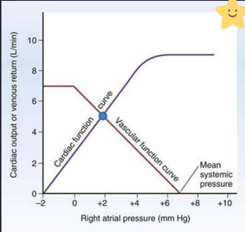
B3LG4.1. What is the venous return equation, and why does increasing RAP reduce venous return when other factors are constant?
Answer:
Venous return = (mean systemic filling pressure − right atrial pressure) / resistance to venous return.
Mean systemic filling pressure represents the upstream pressure available to drive systemic venous flow when the heart is not pumping.
RAP is the downstream pressure.
Increasing RAP while mean systemic filling pressure and resistance remain constant decreases the pressure gradient → reduces venous return.
Extra Information:
This is the mechanical reason the venous return curve slopes downward.
Increasing mean systemic filling pressure favors greater venous return.
Increasing resistance to venous return reduces flow for a given driving pressure difference.
Source: B3LG4 Cardiovascular Coupling PDF pp. 7–10, 18 | First Aid 2025 p. 291
B3LG4.1. What is the meaning of the right atrial pressure intercept of the Guyton venous return curve?
Answer:
The RAP-axis intercept occurs where venous return equals zero. At that point, RAP equals mean systemic filling pressure, so there is no pressure gradient to drive venous flow.
Increasing stressed blood volume or venous tone raises mean systemic filling pressure and shifts this intercept rightward.
Hemorrhage or venodilation reduces it and shifts the intercept leftward.
Extra Information:
Mean systemic filling pressure is not the same variable as measured systemic arterial pressure.
Venodilation can reduce stressed volume without changing total blood volume.
A rightward intercept shift is a useful clue for increased circulating stressed volume.
Source: B3LG4 Cardiovascular Coupling PDF pp. 8–10, 34–37 | First Aid 2025 p. 291

B3LG4.1. What determines the slope and plateau of the venous return curve?
Answer:
The downward slope reflects resistance to venous return, with greater resistance producing less flow for a given driving pressure difference.
At very low or negative RAP, large veins may collapse as surrounding pressure exceeds intravascular pressure.
This prevents further increases in venous return despite additional decreases in RAP and creates a plateau in the venous return curve.
Extra Information:
Changes in venous resistance alter the slope.
Changes in stressed blood volume or venous tone change mean systemic filling pressure.
Do not confuse changes in vascular function with changes in myocardial inotropy.
Source: B3LG4 Cardiovascular Coupling PDF pp. 7–10 | First Aid 2025 p. 291
B3LG4.2. Predict the acute Guyton curve changes caused by an increase in plasma volume.
Answer:
Increased plasma volume increases stressed vascular volume → raises mean systemic filling pressure → shifts the venous return curve rightward and upward.
With cardiac contractility unchanged, the new intersection generally has higher venous return, cardiac output, and RAP.
The heart moves along its existing Starling curve because increased filling permits a greater stroke volume.
Extra Information:
The faculty example increases CO from approximately 5.1 to 5.8 L/min and RAP from 1.9 to 4.2 mmHg.
Renal sodium and water retention can produce this shift.
A diseased heart may show a disproportionately large increase in RAP with limited CO improvement.
Source: B3LG4 Cardiovascular Coupling PDF pp. 18, 32–33 | First Aid 2025 p. 291

B3LG4.2. Predict the acute Guyton effects of hemorrhage before compensation occurs.
Answer:
Hemorrhage decreases circulating blood volume and stressed venous volume → decreases mean systemic filling pressure → shifts the venous return curve leftward/downward.
Venous return falls, lowering ventricular preload and stroke volume → cardiac output decreases.
With unchanged contractility, the operating point moves to a lower cardiac output and lower RAP.
Extra Information:
The faculty example changes CO from 5.1 to 4.3 L/min and RAP from 1.9 to 0.2 mmHg.
This is an acute uncompensated response.
Later sympathetic and RAAS responses can shift the curves again.
Source: B3LG4 Cardiovascular Coupling PDF pp. 30–31 | First Aid 2025 p. 291

B3LG4.2. Explain why venodilation reduces venous return even if total blood volume is unchanged.
Answer:
Venodilation increases venous capacitance, allowing more blood to occupy the unstressed venous reservoir.
Stressed vascular volume and mean systemic filling pressure decrease → the pressure gradient driving venous return falls → the venous return curve shifts left/downward → RAP, preload, and cardiac output generally decrease in a normal heart.
Extra Information:
Nitroglycerin is the faculty's example.
Acute venodilation can resemble mild hemorrhage on a Guyton diagram.
The critical variable is stressed volume, not necessarily total blood volume.
Source: B3LG4 Cardiovascular Coupling PDF pp. 34–37 | First Aid 2025 p. 289

B3LG4.2. Explain how the kidneys regulate the long-term venous return curve through sodium and water handling.
Answer:
Renal sodium retention expands extracellular and plasma volume → increases stressed blood volume and mean systemic filling pressure → promotes greater venous return and cardiac output.
Natriuresis and diuresis reduce volume → decrease filling pressure and venous return.
Hormonal systems such as RAAS respond to perceived low perfusion and can shift the cardiovascular equilibrium through sodium retention, vasoconstriction, and venous tone.
Extra Information:
Renal regulation is especially important over hours to days.
A failing heart may respond to fluid retention with large increases in filling pressure and congestion.
Increased effective arterial pressure can promote pressure natriuresis.
Source: B3LG4 Cardiovascular Coupling PDF pp. 14, 32–33, 44–48 | B3LG3 Antihypertensives PDF pp. 4, 9
B3LG4.3. Identify the four phases of a normal left ventricular pressure-volume loop and the valves involved.
Answer:
Ventricular filling occurs after mitral valve opening, increasing volume at relatively low pressure.
Isovolumetric contraction begins when the mitral valve closes, increasing LV pressure at constant volume until the aortic valve opens.
Ejection decreases ventricular volume as blood enters the aorta.
Isovolumetric relaxation begins after aortic valve closure, lowering LV pressure at constant volume until the mitral valve opens.
Extra Information:
Stroke volume equals EDV minus ESV.
The loop's horizontal width represents stroke volume.
True isovolumetric phases require both inflow and outflow valves to be closed without regurgitant flow.
Source: B3LG4 Cardiovascular Coupling PDF pp. 11–13 | First Aid 2025 p. 292

B3LG4.3. Explain how aortic stenosis alters the pressure-volume loop.
Answer:
Aortic stenosis increases resistance to LV systolic ejection → the LV must generate a larger pressure gradient across the stenotic valve → systolic LV pressure rises.
Greater afterload impairs ejection → ESV increases and stroke volume decreases, often narrowing the PV loop.
Chronic pressure overload promotes concentric LV hypertrophy, which decreases ventricular compliance and raises diastolic filling pressure.
Extra Information:
Mild AS may initially have little EDV change.
AS can produce narrow pulse pressure and delayed systolic ejection.
A stiff hypertrophied LV may generate an S4.
Source: B3LG4 Cardiovascular Coupling PDF p. 20 | First Aid 2025 pp. 293, 295
B3LG4.3. Explain how aortic regurgitation changes the PV loop and distinguish total from effective forward stroke volume.
Answer:
In aortic regurgitation, aortic blood flows backward into the LV during diastole → LV diastolic volume rises → EDV and ventricular preload increase.
Because regurgitation continues during periods that would ordinarily be isovolumetric, true isovolumetric relaxation is lost.
The loop typically becomes wider due to increased total ventricular stroke volume, although effective forward systemic output may fall because part of each ejected volume returns to the LV.
Extra Information:
The faculty's description of reduced stroke volume refers to impaired effective output, while First Aid distinguishes the increased total ejected volume.
Chronic volume overload causes LV dilation and eccentric hypertrophy.
Aortic diastolic runoff produces wide pulse pressure.
Source: B3LG4 Cardiovascular Coupling PDF p. 21 | First Aid 2025 pp. 290, 293
B3LG4.3. Compare the changes in EDV, ESV, LV pressure, and pulse pressure in aortic stenosis versus aortic regurgitation.
Answer:
Aortic stenosis → markedly increased LV systolic pressure, increased ESV, decreased stroke volume, and often reduced pulse pressure.
Chronic pressure overload causes concentric hypertrophy and reduced compliance.
Aortic regurgitation → increased EDV, loss of true isovolumetric phases, increased total ventricular stroke volume, and widened pulse pressure due to high systolic ejection combined with low aortic diastolic pressure.
Extra Information:
AS is a pressure-overload lesion.
AR is a volume-overload lesion.
The effects on effective forward flow and total ejected volume must be distinguished.
Source: B3LG4 Cardiovascular Coupling PDF pp. 20–21 | First Aid 2025 pp. 290, 293
B3LG4.3. Why can aortic stenosis cause myocardial ischemia even without coronary artery obstruction?
Answer: Severe aortic stenosis increases LV systolic pressure and wall stress, raising myocardial oxygen demand. Compensatory concentric hypertrophy increases myocardial mass and may impair diastolic relaxation. Elevated LV end-diastolic pressure reduces the effective coronary perfusion pressure gradient during diastole. Thus, oxygen demand rises while subendocardial perfusion reserve decreases → ischemia or angina can occur without an obstructive epicardial coronary lesion.
Extra Information:
- Coronary perfusion of the LV occurs predominantly during diastole.
- LV hypertrophy reduces compliance and raises filling pressures.
- This mechanism helps explain exertional angina in AS.
Source: B3LG4 Cardiovascular Coupling PDF pp. 17, 20 | First Aid 2025 pp. 288–289, 293
B3LG4.4. Predict changes in Guyton curves after acute hemorrhage versus high salt intake.
Answer: Hemorrhage reduces blood volume and mean systemic filling pressure → venous return curve shifts left/down → CO and RAP decrease. High salt intake with renal sodium/water retention expands blood volume and mean systemic filling pressure → venous return curve shifts right/up → CO and RAP increase, provided cardiac function remains unchanged.
Extra Information:
- Both cases primarily alter the vascular function curve.
- Cardiac output changes through movement along the existing cardiac function curve.
- Heart failure can magnify the RAP increase after salt and fluid retention.
Source: B3LG4 Cardiovascular Coupling PDF pp. 30–33 | First Aid 2025 p. 291
B3LG4.4. Explain how increased systemic vascular resistance changes the Guyton operating point.
Answer: Increased SVR raises ventricular afterload, making ejection more difficult and tending to lower cardiac output. Increased resistance also restricts flow through the systemic circulation and changes the vascular function relationship. The faculty's isolated increased-SVR example demonstrates a lower equilibrium CO with relatively little RAP change.
Extra Information:
- Faculty example: CO falls from 5.1 to 3.7 L/min while RAP is approximately unchanged at 1.8 mmHg.
- Both cardiac and vascular function relationships can change when TPR changes.
- Avoid assuming increased arterial pressure always increases cardiac output.
Source: B3LG4 Cardiovascular Coupling PDF pp. 22–23, 38–39 | First Aid 2025 p. 291

B3LG4.4. Explain how decreased SVR affects CO and RAP during early distributive shock.
Answer:
Systemic arteriolar dilation reduces afterload and resistance to systemic flow.
When the heart can respond, the reduction in resistance can permit increased cardiac output despite low arterial pressure.
On the faculty's Guyton diagram, the new operating point has higher CO with only a modest change in RAP.
Extra Information:
Faculty example: CO rises from 5.1 to 7.1 L/min and RAP changes from 1.9 to 2.3 mmHg.
High cardiac output does not guarantee adequate MAP because TPR may be very low.
Later myocardial depression or volume loss can change the response.
Source: B3LG4 Cardiovascular Coupling PDF pp. 40–41 | First Aid 2025 pp. 290–291

B3LG4.4. Why is the RAP response to a pure change in SVR often less predictable than the CO response?
Answer: Changing SVR influences both ventricular afterload and systemic vascular flow resistance. These competing changes can shift cardiac and venous return curves simultaneously. Therefore, CO often changes predictably with large afterload alterations, but the exact RAP change depends on myocardial function, venous return resistance, venous tone, and other hemodynamic conditions.
Extra Information:
- First Aid explicitly emphasizes that RAP changes with TPR alterations can be unpredictable.
- Consider whether the problem also includes venous constriction or volume retention.
- Do not interpret SVR as identical to mean systemic filling pressure.
Source: First Aid 2025 p. 291 | B3LG4 Cardiovascular Coupling PDF pp. 23, 38–41