lec 21 signal transduction I
 Call Kai
 Learn
 Practice Test
 Spaced Repetition
 Match
 Flashcards
 Knowt Play
1/74

Earn XP
Description and Tags
signaling enzymes, second messengers, modular domains
75 Terms
signal transduction
process by which an extracellular signal is amplified and converted to a cellular response
extracellular signals
molecular signals from other cells
environmental stresses
extracellular matrix
nutrient signals
cellular responses
gene expression
morphological changes
production/secretion
cell growth/division
cell death
signal transduction chemical process steps
reception
transduction
response
signal reception
most signals are chemical and are first sensed by receptors
signal transduction
once signals are detected, they will be recoded, amplified, and transmitted through a signal transduction pathway consisting of discrete chemical reactions
signal response
when acted upon by the relay molecules, cellular targets bring about responses underscored by chemical changes
a limited number of mechanisms mediate signal transduction
proteins and non-protein small molecule
proteins as a on-off switch mechanism
activation or inactivation of signaling enzymes
change in binding interaction between proteins, or between proteins and other biomolecules
change in protein conformation
post-translational modification
change in subcellular localization or local concentration
non-protein small molecule as a on-off switch mechanism
production of intracellular small signaling molecules (second messengers)
part 1: signaling enzymes
roles of enzymes in signal transduction
catalyze chemical reactions which control many key steps in signaling (hella convenient)
write and erase signals in a controlled fashion
provide simple mechanisms for signal amplification
major classes of signaling enzymes
covalent modification of proteins and lipids
effecting conformational change in other proteins
generation and elimination of small signaling molecules
covalent modification of proteins and lipids
kinases, phosphatases
proteases, lipases
effecting conformational change in other proteins
guanine nucleotide exchange factor (GEF), GTPase-activating protein (GAP)
generation and elimination of small signaling molecules
adenylyl cyclase, phosphodiesterase
enzyme catalytic activity can be controlled through input-driven protein conformational change
protein kinases
catalyze protein phosphorylation by transferring gamma-phosphate of ATP to the side chain of selected amino acids
in eukaryotic cells, protein phosphorylation occurs on…
the hydroxyl group of serine, threonine (Ser/Thr protein kinases)
hydroxyl group of tyrosine (Tyr protein kinases)

protein kinases can be…
soluble proteins (floating in cytosol)
membrane proteins (the kinase domain exists as an intracellular domain)
can be localized in the membranes
typically consist of integral transmembrane segments, but could also have a domain hanging out from the membranes
structure of eukaryotic protein kinase catalytic domain
despite the variety in protein kinases, they all have a catalytic domain that has a common overall structure:
two-lobed structure
N-lobe (n terminal)
inbetween lobes is the catalytic site
C-lobe (c terminal)
active site (catalytic)
activation loop (regulatory)
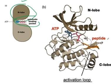
protein kinases function as molecular switches because they can exist in two conformational states: active and inactive
active = protein phosphorylation
inactive = no protein phosphorylation
upstream signals can use a variety of mechanisms to help regulate the equilibrium between the active and inactive conformation → leads to signal transduction
upstream signals activate the kinase → signal propagations
three major mechanisms for regulating protein kinase activity
binding of an activator protein or release of an inhibitor protein
through the production of a small molecule allosteric regulator (ex. second messenger)
through phosphorylation on the activation loop of the protein kinase
kinase cascade
activation of an upstream protein kinase leads to the phosphorylation and activation of a downstream protein kinase
protein phosphorylation changes the chemical property of the modified amino acid
from a polar uncharged amino acid to a neg charged amino acid
change the pI (isoelectric point) of modified protein
(more phosphorylation = more significant change in pI)
phosphorylation leads to these signaling outputs
change in protein conformation and activity
promote or prevent protein binding interaction
change protein subcellular localization
change protein stability (less or more stable)
protein phosphatase
“eraser” of the protein phosphorylation; counteracts the protein kinase
protein phosphatase subfamilies
serine/threonine phosphatases
tyrosine phosphatases
dual specificity phosphatase
protein phosphatase activity is also subject to regulation by upstream input signal
kinase and phosphatase reaction form a unidirectional cycle in which both reaction steps can be controlled and regulated independently because they are independent rxns
true statements about protein phosphorylation in eukaryotic cells
it can be reversed by the action of a protein phosphatase
it is catalyzed by a protein kinase
phosphoryl group usually derives from the gamma-phosphate of ATP
happens on ser, thr, and tyr side chains only
G proteins (guanine nucleotide-binding proteins)
also molecular switches
can exist in two diff states (on or off)
bind and hydrolyze GTP
when bound to GTP = ON
able to interact with downstream effector molecules
when bound to GDP = OFF
G protein functions
signaling: hormonal action, cytoskeletal regulation, and nuclear import and export
non-signaling: translation, translocation, vesicle trafficking
why can G proteins go through conformational switch?
gamma-phosphate on GTP interacts with switch 1 and switch 2 → induces reaction and interactions cause significant conformational rearrangements in switch I and II → enables G protein to interact with downstream effective molecules

to go from off to on conformation in G protein
exchange of GDP with GTP; it is very slow, so it is paired with GEFs
GEFs aka guanine nucleotide exchange factors
paired when G protein needs to go from off (GDP-bound) to on (GTP-bound)
accelerates GDP/GTP exchange to activate G proteins
to go from on to off conformation in G protein
GTP will undergo hydrolysis that is accelerated by GTPase-activator proteins (GAPs) because the reaction is also very slow if not without the catalytic enzyme
leads to inactivation
able to control the molecular switch of G proteins through…
GEF and GAP b/c GEF activates and GAP inactivates
Two main families of G proteins that play key roles in cell signaling
small G proteins: monomeric (only one subunit)
heterotrimeric G proteins: have alpha, beta, and gamma subunits
All G proteins contain the…
20 kDa G domain which bines GTP
small G protein = 1 G domain
heterotrimeric G protein = G domain in alpha subunit
heterotrimeric G proteins
plays role in G protein couple receptor (GPCR) signaling pathway
its beta and gamma subunits form a stable complex and have no enzymatic activity
beta-gamma is tightly associated therefore never dissociate
hetereotrimeric G protein in inactive form
G-alpha bound to GDP
beta-gamma of G forms hetero-trimer bc they associate with each other
heterotrimeric G protein in active form
G-alpha bound to GTP
G-alpha dissociates beta-gamma of G
both Gs can interact with downstream effectors
part 2 second messengers
second messenger
small signaling molecule that acts as an allosteric effector for a protein or an enzyme
bind to receptors and then receptors begin signal transaction pathway
molecules that are generated in response to signal transduction inside the cells
properties of second messengers
small
signaling controlled by synthesis/production and elimination/degradation
second messengers exert their effects by acting as allosteric regulators of downstream effectors
targets include a mide-range of enzymes and chennels
lipid derived-molecules can affect membrain recruitment of target proteins
second messengers lead to fast, distant, and amplified signal transmission
cyclic AMP (cAMP) (adenosine 3’-5’ -cyclic monophosphate)
derived from ATP
soluble second messenger
catalyzed by adenylyl cyclase
cyclic nucleotide phosphodiesterase (degrader)
hydrolyzes cAMP to AMP → eliminating its signlaing function
what is the main intracellular target of cAMP
protein kinase A (PKA)
downstream effector of cAMP
binding of cAMP to PKA activates the enzyme
inositol triphosphate (IP3)
synthesized from PI(4,5)P2
catalyzed by phosphoinositide-specific phospholipase C
water soluble therefore able to do stuff in the cytosol
has gated calcium channel in ER ← when IP3 bounded to gated calcium channel, it opens up and Ca will be released
diacylglycerol (DAG)
synthesized from PI(4,5)P2
catalyzed by phosphoinostide-specific phospholipase C
remains in membrane lipid
binds and activiates protein kinase C (PKC)
PKC
activated by DAG and calcicum conentration
calcium ion
concentration regulated by channels and pumps (degradation and production)
cytosolyc calcium concentration rises through in the presence of hormonal, neural, or other signals
an influx of extracellular calcium through gates calcium channel
the release of calcium from internal storage through gated calcium channel
absence of a signal = cytosylic calcium is removed by…
calcium-ATPase pumps
ca signaling is transient and local
bc high concentration of calcium in cells
effectors of calcium signaling regulated regulatory protein calmodulin (CaM)
why? because effectors of Ca do not typically bind to Ca directly
calmodulin (CaM)
contains four calcium binding sites
undergoes conformational change when Ca binds to it
when bound, hydrophobic surface is exposed which can then interact with downstream effector molecules
regulatory subunit of phosphorylase of beta kinase of muscle, which is activated by calcium
phosphatidylinositol phosphates
3 4 5 hydroxyl groups of it can be phosphorated to form PIPs
diff PIPs are recognized by specific phospholipid-binding protein domains
phosphorylation of phosphatidylinositol is done by phosphatidylinositol (PI) kinases
PI-3 kinases phosphorylates the hydroxyl group on the 3 position
reverse reaction is catalyzed by phosphatidylinositol phosphatases
dephosphorylates PIPs
a phosphatase that removes 3-phosphate from PI (3,4,5) is a tumor suppressor called PTEN
overview of what produces (catalyzes) what second messenger
adenylyl cyclase → cyclic AMP (cAMP)
phospholipase C → diacylglycerol (DAG) and Inositol 1,4,5-trisphosphate (IP3)
phosphatidylinositol-3 (PI-3) kinase → phosphatidylinositol types = PI3,4,5 and PI3 kinase
part 3 modular domains
modular domains
signaling proteins are frequently composed of multiple domains, joined by flexible linker sequences (“loops)
the flexible linker sequences allows the domains to retain their unique structures and unique functions
total function of a signal in proteins is equal to
the linear summation of all of the structure functions of these individual domains
advantages of modular domains
complex signal transduction scheme can be created by combining different domains
complex allosteric regulatory mechanisms can be devised for signaling enzymes
domain recombination can also generate proteins with new functions
two types of modular domains
catalytic and interaction
first class of modular domain: domains that recognize post-translational modification
ex. kinase (protein phosphorylation)
creates a new structure motif (new sites for protein-protein interaction) that can be recognized by other modular domains
ex. Src Homology 2 (SH2) domain: binds phospho-Tyr containing sequence
creates recognition site by an SH2 domain
SH2 recognized protein contains SH2 domain
different SH2 domains show specific preference for residues C-terminal to phospho-tyrosine (achieved through specificity pocket)

second class of modular domain: domains that recognize UNMODIFIED peptide motifs or proteins
protein conformational changes associated with signal transduction often expose unique sequence or structural features, which are recognized by modular protein interaction domains
ex. Src Homology 3 (SH3) domain: binds short, proline-rich sequence (PxxP) motives
proline-rich sequences favor the formation of polyproline type II (PPII) helix, a left-handed helix with three residues per turn
contains binding pocket that disfavors amino acids that do not contain proline
disfavors polar amide group in backbone of amino acid (so all other 19 amino acids) but it can tolerate a more hydrophobic N substitution (proline)
Src Homology 3 (SH3) domain
proline-rich sequences favor the formation of polyproline type II (PPII) helix, a left-handed helix with three residues per turn
contains binding pocket that disfavors amino acids that do not contain proline
disfavors polar amide group in backbone of amino acid (so all other 19 amino acids) but it can tolerate a more hydrophobic N substitution / ring structure(proline)

three class of modular domains: domains that recognize phospholipids
selectively bind phospholipids within biological membrane
target a protein to a particular subcellular organelle
localize a protein to a membrane
Pleckstrin homology (PH) domain
binds the phosphorylated inositol head group of these second messenger molecules → PI (4,5)P2, and PI(3, 4, 5)P3
specificity of phosphoinositide recognition is determined by the subtle differences in binding pocket
scenario: upstream signaling can lead to the activation of a kinase that can phosphorylate phosphatidylinositol molecules → lead to the binding of PH domain
phosphatidylinositol molecule will be recruited from the cytosol to the membrane because the lipids are actually membrane localized

adaptor protein
multi-domain protein that consist of modular domains that only binds to other molecules
proteins with multiple protein-binding domains that mediate the assembly of complexes of three or more components
ex. Grb2 protein links phospho-tyrosine containing receptors to downstream targets that contain proline-rich sequences
in the absence of Grb2, the protein that contains the proline-rich sequences CANNOT interact with the protein containing phospho-tyrosine
in the presence of Grb2, the proline-rich protein & phospho-tyrosine protein can interact with each other
acts as an adaptor, like a circuit and an outlet needing an adaptor

scaffold protein
multi-domain protein that consist of modular domains that only binds to other molecules
function: to organize complex signaling pathways; bind a series of proteins in a given signal transduction pathways
binds all the components within the signal transduction pathway
advantage: bring these components together and minimize the unproductive interaction among other proteins