6.1.5 Recognises common ocular abnormalities and refers when appropriate.
 Call Kai
 Learn
 Practice Test
 Spaced Repetition
 Match
 Flashcards
 Knowt Play
1/6

There's no tags or description
Looks like no tags are added yet.
7 Terms
Before referral for further investigation and suspected diagnosis of Glaucoma, all of the following tests should be performed:
Central VF assessment using standard automated perimetry (full threshold or supra-threshold)
ON assessment and fundus examination using stereoscopic slit lamp biomicroscopy or ONH image if available.
IOP measurement using GAT
Peripheral AC configuration + depth assessment with VH or OCT
Glaucoma
Before deciding to refer, consider:
repeated VFA and IOP measurements on another occasion to confirm a VF defect or IOP of 24mmHg or more
(unless clinical circumstances indicate urgent or emergency referral if needed)
Glaucoma
Refer for secondary care investigation and diagnosis of glaucoma if:
ONH damage on stereoscopic slit lamp biomicroscopy
VF defect consistent with glaucoma
IOP is 24mmHg or more using GAT
What are the visual acuity criteria for cataract referral?
Binocular VA of 6/9 or worse for drivers
OR
Binocular VA of 6/12 or worse for non-drivers
OR
Monocular VA of 6/18 or worse irrespective of the VA of the other eye
What other factors may justify cataract referral?
Pt is experiencing disabling glare from lights even though visual acuity is relatively unaffected
Pt lives alone or is the main carer for a partner or other relative
Significant hearing impairment
Significant impact on the pt’s day to day activities
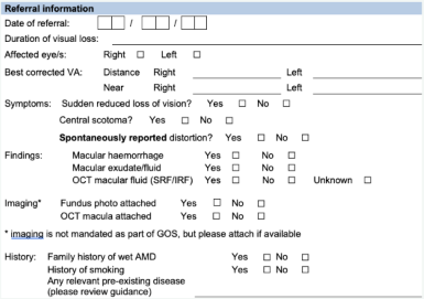
When may AMD patients require an urgent HES assessment?
Subretinal haemorrhage and subretinal fluid suggest choroidal neovascularisation. Urgent referral and assessment
Intraretinal haemorrhage centrally and exudates deposition superiorly. There may be subtle subretinal fluid or thickening. The presence of exudates is an important sign of leakage from choroidal neovascularisation. Urgent referral and assessment
Small areas of intra / subretinal haemorrhage amongst the drusen suggest choroidal neovascularisation. Urgent referral and assessment
AMD
If best corrected visual acuity is worse than 6/96..
These patients may require a HES assessment on a non-urgent basis.
They may benefit from LVA assessment, visual impairment counselling and/or registration
Only patients with wet AMD or CRVO will be offered the 2-week pathway