Animal Science 214 Lab Practical 1
 Call Kai
 Learn
Learn Practice Test
 Spaced Repetition
 Match
Match Flashcards
 Knowt Play
1/353

There's no tags or description
Looks like no tags are added yet.
Name | Mastery | Learn | Test | Matching | Spaced | Call with Kai | Chat |
|---|
No analytics yet
Send a link to your students to track their progress
354 Terms
General Information
X
Dorsal
Back and upper side

Ventral
The underside or abdominal area

Anterior
Front, frontal

Posterior
Back, rear

Medial
Toward the middle or body midline, particularly toward the body midline

Lateral
Away from the body midline; toward the side

Proximal
Near; closer to the point under consideration

Distal
Far or away; farther from the point under consideration

Frontal (or coronal) planes
Divides the animal into dorsal and ventral portions
Transverse or Axial plane
Divides the animal into cranial and caudal (head and tail) portions

Median (or midsagittal) planes
Separate an animal or person into right and left halves
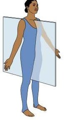
Sagittal planes
Any plane parallel to the midsagittal (median) plane; i.e., divides the animal into left and right portions
When does an organ, gland, or tissue secrete a substance?
If it both produces and exports the secretion out of the tissue or organ.
Give an example of an organ secreting a substance.
The liver secretes bile because the liver both makes bile and exports bile into the bile duct
When does an organ, tissue, or gland excrete a substance?
When a substance is made elsewhere
Give an example of an organ excreting a substance.
The gall bladder excretes bile because the bile is only stored there, but is made in the liver
What is the function of the esophagus in regards to transportation?
Transports food from the pharynx to the stomach
Enzymes
Proteins that catalyze chemical reactions in living systems, i.e., enzymes allow specific reactions to occur.
What are the seven key factors that influence physiology?
Nutrition
Genetics
Age
Environment
Gender
Physical change
Chemical change
Nutrition
Quality and quantity
Genetics
Heritability of traits such as udder size or muscling
Age
Tissues mature as you age and also breakdown
Environment
Highly stressful environments can alter how the body works
Gender
Male and female animals differ on several levels
Physical Change
Disease or injury can alter how a limb moves, if you will
Chemical change
Use of hormones, implants, or antibiotics to change productivity, influence growth or improve health status
Cardiovascular system
X
Arteri-
Artery
Cardi-
Heart
-emia
blood
erythro-
red
hemo-
blood
leuko-
white
vaso-
vessel
What are the functions of the circulatory system?
- Carry nutrients, such as amino acids, glucose, fatty acids and of course O2, to tissues
- Carry waste products from tissues for disposal - the principal ones are urea, NH3 (ammonia), and CO2 (carbon dioxide)
What is the function of the lymphatic system?
Return extracellular fluid to the veins: functions in disease control
Arteries
Carry blood AWAY from the heart
What are the layers of the artery made up of?
connective tissue and smooth muscle
What are the three layers of tissue that arteries are comprised of?
Tunica intima, Tunica media, and tunica adventitia (tunica extrema)
What makes up connective tissue?
Elastin and collagen
Elastin
A protein that allows for greater elasticity of tissues; breakdown overtime, but provides 1.5x more elasticity and provide support to the collagen
Where can elastin be found?
Skin, lung, and artery tissue
Collagen
The main structural protein that provides strength to tissues and gives protection to more fragile structures like organs (ie. kidneys, spleen)
Collagen makes up how much of body protein and has a triple helical structure?
20-30%
Tunica Intima
Innermost coat or layer; consists of a continuous layer of supporting connective tissue
Tunica Media
The middle layer; contains smooth muscle circularly arranged containing elastin. Usually the thickest layer
Tunica Adventitia (tunica extrema)
Outermost layer; connective tissue containing elastin and collagen
Capillaries
Endothelial cells covered with a basement membrane
What is the function of the capillaries?
Function in exchange of materials between the blood and tissues
What are the three types of capillaries?
Continuous, fenestrated, and sinusoidal
Continuous
Endothelial cells are held together by tight junctions.
Where are the continuous cells located?
Most tissues such as skeletal muscles
Fenestrated
The endothelial cell layer has numerous holes, resulting in porous capillaries that allow for rapid absorption and filtration.
Where are the fenestrated cells located?
Kidney and intestines
Sinusoidal
The endothelial cell layer has numerous layers has numerous large intercellular gaps and an incomplete basement cell layer. This allows for the exchange of large molecules and materials, such as red blood cells.
Where are the sinusoidal capillaries located?
Liver, spleen, lymph nodes, bone marrow, and endocrine glands
Veins
Blood vessels that carry blood back TO the heart; smooth muscle circularly arranged, more collagen, less elastin
Venules
These are much like capillaries but of larger diameter
Lymph Ducts
Lymph is extracellular fluid, which is returned to the blood via the lymphatic system
Know the parts of the heart

Right atrium
Receives blood From the vena cava and moves it TO the right ventricle
Right ventricle
Pumps blood FROM the right atrium TO the pulmonary artery
Left Atrium
Receives blood FROM the pulmonary veins and pumps it TO the left ventricle
Left Ventricle
The largest heart chamber. Pumps blood TO the aorta FROM the left atrium
Atrioventricular valves
Valves located between the atria and ventricles; prevent blood from flowing from the ventricles back into the atria
Semilunar valves
Valves located between the ventricles and the arteries leaving the heart. Prevent blood from flowing back into the ventricles
Vena cava
The largest vein in the body; has two parts. The superior vena cava carries blood from the upper body and the inferior vena cava brings blood from the lower body to the right atrium.
Aorta
The largest artery in the body. Carries blood FROM the left ventricle TO the body
Coronary Arteries
multiple arteries running on both sides of the heart that supply nutrients and O2 to the heart muscle
Pulmonary Artery
Carries low-oxygen blood FROM the right ventricle TO the lungs.
Pulmonary vein
Carries high-oxygen blood FROM the lungs TO the left atrium
What are the two types of heart valves?
Atrioventricular and semilunar
Atrioventricular valves
These permit blood flow from the atria into the ventricles but prevent blood from flowing back into the atria when the ventricles contract. The right atrioventricular valve is tricuspid, meaning that it has three leaflets or cusp
Semilunar valves
These are located where blood leaves the heart and are called the aortic semilunar valve and pulmonary semilunar valve. The semilunar valves permit blood to leave the heart but not return.
Lub
The heart sound made when the atrioventricular valve snaps shut
Dub
The heart sound made when the semilunar valve snaps shut
Pulmonary circulation
Blood circulates from the heart (right ventricle) to the lungs and back to the heart (left atrium). Pulmonary is a term referring to the lungs.
Systemic Circulation
Blood circulates from the heart (left ventricle) to an organ and back to the heart(right atrium)
Portal Systems
Portal circulation is a special case of systemic circulation. Portal circulation refers to blood passing from one organ to another organ that is not the hear
Hepatic portal system
Hepatic is a term referring to the liver. This portal system transports blood collected from the absorptive areas of the digestive system to the liver. Blood from the stomach and the small intestine is taken directly to the liver. From there it is returned to the heart
Hypothalamic-hypophyseal portal system
Blood from the hypothalamus goes directly to the hypophysis (pituitary) before returning to the heart. This system carries hormones, such as the releasing hormones, from the hypothalamus to the hypophysis. This portal system prevents degradation and dilution of the small amounts of hormones produced
What is the blood circulatory pattern?
(Superior and inferior) vena cava --> right atrium --> right ventricle --> pulmonary artery --> Lungs --> pulmonary vein --> left atrium --> left ventricle --> aorta --> organ --> vena cava
How many chambers do reptiles and amphibians have?
3 chambers- 2 atria and 1 ventricle
How heavy is the equine heart?
7-10 pounds
How heavy is the bovine heart?
5 pounds
How heavy is the human heart?
7-15 ounces
How heavy is the porcine heart?
10 ounces
How heavy is the ovine heart?
5 ounces
Internal Anatomy of the Pig
X
Brachi-
arm
Cauda-
tail
cervic-
neck
corpus
body
ped, pod
foot
pharyngo-, laryngo-
throat
nas-, rhin-
nose
-plasty
plastic surgery
pleur-
chest lining
pneum-, pulm-
lungs