Parasitology Practical Prep
 Call Kai
 Learn
 Practice Test
 Spaced Repetition
 Match
 Flashcards
 Knowt Play
1/28

There's no tags or description
Looks like no tags are added yet.
29 Terms

Name this intestinal amoeba
Entamoeba histolytica
12–15 µm
1–4 nuclei, central karyosome, even chromatin, and cigar-shaped chromatoid bodies with blunt/rounded ends
Troph ingests RBCs
stool O&P
dysentery, extraintestinal amebic liver abscesses

Name this intestinal amoeba
Entamoeba dispar
>10 µm
1–4 nuclei, rounded chromatoid bars
NEVER ingests RBCs
Reported as "Entamoeba histolytica/dispar" unless separated by PCR/antigen testing
Non-pathogenic commensal

Name this intestinal amoeba
Entamoeba hartmanni
5–10 µm
1–4 nuclei and rounded chromatoid bodies
Non-pathogenic commensal
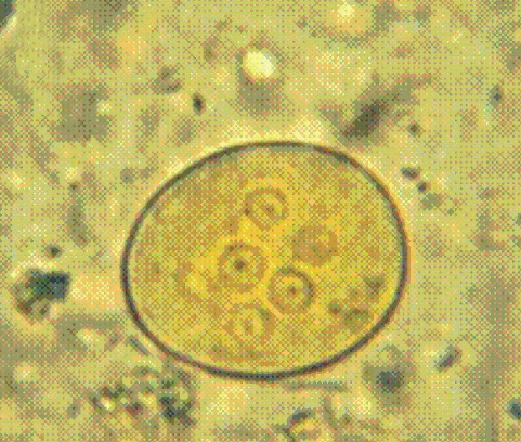
Name this intestinal amoeba
Entamoeba coli
10–35 µm
1–8 nuclei (mature = 8), eccentric karyosome, coarse/uneven peripheral chromatin, and splintered/sharp/pointed chromatoid bodies
Non-pathogenic commensal
Troph = "junky cytoplasm"
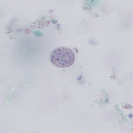
Name this intestinal amoeba
Endolimax nana
5–10 µm
oval cysts with up to 4 blot-like/clumped nuclei and NO peripheral chromatin
Non-pathogenic commensal

Name this intestinal amoeba
Iodamoeba bütschlii
5–20 µm
1 nucleus with a large karyosome and a prominent, large glycogen vacuole that stains dark brown with iodine
Non-pathogenic commensal
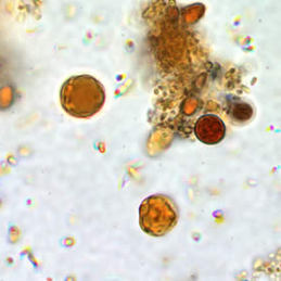
Name this intestinal amoeba
Blastocystis hominis
Spherical cell (5–30 µm, avg 8–10 µm) with a large central vacuole/body compressing 1–4 dark nuclei into a thin outer rim of cytoplasm
Diarrhea/abdominal discomfort

Name this flagellate
Giardia duodenalis
9–12 µm
Oval with 4 nuclei and visible internal fibrils/axonemes
Trophozoite (10–20 µm × 12–15 µm): Pear/pyriform shape with 2 nuclei ("monkey face")
"Beaver Fever" diarrhea
Case clues: outbreaks from drinking untreated water or daycare contact

Name this flagellate
Dientamoeba fragilis
Flagellate with NO CYST STAGE
9–12 µm
contain 1 or 2 nuclei (60–70% are binucleate) with karyosomes fragmented into 4–8 granules; no peripheral chromatin
diarrhea, abdominal pain
Case Clues & Sample: Transmitted via helminth eggs (Ascaris / Enterobius), PVA-fixed trichrome stain

Name this flagellate
Chilomastix mesnili
Cyst (6–10 µm)
lemon-shaped with a clear knob at one end
Trophozoite (10–15 µm) is pear-shaped with a curved cytostomal fibril ("shepherd's crook")
Non-pathogenic commensal

Name this ciliate
Balantidium coli
Cyst (50–70 µm) is round with a thick wall and visible macronucleus
Trophozoite (50–100 µm) is ciliated with a kidney bean-shaped macronucleus
Case Clues & Sample: Exposure to swine/pigs
Specimen: Stool wet mount/O&P
dysentery, intestinal ulceration
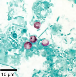
Name this coccidia
Cryptosporidium
Small, spherical oocysts (4–6 µm)
Modified Acid-Fast positive (stains bright pink/red)
Autoinfective and chlorine-resistant
Case Clues & Sample: Daycare outbreaks, contaminated swimming pools/water supplies
diarrhea; life-threatening in AIDS/immunocompromised patients

Name this coccidia
Cyclospora cayetanensis
Spherical oocysts (8–10 µm — twice the size of Cryptosporidium)
Modified Acid-Fast positive (variable/ghost staining)
Autofluoresces intense blue under UV light
Case Clues & Sample: Summer outbreaks associated with imported fresh produce
relapsing watery diarrhea

Name this coccidia
Cystoisospora belli
Large, ellipsoidal/spindle-shaped oocysts (25–30 µm × 10–19 µm) containing 1–2 sporoblasts
Modified Acid-Fast positive and autofluoresces blue under UV light
Severe watery diarrhea in AIDS patients

Name this free-living amebae
Naegleria fowleri
Amoeboid trophozoites with blunt pseudopods in CSF wet mount
Case Clues & Sample: Water-skiing or swimming in warm fresh water; nasal entry through cribriform plate
Primary Amoebic Meningoencephalitis (PAM) — rapidly fatal CNS infection

Name this free living amebae cyst form
Acanthamoeba
double-walled wrinkled/star-shaped cysts
Case Clues & Sample: Contact lens wearers using homemade saline or tap water
Specimen: Corneal scrapings, tissue biopsy, CSF
Granulomatous Amoebic Encephalitis (GAE); Acanthamoeba keratitis

Name this flagellate troph
Trichomonas vaginalis
Pear-shaped trophozoite (7–23 µm) with 4 anterior flagella, axostyle, short undulating membrane, and jerky motility
NO CYST STAGE
Case Clues & Sample: Sexually transmitted, Vaginal swab wet mount, urine sediment wet mount during routine urinalysis

Name this hemoflagellate
Leishmania spp.
Diagnostic human stage = amastigote (intracellular in macrophages, 2–4 µm, featuring a nucleus and a rod-like kinetoplast) Vector stage = promastigote
Case Clues & Vector: Vector = Phlebotomine Sandfly
Specimen: Tissue biopsy, bone marrow
Skin ulcers, nasal septa destruction, donovani kala azar severe anemia

Name this hemoflagellate
Trypanosoma brucei
14–33 µm, with a small terminal kinetoplast, undulating membrane, and free flagellum
Case Clues & Vector: Vector = Tsetse fly
Specimen: Giemsa-stained blood smear, lymph node aspirate, CSF
African Sleeping Sickness
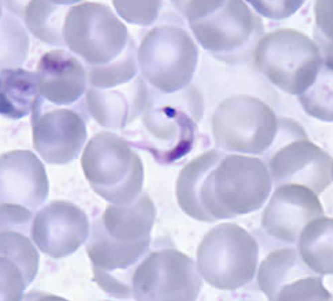
Name this flagellate
Trypanosoma cruzi
rigid 'C' or 'U' shape on blood smear with a very large terminal kinetoplast
Chagas Disease
Vector = Triatomine / Reduviid bug ("Kissing bug")

Name this plasmodium
Plasmodium falciparum
Infected RBCs are NORMAL size
Rings are small/delicate with double chromatin dots ("headphones"), appliqué/accolé forms, and multiple rings per RBC
Banana- or crescent-shaped gametocytes
Vector = female Anopheles mosquito
Most severe/deadly malaria

Name this plasmodium
Plasmodium vivax
Infected RBCs are ENLARGED
Trophozoite is amoeboid; Schüffner's dots present in RBC
Schizont contains >12 merozoites (12–24)
most widespread geographically (80% of cases)
Relapses due to hypnozoites in liver

Name this plasmodium
Plasmodium ovale
Infected RBCs are ENLARGED
OVAL-shaped with fimbriated/fringed/jagged edges and Schüffner's dots
Schizont contains 6–14 (avg 8) merozoites
liver hypnozoites cause malaria relapses

Name this plasmodium
Plasmodium malariae
Infected RBCs are NORMAL or SMALL size
Trophozoite forms a distinct band or basket form across the RBC
Schizont forms a symmetrical "rosette" pattern (6–12 merozoites surrounding central dark pigment)
Quartan malaria (72h cycle periodicity)

Name this plasmodium
Babesia microti
Small pleomorphic rings in normal RBCs
Classic hallmark: Maltese Cross (tetrad form of 4 merozoites)
NO MALARIAL PIGMENT (hemozoin) or gametocytes
Case Clues & Vector: Vector = Ixodes scapularis tick
fever, chills, hemolytic anemia

Name this nematode (roundworm)
Enterobius vermicularis (Pinworm)
Egg (50–60 × 20–30 µm) is D-shaped (flattened on one side) with a thick, smooth double-layered shell and larva inside
Adult female worm (8–13 mm) has a pointed tail
Case Clues & Sample: Gravid female migrates to perianal skin at night to lay eggs
Specimen: Cellophane Tape / Swube tube prep
intense perianal itching

Name this nematode (roundworm)
Trichuris trichiura (Whipworm)
Egg (50–55 × 20–25 µm) is barrel- or lemon-shaped, yellow-brown/bile-stained, with a thick shell and prominent clear mucoid plugs at BOTH poles (bipolar plugs)
Adult worm has a thin whip-like anterior end
Case Clues & Sample: Ingestion of eggs from soil/food
bloody diarrhea, rectal prolapse in children

Name this nematode (roundworm)
Ascaris lumbricoides (Large Roundworm)
Fertile egg (45–75 × 35–50 µm) is golden-brown with a thick, bumpy/mammillated (albuminous) outer shell
Unfertilized egg is larger, elongated, with irregular mammillations
Adult worm is large (6–12 inches / 15–35 cm)
Case Clues & Sample: Ingestion of embryonated eggs from soil/food
most common helminth infection worldwide
Larval migration through lungs causes pneumonia
adult worms cause bowel/biliary obstruction
