Construction of Neuronal Circuits during development + Neural regen, Stem cells
 Call Kai
 Learn
 Practice Test
 Spaced Repetition
 Match
 Flashcards
 Knowt Play
1/71

There's no tags or description
Looks like no tags are added yet.
72 Terms
WHy are neurons really just highly specialized POLARIZED epithelial cells (structure/function of each)
Epithelial cells have apical (secretion) surface and basal (junctions, intracellular comms( surfaces
Neurons:
axons → secretion
Dendrites → Transuction + Communication
what is a protein that is required for axonal development?
Par 3 Protein

What does having more dendrites mean for signal integration?
More dendrites = Inc input signal integration
How do microtubules betwee axons and dendrites differ?
Axons → Axonal microtubules = uniform, parallel, specific orientation to target
PLUS end faces axon tip
Dendritic microtubule → mixed orientation
PLUS ends face cell body or dendritic tip
What is MAP2 and Tau
MAP2 → molecule mostly found in dendrites
Tau → Molecules mostly found in axons
What are TROPIC factors vs TROPHIC factors
Tropic → For axon growth
Trophic → For Supporting survival of AXONS AND growth for DENDRITES
What are the 3 molecules required for dendrite growth (dendrite growth molecules) (AND ALSO TROPHIC FACTORS)
NT-3 (Neurotrophin 3)
BDNF (brain-derived Neurotrophic factor)
Nerve Growth Factor (NGF)
NT4/5
What are the 3 stages of axon outgrowth?
Protrusion
Engorgement
Consolidation
What is filipodia and lamellipodia respectively, what do they do? What are they a part of?
Filipodia → Actin fingers → Guides developement
Lamellipodia → Webbing → Protrusions
!!! PART OF AXON GROWTH CONE
Depolymerization and polymerization of actin during growth cone movement
What did (Ramon y Cajal) propose (what model) and what is it?
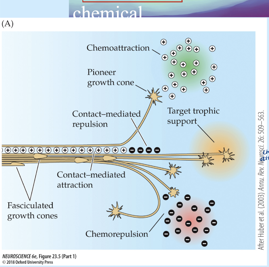
Chemoattraction model
Axon growth is guided by chemicla attraction of molecules that were secreted by the target cells (chemotaxis = movement towards the chemicals)
What is chemotaxis?
Movement twoard a chemical
What are the tropic factors that are used for chemo attraction and cemo repulsion? FOR AXON GUIDANCE
Netrin →
Slit
Semaphoris
Netrin is mostly located in ___ and makes sense b/c ____
Mostly locaated in floor plate and makes sense b/c it guides commissural axons expressing DCC receptor across midline in spinal cord
What does SLIT do for axon growth aloneE?
Has commissural axons stay on same sid (NO crossing over to other side)
prevents crossing over before and after if it has occurred.
What do semaphorins do for axons?
Tell them where NOT TO GROW (chemorepellent)
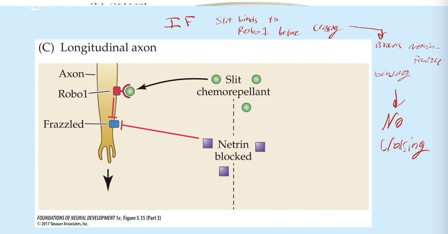
What is essential proteins receptor involved in neuron projection
Frazzled protein
Robo1
Mechanism of Netrin and how it works with Frazzle and Slit-Robo-1 to cause growing to dorsal side?
Netrin binds to Frazzle protein on VENTRAL side of nervous system → Guides the axon to cross over to the orther side and grow (away from netrin, grows toawrds the DORSAL side)
To keep on dorsal side:
Frazzle stops interaction with Netrin → ROBO1 receptor on axon binds to SLIT on dorsal side → SLIT-ROBO1 interaction prevents crossing back over.

Why does the mechanism for netrin have to be specific (why does netrin frazzle. binding have ot happen first)
IF Slit were to bind to ROBO1 before Netrin-Frazzle biinding to cause intial cross over to dorsal side → Netrin-Frazzle is blocked from binding → No crossing to dorsal side in first place
once cross border don’t come back
What happens after corssing generally?
diff receptors are expressed which is what keeps the axon in place and crossing over again
How is growth of the axon happening and with what? (what specificallt is being used to grow it)
polymetization and de-polymerization of F-actin

Nerve pathways with robo KO, slit KO, and constant ROBO expression

why is adhesion important for growth cone progress?
Allows adherence to other cells and to the ECM
What are 3 cell adhesion molecules (CAMs)
neuro cell adhesion molecule (NCAM)
L1
Cadherins (Calcium-dependent cell adhesion proteins)
What is corpus callosum agenesis, what is it caused by?
Happens because of L1 mutation, corpus callosum never forms properly b/c no adhesion
In roger sperry frog experiment, what was he looking at?
Topographic mapping in frogs
projections in frogs and functions of components? (tectum)
Axons in ANTERIOR retina project to POSTERIOR tectum
^and vise versa
Anterior retina → Posterior tectum
Posterior retina → Anterior tectum
Tectum → receives visual info about the fly and will direct tongue movement toward the fly
What did Roger sperry specifically do to the frog’s eye, what happened as result?
Rotation of the frog eye. When fly presented in specific direction, frog shot it’s tounge out in exact opposite direction of the fly.
why?
Eye rotated 180° → Retinal axons retain their original position → Eph gradient guides axon to their original tectal target, and don’t establish new ones even tho eye was flipped → Visual map becomes misaligned → Frog perceives fly in the opposite direction → Tongue strikes in the wrong direction.

What is Epherine (EPH) and epherin receptor? What happens when binding?
epherine binds to it’s receptor → repuslive signal causing growth cone to grow away from epherine source

What does decrease in ephrin lead to?, can also be a result of ___
Agenesis of corpus callosum → leads to visual issues
fetal alcohol syndrome
What is Nerve Growth Factor (NGF)
Factor that promotes growth of neurons, ipmplications in development but also cancer and neurodegenerative disease
What does the neurotrophic hypothesis entail?
During development more neurons are produced than what will end up being used.
Neuronns compete for survival factors, trophic factors, (limited supply)
Winners live loosers due. Target cells produce limited trophic substance
What is one motor unit:
motor neuron and the muscle fiber it innervates. Only ONE motor neuron innervates a muscle fiber, and that one motor neuron can innervate other muscle fibers as long as it is the only one doing so.

number of cells that survive is dependent on ____. compteition for ____
number of cells that survive is dependent on INTERACTIONS WITH CELLS IN THE TARGET. compteition for TROPHIC SUPPORT


Does receptor matter for neurotrophins for it’s responses
Yes. Diff receptors = diff responses

what are the 3 types of neural “repair” after an injury?
Axon regrowth
Restoration of damanged central nerve
New neuron generation
Dmg to peripheral target cause for axon regrowth?

How is restoration of damanged central nerves affected by glia?
After injury, there is overgrowth of glia, restricting some restoration

Where does generation of new neuron happen and not happen, How are they generated?
CANNOT do in erebral cortex, only some regions of brain
Same process as before using neural stem cell (proliferate, migrate, differentiate)

What did Henry Head do with his own nerves to demonstrate healing?
Cut his cutaneous and radial nerve → Stitched them back together → Some crude functions recovered after 6-13 weeks. Fine motor control slowly recovered after 2 years.

What key cels are involved in Peripheral nerve regeneration (where nerve regeneration is mailny constricited to, doesn’t happen much in CNS)
Schwann cells
Macrophages
Axon promoting growth signals
What are the axon promoting growth signals?
Laminin
Fibronextin
Collagen
NCAM
L1
N-Cadherin
NGF
BDNF
Steps for regeneration of peripheral nerve
Peripheral nerve damaged → Macrophages remove damaged myelin → Axons regenerate with schwaan cells due to secretion of the axon promoting growth factors → Regenrating axon growth cone grows in direction of those promoting molecules
What is needed for successful recovery (with proximal and distal ends of nerve)
PERCISE re-apposition is NEEDED for a good recovery

Do crushed or cut nerves have a more successful recover?
Crushed nerves
How do undamaged nerves contribute to regeneration of damaged nerves. What are they releasing to help?
they help regeneration of damaged nerves, upregulate guidance molecules like integrins in sensory and motor neurons.
What is special about optic nerve?
It is only cranial nerve NOT part of PNS, and thus therefor is a part of CNS. Therefor cannot regenerate

For example of reinnervation at NMJ, what molecules are upregulated and what are down regulated?
BDNF and NGF upregulated, NT3/4 downregulated in this instance
What is the Dorsal Root Entry Zone (DREZ)
Transition zone where sensory neuron from dorsal root ganglia enters the spinal cord (interface between PNS and CNS)
What is a cell heavily involved in repairing dorsal root ganglia? How do they function
Astrocytes (type of glia)
Dorsal root ganglia injury → influx of astrocytes into DREZ, non-permisive environment created → astrocytes form barrier at DREZ, blocking injured fibers from regrowing into spinal cord
Why does the CNS not regenerate as well ad PNS? (5 main reasons)
Glial cells inhibit growth (secrete growth inhibitory factors), astrocytes and microglia secrete
Extensive neuronal death
Presence of inhibitory molecules in CNS myelin debris
Many classes of nerve cells and complex connections
Neuronal stem cells have limited ability to divide
What happens with debris in CNS with injury to axon?
Nothing, debris is not removed. Makes the environment toxic and unable to regenerate
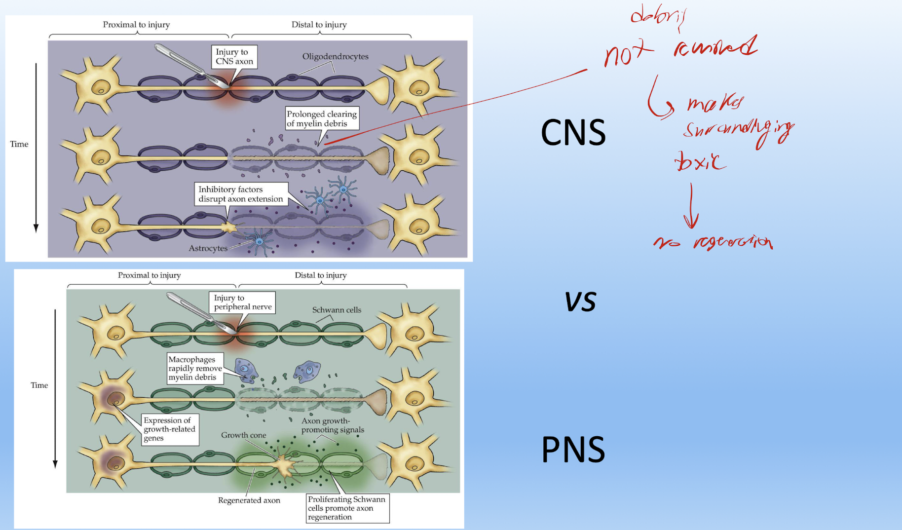
What do oligodendrocytes do to CNS axons that deter axonal regeneration. What are the inhibitory molecules
They limit plasticity and also secrete the inhibitory stuff and toxins.
MAG
Nogo-A
Omgp
Ephrin B3
Semaphorin 4D
What is a glial scar, and how does it prevent regrowth of CNS axons
Scar made of glial cells, rich with inhibitory molecules:
Chondroitin
Suldate
Proteoglycan
What else do astrocytes produce after injury and with glial scars
Produce inhibitory molecules semaphorine, epherins, and slit (DIRECT WHERE NOT TO GO)
What are more detrimental effects of reactive astrocytes?
Loss of homeostatic function
More gain of detrimental function with potential toxic effects

how can the glia scar be protective?
The glial scar helps contain the injury, remove cellular debris, limit inflammation, and protect surrounding healthy neurons. However, excessive scarring can inhibit axonal regeneration.
how does myelination/remyelination of axons affect multiple sclerosis
more remyelination → less severe for multiple sclerosis
Some examples of CNS damage that lead to cell death?
TBI
Neurodegenerative disease (AZD, ALS, Parkinson’s, Alzheimer (Tau protein accumulation)
Tumors
Hypoxia (stroke)
Exitotoxicity - glutamate overstim caused by abnormal activity of neurons after brain damage
What is capase-3 and what is it imporant for__
Capase-3 → leads to apoptosis and necrosis
What are some stimuli for apoptosis?
DNA dmg
Hypoxia
Stress
Growth Factor WIthdrawal
What are 4 examples of neural regeneration in the CNS accross 4 diff species?
Fish retina
Male Canary new neuron generation
Neuron generation in mammalian olfactory bulb
Dentae gyrus of hippocampus in mammals
For each of the 4 examples list what each of them is about and what the new neuronal generation is used for?
Fish retina → Growth of Retin, continual generation from retinal precursors
Male Canary new neuron generation → Male birds have new neurons generating for changes in their songs due to increased testosterone. Injecting testosteron makes female birds sing too (Testosterone = singing)
Neuron generation in mammalian olfactory bulb → new neurons to replace old ones as they adapt to sensory input, happens w/ increased interneurons in later ventricle
Dentae gyrus of hippocampus in mammals → increased interneurons, hippocampus uses for helping in forming memories
Remember: SVZ = where new neurons are produced; RMS = where they migrate; olfactory bulb = where they differentiate and integrate.
What are two ways in labeling dividing neurons?
BrdU technique (only works with molecules in S phase)
atmospheric 14C
How is 14C used to establish that most neurons are born before birth
14C is increased in atmosphere because of nuclear testing
ppl born before nuke → their DNA had the C14 levels that represented the atmosphere BEFORE the nuke. The few neurons that were produced after birth in adulthood had the new high cortical C14 levels.
^^^^^^^^^^BUT THERE WAS MUCH MORE LOWER C14 LEVEL DNA THAN HIGH C14 LEVEL
This proved that most neurons are born before birth

Benefit of Stem cells for neurons after transplantation?
Added trophic support to prevent death. may require addition of cells continually supplying trophic support.
Does bmp have same effect for neural induction early on and later
No
Early on → leads to skin development
Later → (if noggin chordin block it’s activity so neuroectoderm can form) Leads to sensory neuron development in dorsal region
What is the gradient and signaling of molecules at different times leading to diff outcomes called?
Neural Patterning
gradients activate transcription factors at diff places at diff times
Relationship between ectoderm becoming neuroectoderm (nervous tissue) instead of the epidermis (skin). Which molecules lead to each and by what mechanism for INITIAL NEURAL INDUCTION
Default → Ectoderm wants to become neuroectoderm (nervous tissue)
BMP signaling prevents that and causes ectoderm to become Skin
Noggin/Chordin block BMP activity → Allow ectoderm to neuroectoderm
BMP and SHH function after neuroectoderm is alr formed?
BMP → Dorsal → Sensory neurons
SHH → Ventral → Motor neurons