Exam 3 Ch 5-6
 Call Kai
 Learn
Learn Practice Test
 Spaced Repetition
 Match
Match Flashcards
 Knowt Play
1/86

There's no tags or description
Looks like no tags are added yet.
Name | Mastery | Learn | Test | Matching | Spaced | Call with Kai | Chat |
|---|
No analytics yet
Send a link to your students to track their progress
87 Terms
Integumentary System consists of
Skin
Sudoriferous (sweat) glands
Sebaceous (oil) glands
Hairs
Nails
Subcutaneous tissue

skin consists of two layers
epidermis
dermis
hypodermis (not part of skin)
epidermis is the outermost layer of the skin, avascular, and consists of
epithelial tissue
dermis underlies the epidermis, vascular, and consists of
dense connective tissue
hypodermis or subcutaneous tissue is not part of the skin but still functions as ___ in the integumentary system
loosely anchors skin to muscles
shock absorption
insulation
epidermis consists mostly of
keratinized stratified squamous epithelium
types of cells that make up the epidermis
Keratinocytes
protection
Melanocytes
pigment- melanin
Dendritic cells
macrophages- ingest foreign substances and activate immune system
Tactile epithelial cells
sensory receptors
4-5 layers of epidermis (come lets get sun burnt)
Stratum basale
Stratum spinosum
Stratum granulosum
(Stratum lucidum)- palms/soles
Stratum corneum
cells above stratum granulosum are _____ while cells below are ____
dead, alive
dermis consists of what cell types
fibroblasts, macrophages, and occasionally mast cells and white blood cells

two layers of dermis
papillary dermis
reticular dermis
papillary dermis is the thin superficial layer of _____ consisting of loose, interlacing collagen and elastic fibers and blood vessels
areolar connective tissue
dermal papillae is the superficial region of dermis that sends fingerlike projections up into epidermis that contains
capillary loops, free nerve ending, and touch receptors
friction ridges (occur in thick skin are a collection of ridges formed by dermal papillae on top of dermal ridges) function as
enhance gripping ability
sense of touch-vibrations
reticular dermis consists of coarse _____ that provide stretch/recoil and strength and resilience
dense irregular connective tissue
dermal vascular plexus (reticular dermis)
network of blood vessels between reticular layer and subcutaneous tissue
cleavage tension lines (reticular dermis)
reticular layer are caused by many collagen fibers running parallel to skin surface
invisible
flexure lines (reticular dermis)
dermal folds at or near joints
(hands, wrists, joints)
Extreme stretching of skin can cause dermal tears, leaving silvery white scars called
striae
Acute, short-term traumas (e.g., burn or repeated friction) to skin can cause ___ , fluid-filled pockets that separate epidermal and dermal layers
blisters
what 3 pigments contribute to skin color
melanin carotene and hemoglobin
melanin is the only pigment made in skin by
melanocytes
two forms of melanin
reddish yellow or brownish black
excessive sun exposure can destroy ___ that causes alterations in D N A that may lead to skin cancer
folic acid (Necessary for DNA synthesis)
carotene has a yellow orange pigment most obvious in palms and soles because it accumulates in what tissues
stratum corneum and subcutaneous tissue
hemoglobin has what color
pinkish
What other factors cause alterations in skin color
Blueness, or cyanosis: Low oxygenation of hemoglobin; sign of respiratory or cardiovascular issues
Pallor, or blanching: Emotional stress, low blood pressure, anemia (decreased ability of blood to carry oxygen),
Redness, or erythema: Embarrassment, fever, inflammation, allergy
Yellowness, or jaundice: Liver disorders
Red/purple/green/yellow marks, or bruises, or ecchymoses, or hematomas: Result of blood leakage from damaged blood vessel beneath skin
Brown or black patches: Hyperpigmented, thickened skin in folds may be a sign of endocrine disorder
hair consists of
dead keratinized cells
what are the functions of hair
Sense insects on skin before bite or sting
Hair on head guards against physical trauma
Protect from heat loss
Shield skin from sunlight
Eyelashes shield eyes
Nose hairs filter particles from inhaled air
list two regions of hair
Root: portion within scalp, where keratinization is still going on
Shaft: portion that extends above scalp, where keratinization is complete

what are the 3 parts of the hair shaft
Medulla: central core of large cells and air spaces
Cortex: several layers of flattened cells surrounding medulla
Cuticle: outer layer consisting of overlapping layers of single cells
hair pigmentation is made by ___ in hair follicles
melanocytes
hair follicle
extends from epidermal surface to dermis (sometimes into subcutaneous tissue)

hair bulb
•Hair bulb: expanded area at deep end of follicle
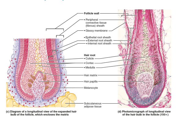

hair follicle receptor
sensory nerve endings that wrap around bulb

wall of the hair follicle is composed of what
Peripheral connective tissue sheath, or fibrous sheath
Derived from dermis, forms external layer of follicle wall
Glassy membrane
Basement membrane of follicle epithelium
Epithelial root sheath
Derived from epidermis, external root sheath and internal root sheath
•Papilla of a hair follicle, or hair papilla
–Dermal tissue containing a knot of capillaries that supplies nutrients to growing hair
if the papilla of a hair follicle is destroyed what happens
the follicle stops producing hair
hair matrix
•Hair matrix: actively dividing area of bulb that produces hair cells
–As matrix makes new cells, it pushes older ones upward
arrector pili
small band of smooth muscle attached to follicle
Responsible for “goose bumps”
name two types of hair
Vellus hair- pale, fine body hair of children and adult females
Terminal hair- coarse long hair
scalp, eyebrows, face and neck of males, pubic regions

what affects hair growth
nutrition and hormones
follicles go through ___ between active and resting phases
growth cycles
hirsutism
condition in which females develop coarse, terminal hair in masculine distribution
by 60-65 years old most experience balding as terminal hair is replaced with
vellus hair
true balding is a pattern commonly found in
men
▪Hair follicles respond to androgens with decreasing growth cycles
Telogen effluvium
abrupt hair thinning caused by an abundance of hair follicles entering resting phase at same time
caused by stress (hormonal changes with childbirth, surgery, severe emotional stress, or crash dieting) or low thyroid hormone levels, drugs, Severe dietary deficiencies of proteins or minerals
nails are scale-like modifications of epidermis that contain
hard keratin
nails act as
protective cover for distal, dorsal surface of fingers/toes
nails consist of
Root embedded in skin
Nail plate or body visible attached portion
Free edge

nail bed
•epidermis underneath keratinized nail plate

nail matrix
thickened portion of bed responsible for nail growth

nail folds
skin folds that overlap border of nail
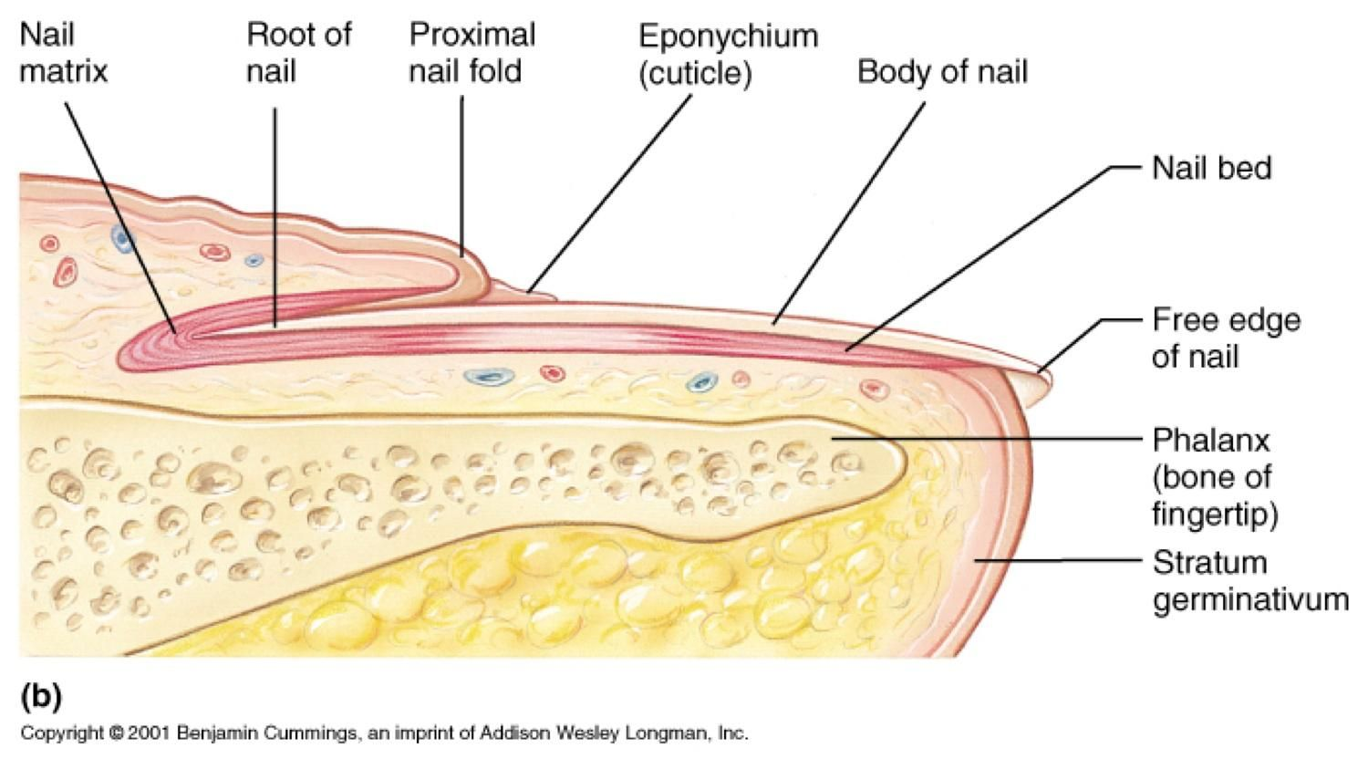
cuticle or eponychium
nail fold that projects onto surface of nail body

hyponychium
area under free edge of plate that accumulates dirt
nails appear pink bec of
underlying capillaries

lunule
thickened nail matrix
yellow tinged nails indicate
respiratory or thyroid gland disorder
thickened yellow nails are caused by
fungal infection of nail
Koilonchya, also called “spoon nail,” is an outward concavity of nail may signal
iron deficiency
Beau’s lines are horizontal lines across nails may indicate
severe illnesses such as uncontrolled diabetes, heart attack, or cancer chemotherapy
sudoriferous glands help control
body temp
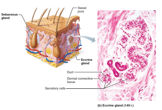
two types of sweat glands
Eccrine sweat glands- no odor
Also called merocrine sweat glands
Apocrine sweat glands- odor
Despite name, are merocrine glands
sweat glands contain what cells
•myoepithelial cells
eccrine glands location, function, secretion
Location: palms, soles, forehead
Function: thermoregulation
secretion: 99% water, salts, metabolic wastes (urea, uric acid, and ammonia), and dermcidin (microbe-killing peptide)
apocrine glands location, function, secretion
location: axillary and anogenital areas (armpit and genitals)
function: Function unknown but may act as sexual scent gland
secretion: viscous milky or yellowish sweat that contains fatty substances and proteins
Bacteria break down sweat, leading to body odor
types of modified apocrine glands
Ceruminous glands and Mammary glands:
Ceruminous glands - modified apocrine glands
location: lining of external ear canal
secretion: cerumen - earwax
Mammary glands - modified apocrine glands
location: boob
secretion: secrete milk
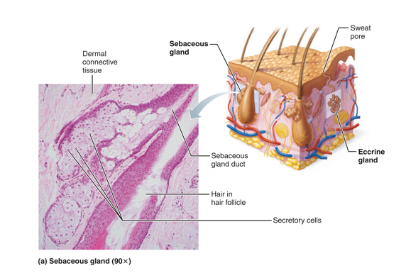
sebaceous glands location, function, secretion
location: across body, develop from hair follicles
Function: producing and secreting sebum which protects from protects your skin from: friction, Moisture loss or brittle hair, Infections caused by bacteria or fungi
Secretion: Secrete sebum
Oily secretion
Bactericidal (bacteria-killing) properties
Softens hair and skin
eccrine vs. apocrine vs. sebaceous glands

acne
an infectious inflammation of the sebaceous glands, resulting in pimples (pustules or cysts)
Whiteheads, or closed comedones,
blocked sebaceous glands
blackheads
If secretion is oxidized, whitehead becomes blackhead, or open comedones
Overactive sebaceous glands in infants can cause seborrhea
–Begins as pink, raised lesions on scalp that turn yellow/brown and slough off
what are the main functions of skin
Protection
Body temperature regulation
Cutaneous sensations
Metabolic functions
Blood reservoir
Excretion
protection - skin function
skin has 3 barriers:
Chemical barriers: skin secretes chemicals (sweat and sebum), low pH, Melanin protects against UV
Physical barriers: keratinized cells of stratum corneum block most water and water-soluble substances
Biological barriers:
Dendritic cells (epidermis)- engulf foreign invaders and active immune system
Macrophages (dermis)- activate immune system
temperature regulation- skin function
insensible to sensible perspiration - more sweat produced to cool body
cold temp: –Dermal blood vessels constrict
insensible perspiration
Under normal, resting body temperature, sweat glands secrete about 500 m l/day of unnoticeable sweat
sensible perspiration
If body temperature rises, dilation of dermal vessels can increase sweat gland activity to produce 12 L (3 gallons) of noticeable sweat to cool the body
cutaneous sensations- skin function
part of nervous system
respond to stimuli outside of the body
metabolic functions- skin function
Skin can synthesize vitamin D needed for calcium absorption in intestine
Chemicals from keratinocytes can disarm some carcinogens
Keratinocytes can activate some hormones
Skin makes collagenase, which aids in natural turnover of collagen to prevent wrinkles
blood reservoir- skin function
Skin can hold up to 5% of the body’s total blood volume
Skin vessels can be constricted to shunt blood to other organs, such as an exercising muscle
excretion- skin function
Skin can secrete limited amounts of nitrogenous wastes, such as ammonia, urea, and uric acid
Sweating can cause salt and water loss
skin burns definition rule of nines complications
definition: tissue damage caused by heat, chemicals, electricity, radiation, or friction. Burns vary in severity based on depth and total body surface area (TBSA) involved.
rule of nines: a quick method used in emergency care to estimate what percentage of the body is burned and to estimate fluid loss
complications: infection, fluid loss, hypothermia, respiratory complications, scarring, organ dysfunction,

skin cancer definition rule of nines complications
basal cell carcinoma: Most common; least malignant; arises from stratum basale; pearly nodule; slow‑growing.
squamous cell carcinoma: Arises from stratum spinosum; scaly red papule; can metastasize.
malignant melanoma: Cancer of melanocytes; highly metastatic; dangerous; irregular dark lesion.
abcd rule: Asymmetry, Border irregularity, Color variation, Diameter > 6 mm.
skin burn severity
first: Epidermis only; red; painful; no blisters; heals without scarring.
second: Epidermis + dermis; blisters; very painful; moist; may scar.
third: Full thickness; charred/leathery; painless; requires grafting; severe scarring.