ECU BIOL 2141 Unit Two "Structures to Know"
 Call Kai
 Learn
 Practice Test
 Spaced Repetition
 Match
 Flashcards
 Knowt Play
1/93

There's no tags or description
Looks like no tags are added yet.
94 Terms
stratum corneum
outermost layer of the epidermis & first line of defense
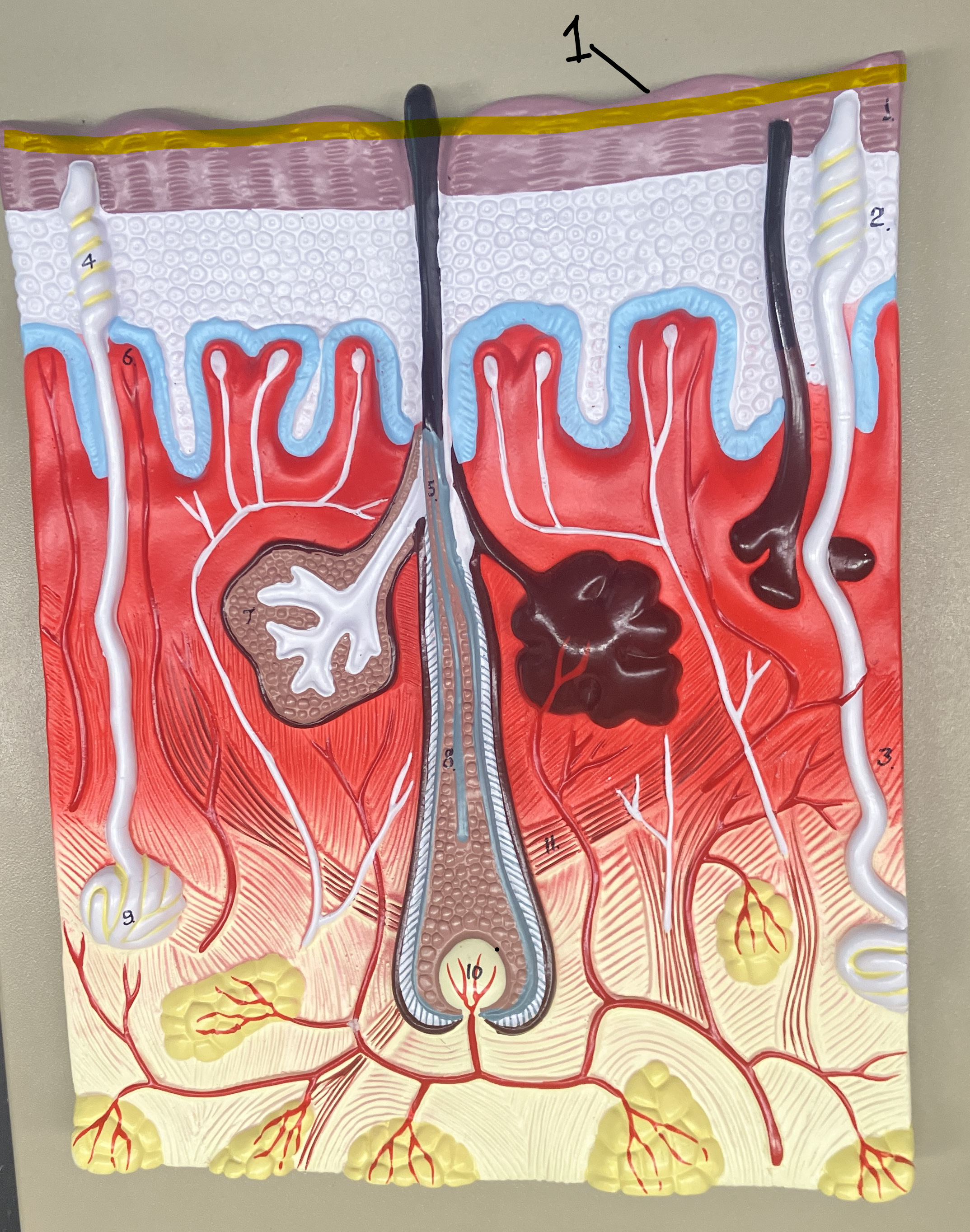
dermal papillae
finger-like projections located within the dermis that contribute to the structural and functional characteristics of the skin
hair shaft
visible, nongrowing/nonliving portion of the hair

hair follicle
small, tunnel-like organs that grow the hair

hair root
living part of a hair inside of the hair follicle

hair shaft
visible, nongrowing/nonliving portion of the hair

hair follicle
small, tunnel-like organs that grow the hair

hair root
living part of a hair inside of the hair follicle

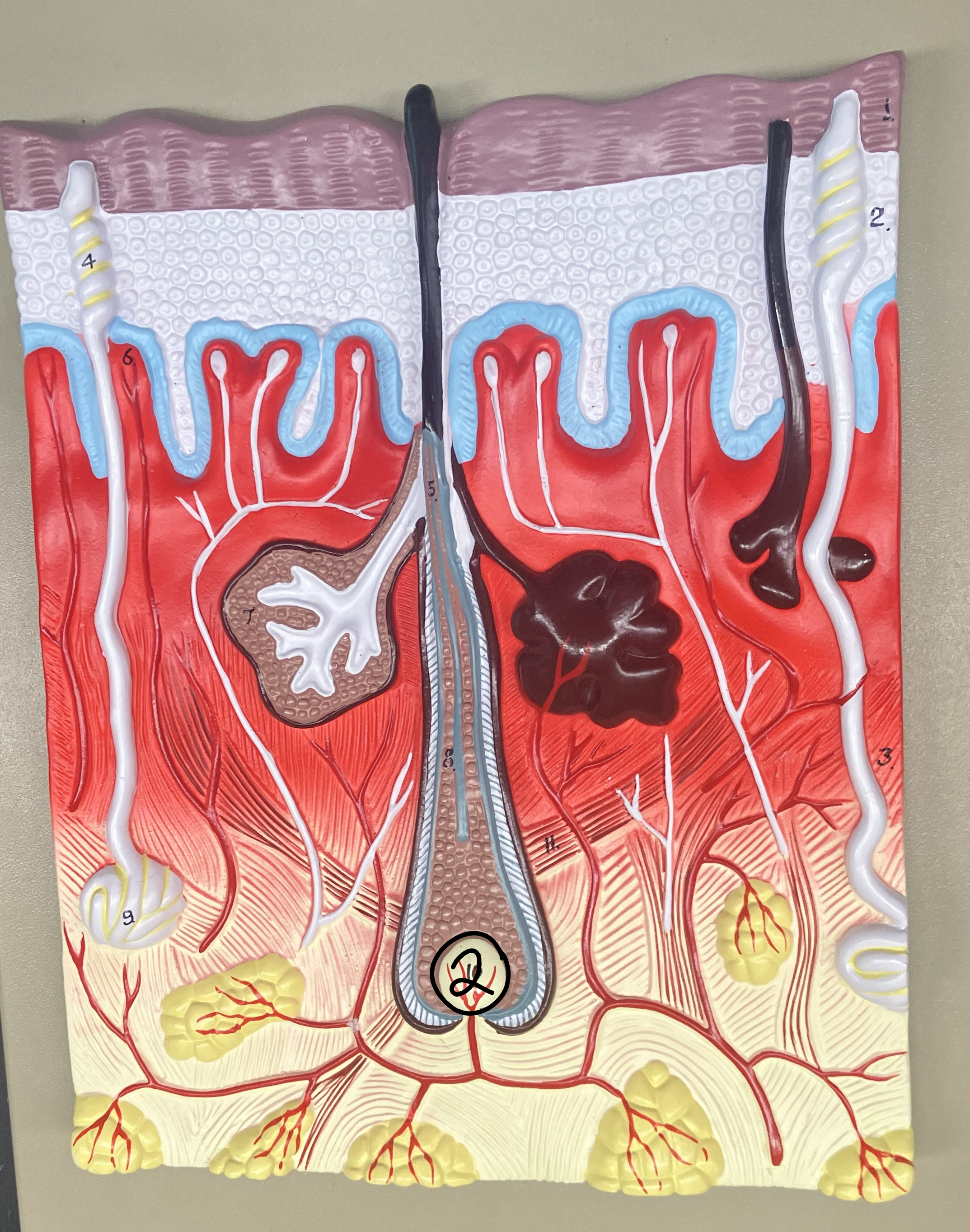
hair papilla
a specialized mesenchymal compartment that forms at the base of the hair follicle
arrector pili muscle
small smooth muscles which attach to hair follicles (make your hair stand on end)

sebaceous gland
microscopic glands found in your hair follicles that secrete sebum (oily, protective substance)

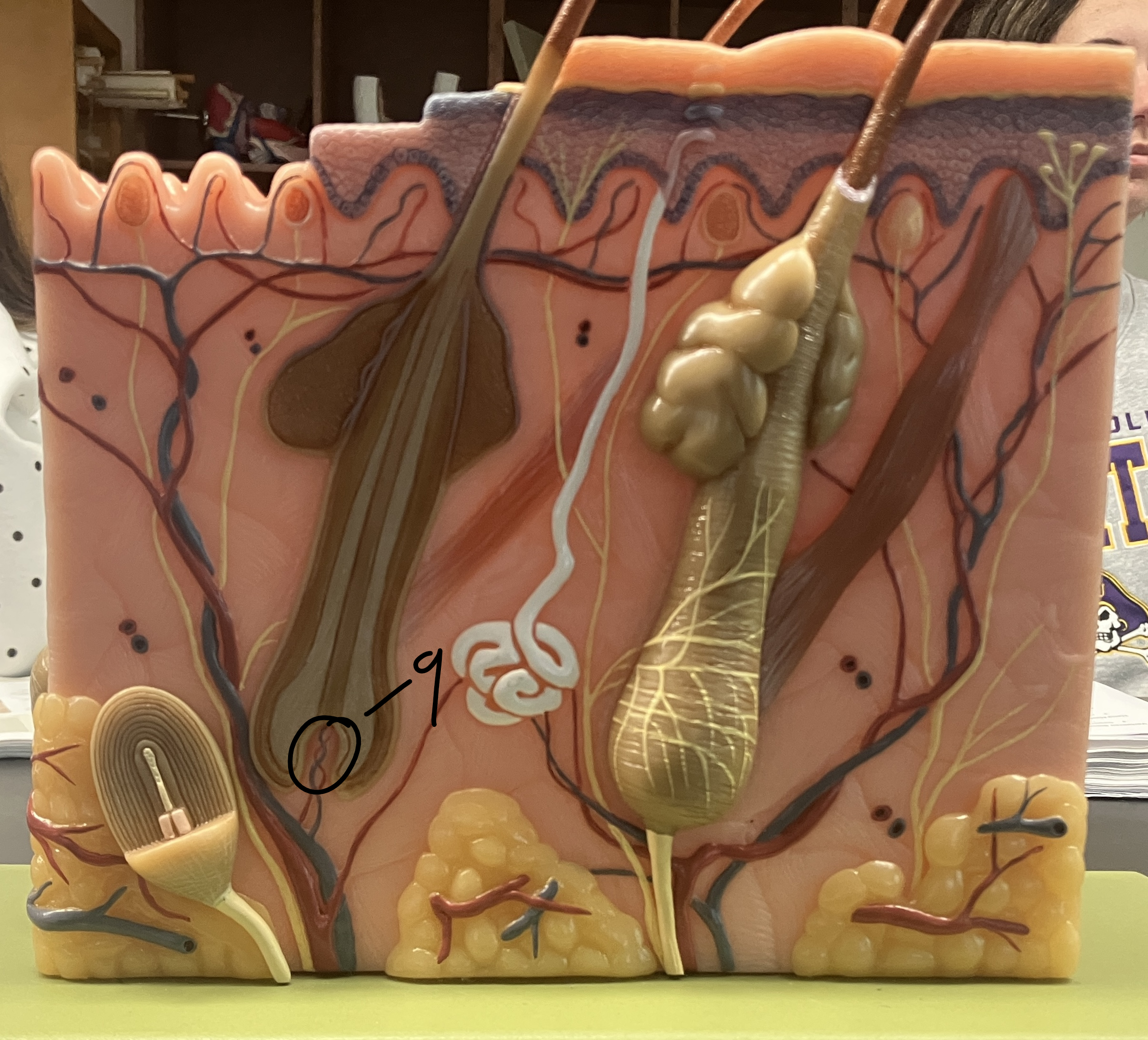
eccrine gland
coiled sweat gland
meissners corpsucle
a cutaneous nerve ending responsible for transmitting the sensations of fine, discriminative touch and vibration

free nerve ending
unspecialized, afferent nerve fiber sending its signal to a sensory neuron.

pacinian corpuscle
sensory nerve ending in the skin and tissues that acts as a rapidly adapting mechanoreceptor for deep pressure and high-frequency vibration
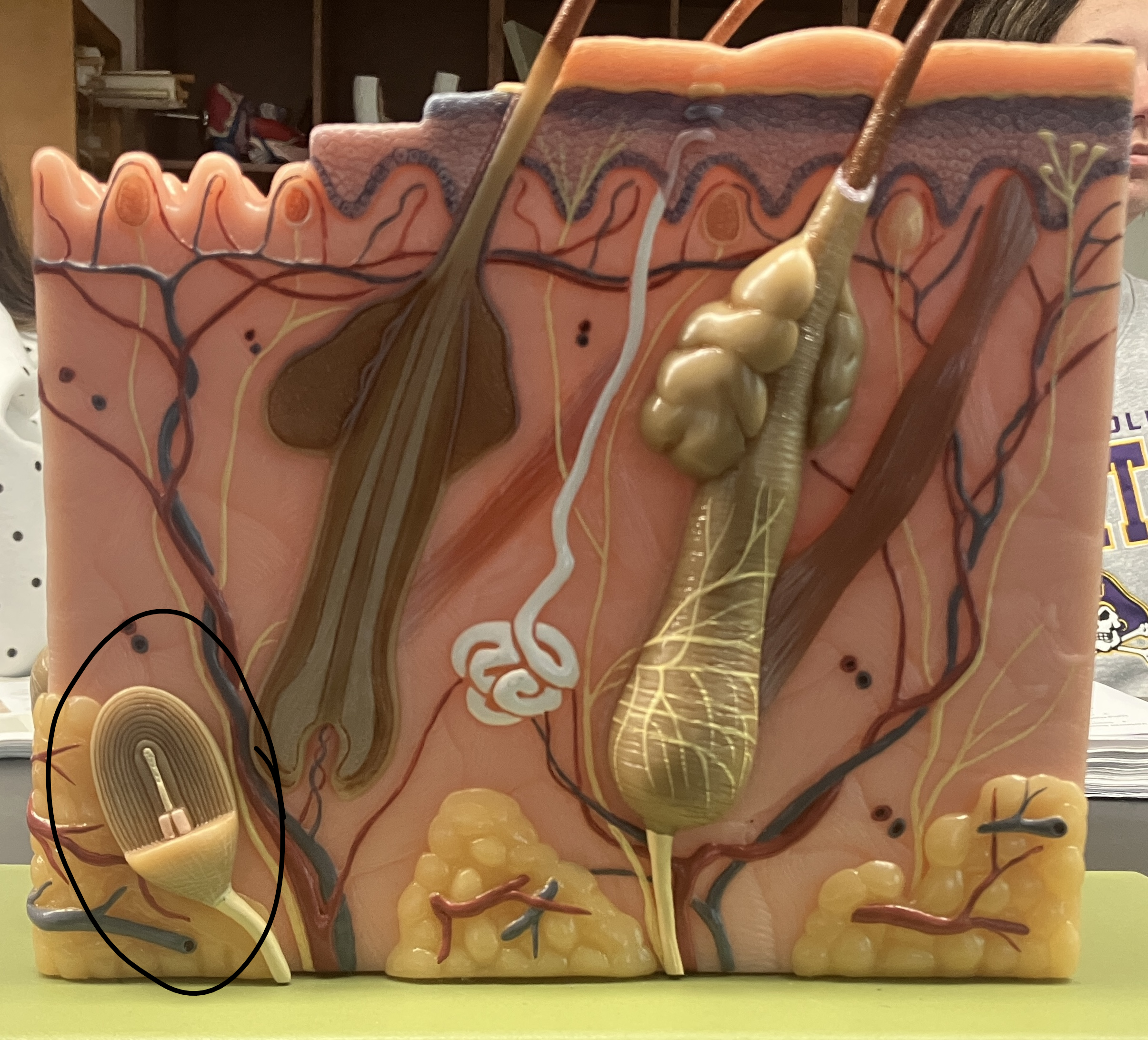
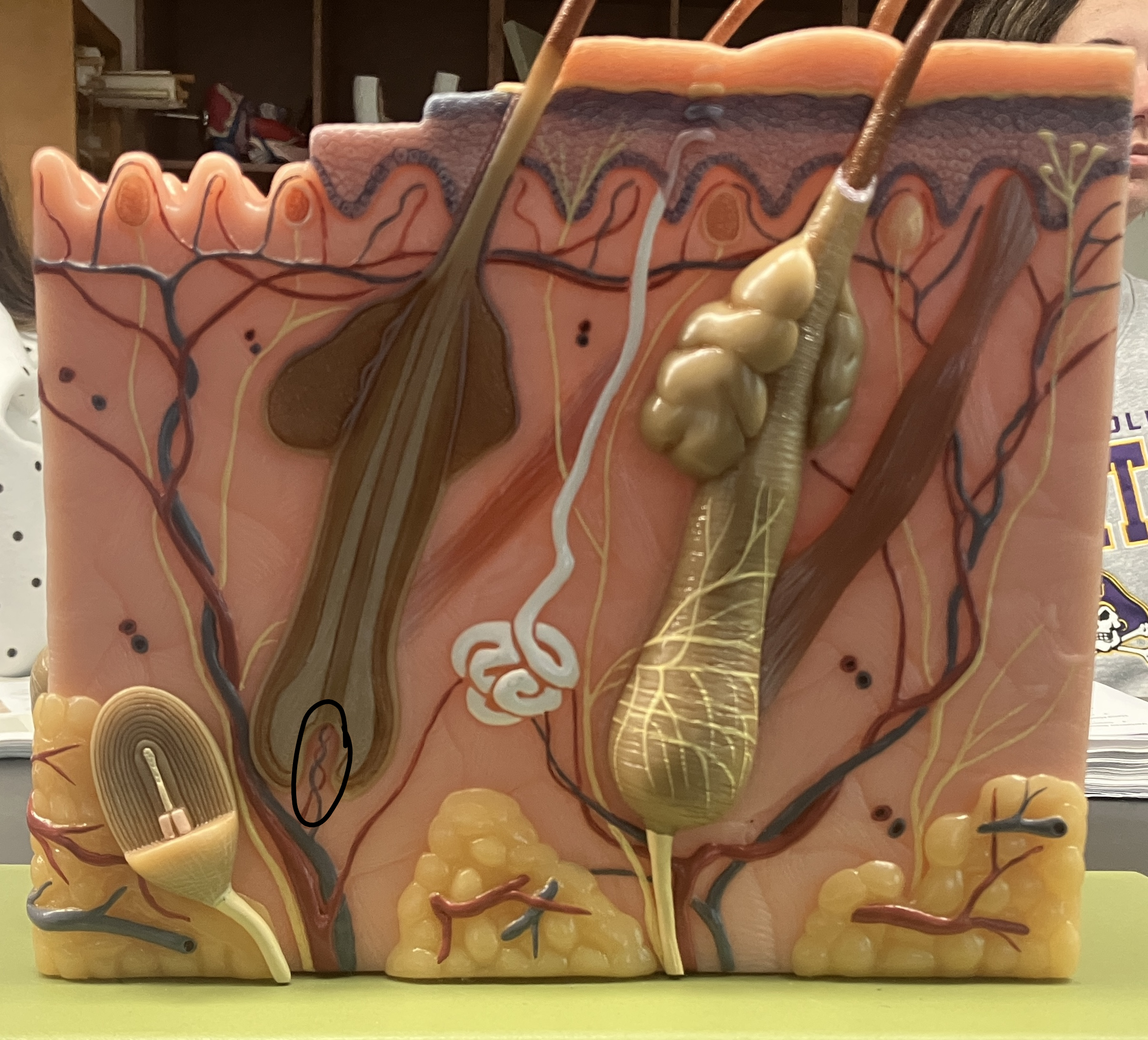
sensory nervie fibers / nerves
[Blue Lines] specialized nerve endings that detect touch, pain, temperature, and pressure, and send these signals to the brain

epidermis
outer most layer of the cutaneous membrane

dermal papillae
finger-like projections located within the dermis that contribute to the structural and functional characteristics of the skin
stratum corneum
outermost layer of the epidermis & first line of defense

stratum granulosum
a thin layer of cells in the epidermis lying above the stratum spinosum and below the stratum corneum

stratum spinosum
a layer of the epidermis found between the stratum granulosum and stratum basale, used in immune defense

stratum basale
deepest layer of the epidermis

hair papilla
a specialized mesenchymal compartment that forms at the base of the hair follicle

sebaceous gland
microscopic glands found in your hair follicles that secrete sebum (oily, protective substance)
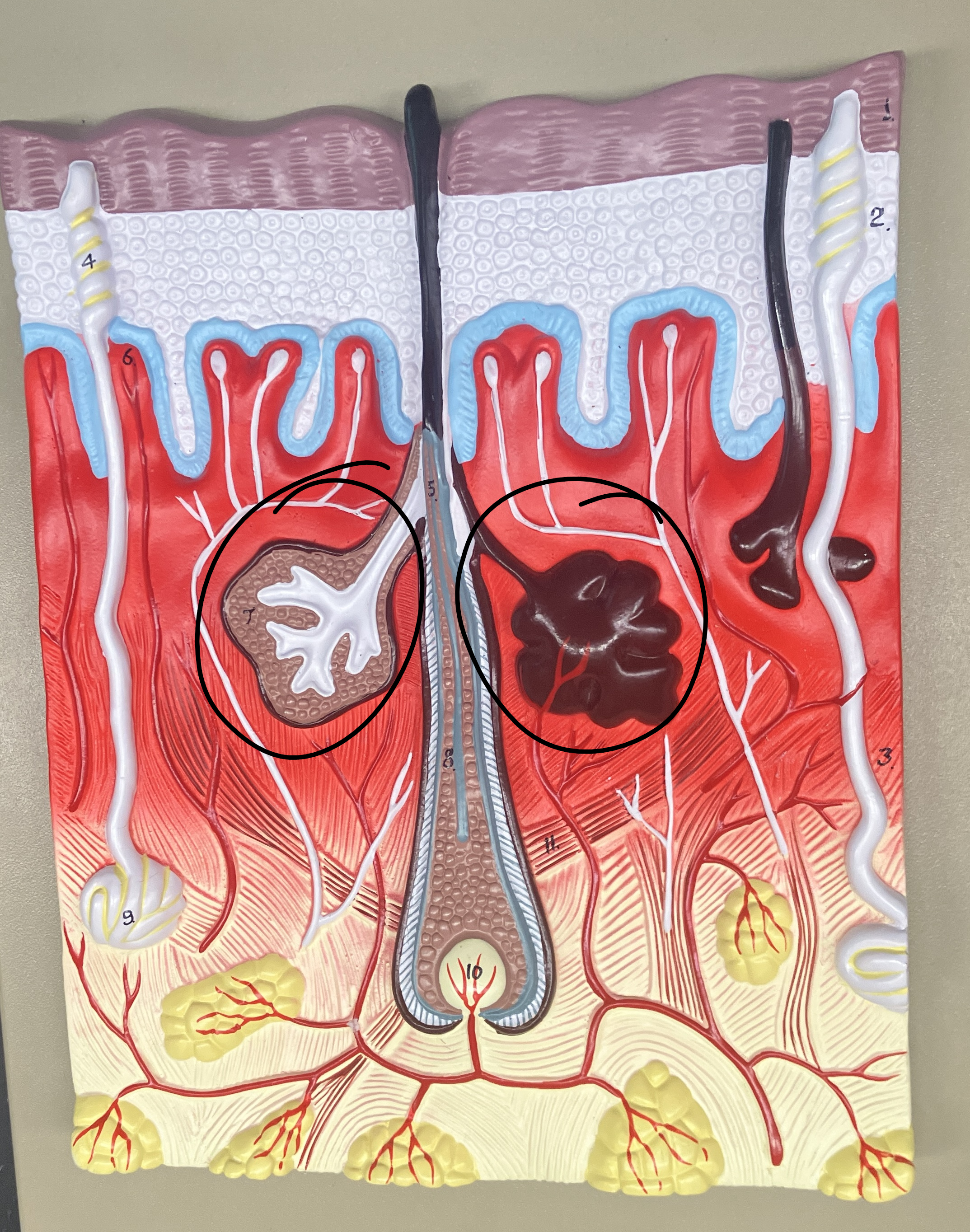
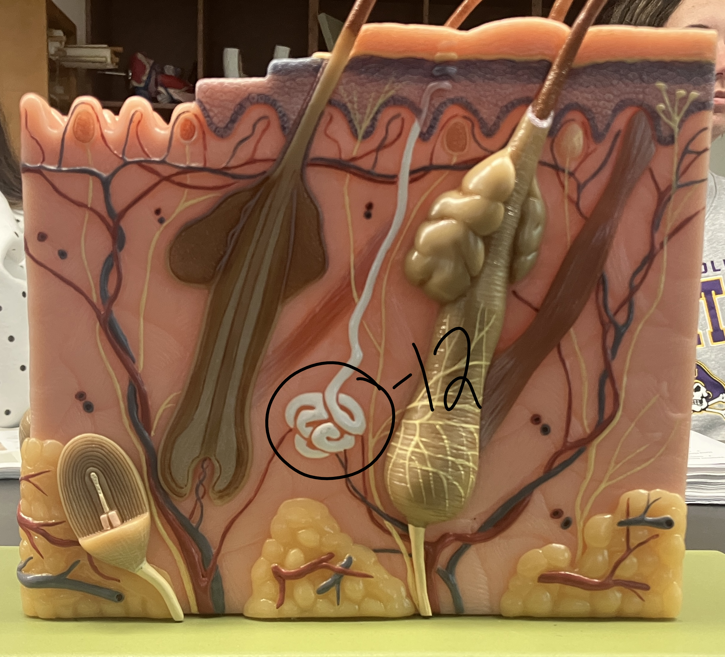
eccrine gland
coiled sweat gland

meissner’s corpuscle
a cutaneous nerve ending responsible for transmitting the sensations of fine, discriminative touch and vibration

free nerve ending
unspecialized, afferent nerve fiber sending its signal to a sensory neuron.

suture (structural & functional types)
joints between bones
structural → fibrous or suture
functional → synarthrotic

temporomandibular (structural & functional types)
a sliding hinge that connects your lower jawbone (mandible) to the temporal bone of your skull on each side of your head
structural → synovial or modified hinge
functional → diarthrotic

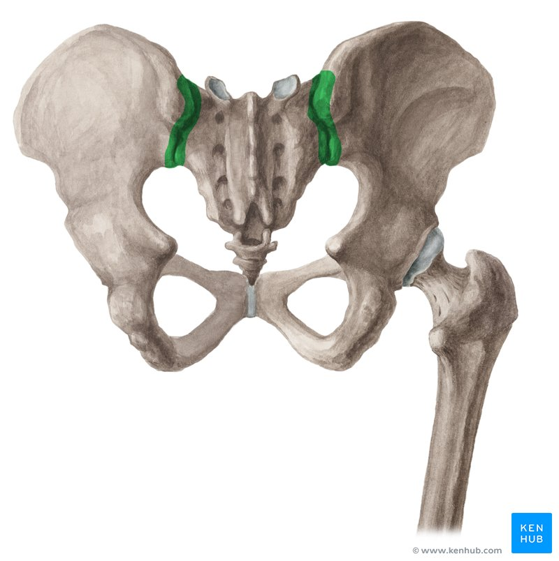
sacroiliac joint (structural & functional)
connects the spine to the pelvis
structural → synovial
functional → diarthrotic
pubic symphysis
nonmoving, connects left & right pelvic bones
structural → cartilaginous
functional → amphiarthrotic

intervertebral (structural)
connection between spinal vertebrae, small movement & shock absorption
structural → cartilaginous or symphysis

carpometacarpal joint (Structural & functional types)
connects the carpals to the metacarpals
structural → synovial
functional → diarthrotic

metacarpophalangeal joint (structural & functional)
connects the metacarpals to the phalanges
sctructural → synovial
functional → diarthrotic
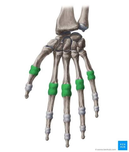
interphalangeal (structural & functional)
connects the phalanges
structural → synovial
functional → diarthrotic

iliofemoral ligament
strongest ligament in the body, connects the pelvis to the femur

pubofemoral ligament
connects the pelvis & femur

patellar tendon
connects the bottom of the patella to the tibia

quadriceps tendon
connects the quadriceps muscle to the top of the patella

fibular collateral ligament
connects the fibula (thin bone) to the femur
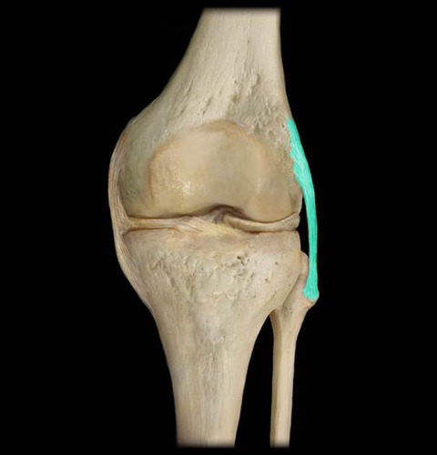
tibial collateral ligament
connects the fibula (thick bone) to the femur

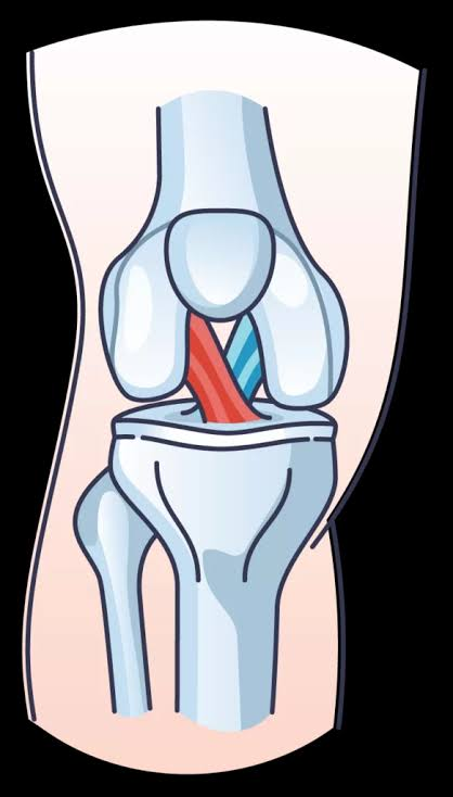
posterior cruciate ligament
POSTERIOR ligament that connects the back of the tibia to the femur

anterior cruciate ligament
ANTERIOR ligament that connects the tibia to the femur
lateral meniscus
C-shaped fibrocartilage between the femur and tibia
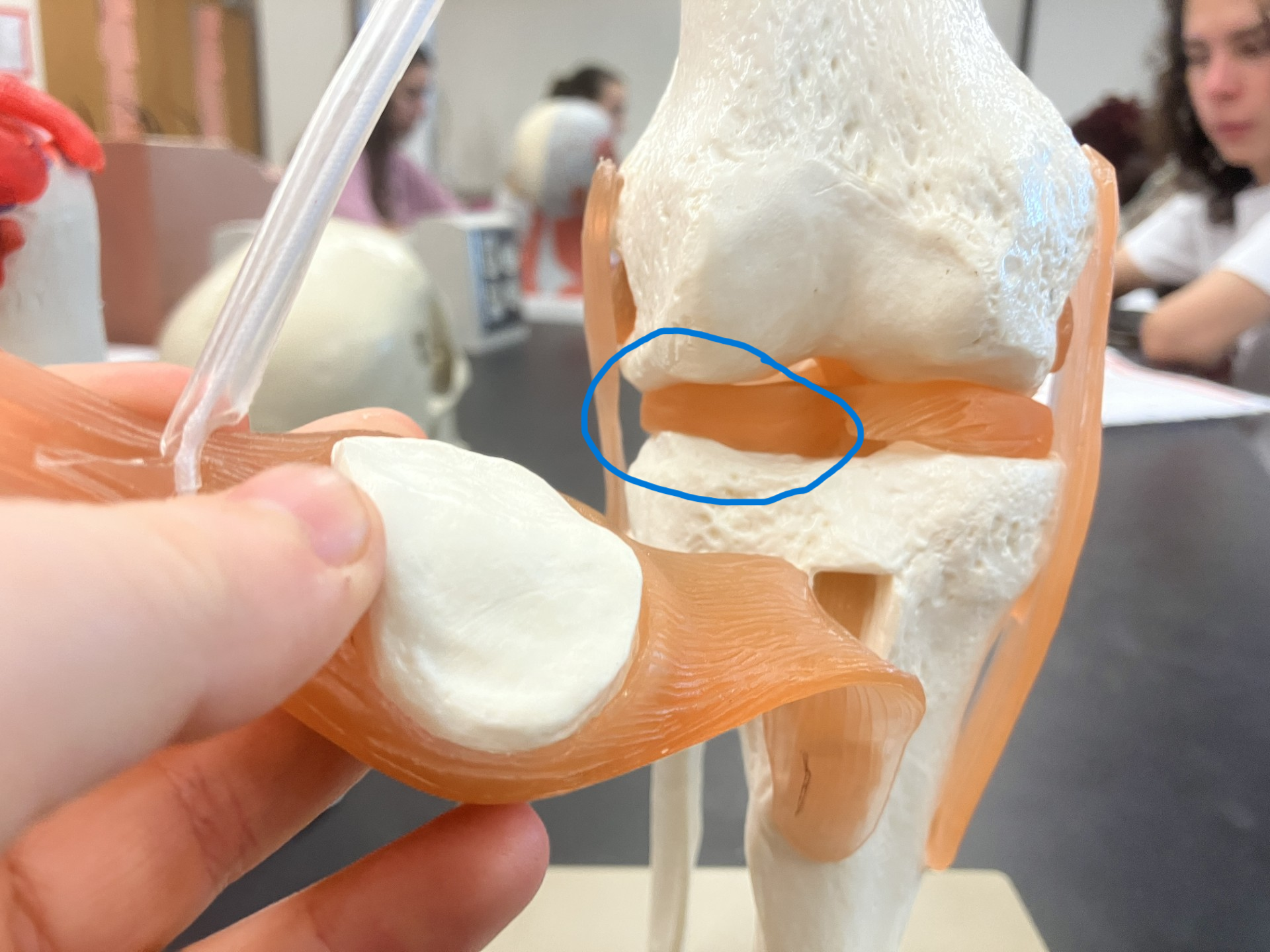
medial meniscus
C-shaped fibrocartilage between the femur and tibia

caroacromial ligament
connects the acromion and coracoid process of the scapula

coracohumeral ligament
a broad, strong intrinsic ligament that strengthens the superior part of the shoulder joint capsule

glenohumeral ligament
three thickened bands of the shoulder joint capsule that connect the glenoid cavity of the scapula to the humerus

buccinator
“cheek muscle”, lateral wall of oral cavity

temporalis
sits against the temporal bone

orbicular oris
articulation of the mouth

frontalis OR frontal belly
sits on the frontal bone, articulates forehead

orbicularis oculi
articulates around the eyes

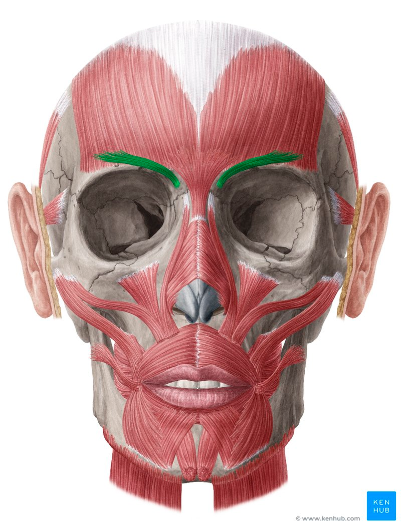
corrugator supercilii
articulates the eyebrow
zygomaticus
located in the cheek, articulate smiling & upper lip movement

depressor anguli oris
lateral to the chin, articulates the mouth downward

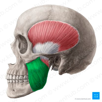
masseter
primary role in chewing & closing the jaw
mylohyoid
forms the muscular floor of the oral cavity, under the chin

digastric anterior belly
beneath the jaw in the ANTERIOR neck, helps with chewing/swallowing/speech

digastric posterior belly
beneath the jaw in the POSTERIOR neck, helps with chewing/swallowing/speech

stylohyoid
swallowing, speech, airway support

sternocleoidmastoid
connects breastbone/collarbone to base of the skull

sternohyoid
depression, stabilization, neck flexion
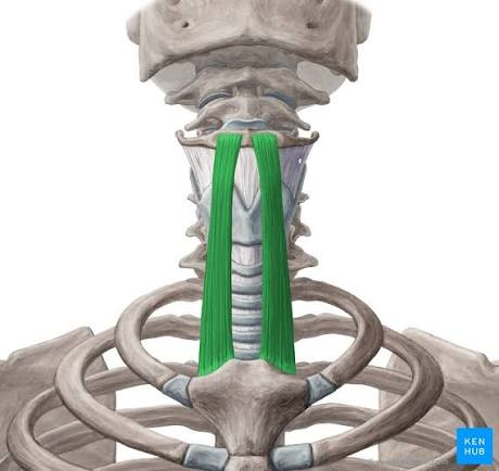
omohyoid
vascular protection, depression of hyoid bone

thyrohyoid
swallowing & speech

trapezius
shrugging, retraction, depression, head+neck movements

risorius
artciulation & expression of the mouth

rhomboid minor
under the trapezius, upper of two rhomboids

infraspinatus
main external rotator of the shoulder joint
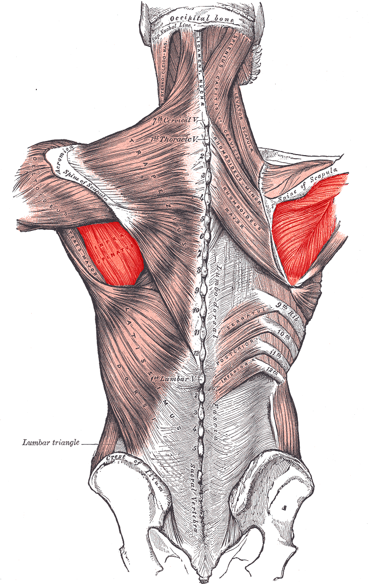
supraspinatus
abducts the arm (move away)

teres major
below the deltoid, extending arm

subscapularis
sits on the spacula, stabilization, rotates the arm inward

acromioclavicular ligament
bulbous joint connecting the collarbone (clavicle) to the shoulder blade (acromion)

coracoclavicular ligament
connects the clavicle (collarbone) to the scapula (shoulder blade)

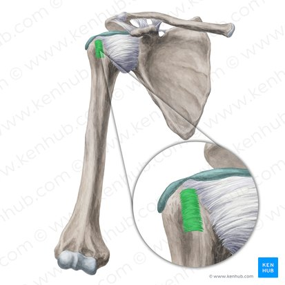
transverse humeral ligament
between the greater and lesser tubercles of the proximal humerus
gluteus maximus
a s s

glutus medius
anterior & superior to glutus maximus, smaller

psoas major
hip flexion, spinal stabilisation

iliacus
sits on the ilium

erector spinae
GROUP of muscles running vertically from pelvis/sacrum to head/back of neck

longissmus
longest muscle in the errector spinae group

lumbricals
four muscles in the palm of the hand, attach to tendons NOT bones

pronator quadratus
square shaped muscle near the wrist

palmaris longus
slender muscle in the forearm (assists in flexion)

extensor digitorum
POSTERIORLY extends over fingers, wrists, & forearm

deltoid / deltoideus
triangular muscle in the shoulder named after the greek delta

flexor carpi ulnaris
pinky side of the anterior forearm, flexes and abducts

flexor digitorum superficialis
ANTERIORLY extends over fingers, wrists, forearm

triceps brachii
three headed muscle on the back of the upper arm
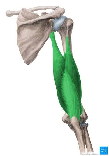
biceps brachii
two headed muscle on the front of the upper arm
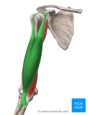
brachialis
deep, upper arm, strongest flexor of elbow
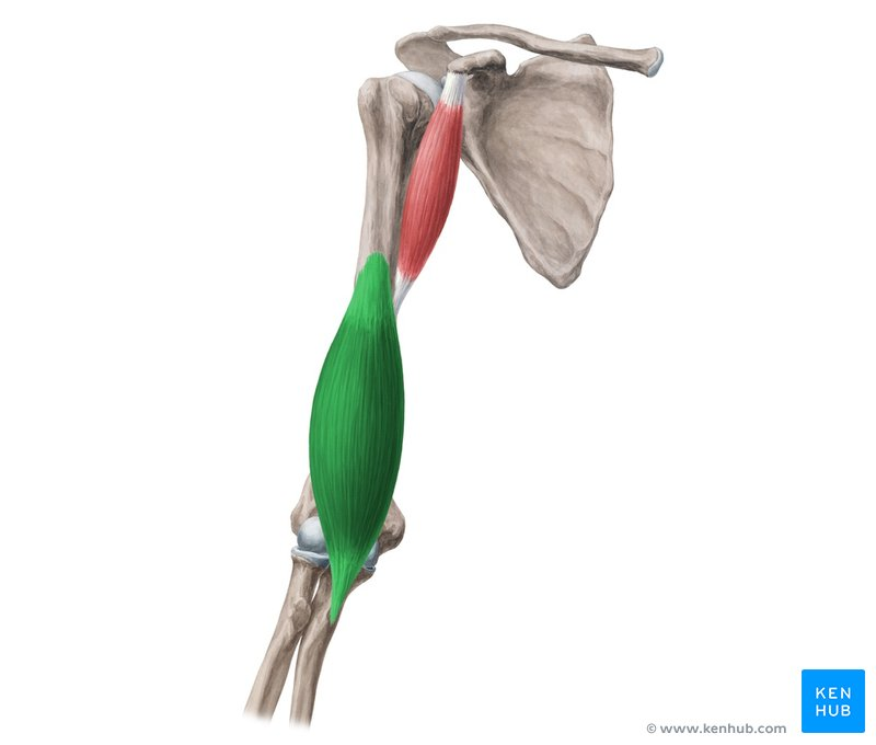
brachioradialis
located on the thumb side of the forearm, flexes the elbow
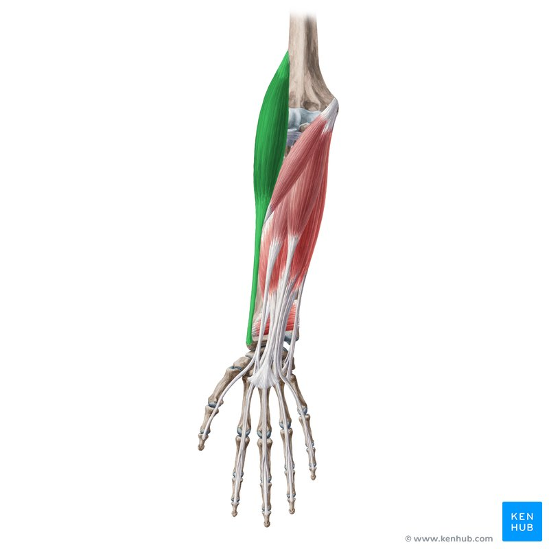
anconeus
small, triangular muscle on the back of the elbow
