Ch 14 Cardiac Output Blood Flow and Blood Pressure
 Call Kai
 Learn
 Practice Test
 Spaced Repetition
 Match
 Flashcards
 Knowt Play
1/51

There's no tags or description
Looks like no tags are added yet.
52 Terms
What is cardiac output (CO)?
Volume of blood pumped by each ventricle per minute
CO = SV × HR
How is CO related to SV and HR?
Directly related (directly proportional)
SV or HR goes up → CO goes up
SV or HR goes down → CO goes down
What is stroke volume (SV) vs. heart rate (HR)?
SV: blood pushed out per contraction
HR: beats per minute
What is the chronotropic effect?
The ANS changing heart rate (chrono = timing)
Acts on the SA node
What do sympathetic vs. parasympathetic activity do to HR and CO?
Sympathetic: ↑HR → ↑CO
Parasympathetic: ↓HR → ↓CO
What is the SA node pacemaker potential at rest (graph a)?
At −60 mV, HCN channels let Na⁺ in and the cell slowly depolarizes. At −40 mV (threshold), Ca²⁺ channels open and fire the action potential. K⁺ channels then repolarize the cell back to −60 mV and the cycle repeats (~75 bpm).

How does the parasympathetic division slow HR (graph b)?
ACh binds muscarinic receptors and lets K⁺ out. This makes the SA node more negative and farther from threshold, so it takes longer to fire. HR and CO go down.

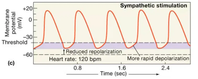
How does the sympathetic division raise HR (graph c)?
NE/Epi bind β1 receptors and let Ca²⁺ in. This makes the SA node more positive and closer to threshold, so it fires sooner. HR and CO go up.
What controls HR, and how?
Cardiac control center in the medulla oblongata
↑HR: sends signals through the sympathetic division
↓HR: sends signals through the parasympathetic division
What are the two ways the sympathetic division raises CO?
↑HR (chronotropic)
↑Contractility of the atria and ventricles → ↑SV
What do sympathetic and parasympathetic activity do to each region of the heart?
SA node: sympathetic speeds up depolarization and HR;
parasympathetic slows them
AV node: sympathetic speeds up conduction;
parasympathetic slows it
Atrial & ventricular muscle: sympathetic makes contractions stronger;
parasympathetic has no significant effect
Takeaway: parasympathetic changes HR only, not SV
What are the 3 variables that determine SV?
End diastolic volume (EDV): blood volume in the ventricles at the end of diastole (ventricular filling)
Total peripheral resistance (TPR): resistance to blood flow in the arteries
Contractility: strength of ventricular contraction
What changes TPR?
The diameter of the arteries
Vasodilation lowers TPR, so more blood flows
Vasoconstriction raises TPR, so less blood flows
How do EDV and contractility affect SV and CO?
Both are directly related
Higher EDV (more blood in) means higher SV, which means higher CO
Higher contractility (stronger squeeze) means higher SV, which means higher CO
The same holds in reverse when they go down
How does TPR affect SV and CO?
TPR and SV are inversely related (the only exception)
Higher TPR means lower SV, which means lower CO
Lower TPR means higher SV, which means higher CO
Why: constricted arteries resist flow, so the ventricle pushes out less blood
Vasoconstriction = higher TPR
Vasodilation = lower TPR
What are the other names for EDV and TPR?
EDV = preload = ventricular filling (the workload on the heart before it contracts)
TPR = afterload (it impedes ejection from the ventricle)
What is ejection fraction?
SV / EDV: the percentage of the starting blood that gets pushed out
Normal is 60%
Below 60% may mean a heart problem (a useful clinical diagnostic tool)
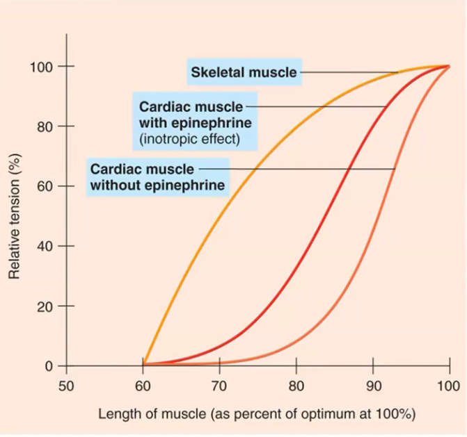
What is the Frank–Starling law of the heart?
As EDV goes up, contractility goes up
Stronger contraction means higher SV
Why does more EDV make the heart contract harder (Frank–Starling mechanism)?
More blood stretches the ventricle, which lengthens the sarcomeres toward their optimal length.
At that length, more cross-bridges form between actin and myosin, so the contraction is stronger (more tension) and SV goes up.

What is the positive inotropic effect?
Sympathetic Epi/NE raise Ca²⁺ inside cardiac muscle cells
More Ca²⁺ means more cross-bridges, so contraction is stronger and contractility goes up
What are the 2 ways contractility increases?
↑EDV: stretch brings sarcomeres to optimal length (Frank–Starling)
Sympathetic stimulation: more Ca²⁺ (positive inotropic effect)
Summarize what controls CO.
HR: sympathetic raises it;
parasympathetic lowers it
SV:
EDV raises it (directly, and through Frank–Starling stretch)
Contraction strength raises it (sympathetic and stretch)
TPR lowers it (inverse)

Congrats you finished the first video fo Ch 14
What is venous return, and why does it control EDV?
The amount of blood returning to the heart through the veins
The veins hold most of the blood at any given time
More venous return means higher EDV, which raises SV and CO
What are the factors that raise venous return?
Blood volume: controlled by urine volume and tissue-fluid volume
Breathing: inhaling drops thoracic pressure, creating a vacuum that pulls blood toward the heart
Venous pressure:
Venoconstriction (from sympathetic stimulation) narrows the veins and pushes blood back
The skeletal muscle pump: moving muscles compress the veins

What affects blood volume?
Urinary system
Lungs
Skin (sweat glands)
Capillary exchange
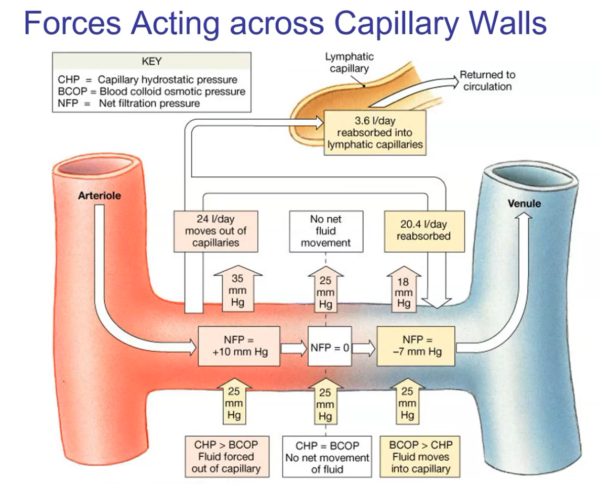
What happens to water at the proximal vs. distal end of a capillary?
Proximal (arteriole side): blood pressure pushes water out
Distal (venule side): osmotic pressure pulls most of it back in
The lymphatic system reclaims the water that isn’t reabsorbed

What are CHP and BCOP?
CHP (capillary hydrostatic pressure): the blood pressure in the capillary; pushes water out
BCOP (blood colloid osmotic pressure): osmotic pressure from solutes (colloids) in the blood; pulls water in
What is net filtration pressure (NFP)?
NFP = CHP − BCOP
Positive: CHP wins, so water leaves the capillary
Negative: BCOP wins, so water is reabsorbed
What are the CHP, BCOP and NFP values along the capillary?
Proximal: 35 − 25 = +10 (fluid forced out)
Middle: 25 − 25 = 0 (no net movement)
Distal: 18 − 25 = −7 (fluid moves in)
BCOP stays at 25 because solutes aren’t removed
CHP drops because BP falls the farther blood is from the heart
Why is lymph needed at the capillary?
More water leaves (+10) than comes back (−7)
The lymphatic system reabsorbs the leftover water (about 3.6 L/day)
It returns that water to circulation through the subclavian vein
What is edema, and what causes it?
Too much water building up in the tissues, because the water lost from the capillaries isn’t reclaimed
Causes:
Lymphatic problems
High BP
Too much salt
Leaking plasma proteins (e.g. albumin), which lowers BCOP
What determines urine volume?
Blood is filtered at the glomerulus; what enters the nephron is the filtrate
Urine volume depends on how much filtrate water is reabsorbed
Hormones regulate this at the distal convoluted tubule
How does ADH restore blood volume?
Dehydration or salt intake raises blood osmolality, which is detected by osmoreceptors in the hypothalamus. The hypothalamus signals the posterior pituitary to release ADH, so the kidneys retain water. It also triggers thirst, so we drink. Together, these raise blood volume and lower osmolality (negative feedback). More blood volume increases venous return, EDV, SV and CO.

What is aldosterone, and what triggers it?
Released by the adrenal cortex
Kidneys retain Na⁺, Cl⁻ and water but lose K⁺
Triggers:
Low salt
Low blood volume
Low BP
Overall job: controls BP by controlling blood volume
What is the renin–angiotensin–aldosterone system (RAAS) pathway?
Low BP and low blood flow to the kidneys trigger the juxtaglomerular apparatus to release renin. Renin converts angiotensinogen into angiotensin I. ACE (in the lungs) converts angiotensin I into angiotensin II. Angiotensin II signals the adrenal cortex to release aldosterone, so the kidneys retain salt and water and blood volume rises. Angiotensin II also constricts arterioles, which raises BP.

What are the 3 effects of angiotensin II?
Triggers aldosterone release, so salt and water are retained
Constricts arterioles, raising BP
Activates thirst centers, so we drink and blood volume rises
What is ANP?
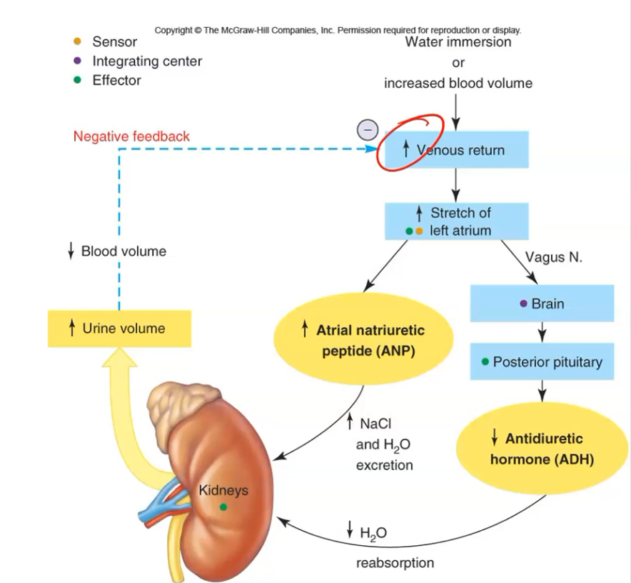
Atrial natriuretic peptide, released when the left atrium is stretched
The antagonist of ADH and aldosterone
Kidneys excrete NaCl and water, so urine volume goes up
How does the body lower blood volume when it’s too high?
High blood volume increases venous return, which stretches the left atrium. The atrium releases ANP, so the kidneys excrete more salt and water. The stretch also signals the brain through the vagus nerve, so the posterior pituitary releases less ADH and the kidneys reabsorb less water. More urine lowers blood volume and venous return (negative feedback), which then lowers EDV, SV and CO.
How does BP relate to blood flow?
BP is the driving force for flow
Blood flows from high pressure to low pressure: from the heart to the capillaries, then from the capillaries back to the heart
How do arterioles control BP?
Constricting arterioles makes blood back up, so BP rises before the constriction and drops after it
Dilating arterioles lowers BP
Arterioles control systemic BP

Why is BP low in the capillaries?
Pressure drops sharply at the arterioles
Low pressure slows blood flow, so blood spends more time in the capillaries, which optimizes exchange
What determines BP?
HR, SV and TPR, which simplifies to CO and TPR
BP is directly related to both
If CO or TPR goes up, BP goes up; if either goes down, BP goes down
What are the 2 main ways the body controls BP?
Sympathetic division:
Constricts arterioles, which raises TPR
Raises HR and contractility, which raises CO
Kidneys: regulate blood volume, which changes venous return, EDV, SV and CO
Where are the baroreceptors, and what do they detect?
Aortic arch and carotid sinuses
They are pressure (stretch) receptors: more stretch means more action potentials

What are the 2 centers in the medulla oblongata, and what do they control?
Cardiac center: controls HR
Vasomotor center: controls arteriole diameter
What is the baroreceptor reflex when BP drops?
When BP drops (e.g. standing up), the arteries stretch less, so the baroreceptors fire fewer action potentials. The medulla oblongata responds by raising sympathetic and lowering parasympathetic activity. The vasomotor center constricts arterioles, raising TPR. The cardiac center raises HR and contractility, raising CO. Both raise BP back up (negative feedback).

How does the body respond when BP is too high?
High BP increases venous return, which:
Slows HR (reflex bradycardia), lowering CO
Stretches the atria, releasing ANP, so more urine is excreted and blood volume drops
Both lower BP
How is BP measured by auscultation?
The cuff compresses the brachial artery, so no sound is heard
As the cuff deflates:
First Korotkoff sound marks systolic pressure (contraction)
Last Korotkoff sound marks diastolic pressure (relaxation)

Which ventricle’s pressure does a BP cuff measure?
The left ventricle
It’s systemic pressure, not pulmonary
What are the BP ranges?
Normal: 120/80
Marginal high: systolic 130 or above
High: systolic 140 or above
Too low: systolic below 100
Why are high and low BP bad?
High: damages vessels (they can burst) and the heart
Low: reduces systemic blood flow