A&P lab muscles - head/neck and trunk/shoulder
 Call Kai
 Learn
Learn Practice Test
 Spaced Repetition
 Match
Match Flashcards
 Knowt Play
1/54

There's no tags or description
Looks like no tags are added yet.
Name | Mastery | Learn | Test | Matching | Spaced | Call with Kai | Chat |
|---|
No analytics yet
Send a link to your students to track their progress
55 Terms
Head muscles
Facial expression
Epicranus
Buccinator
Platysma
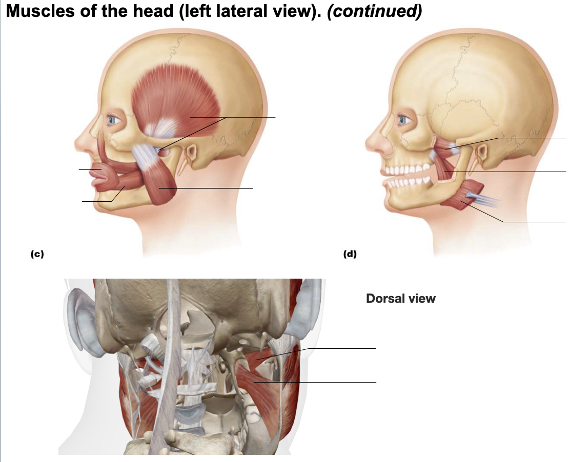
Muscles of mastication
Masseter
Temporalis
Buccinator
Medial pterygoid
Lateral ptergoid
Epicranius - frontal and occipital bellies
Comments - bipartite muscle consisting of frontal and occipital bellies, which covers dome of skull
Origin - frontal belly - epicranial aponeurosis; occipital belly - occipital and temperal bones
Insertion - frontal belly - skin of eyebrows and root of nose; occipital belly - epicranial aponeurosis
Action - frontal belly raises eyebrows; occipital belly fixes aponeurosis and pulls scalp posteriorly
Buccinator
Comments - principle muscle of cheek; runs horizontally, deep to the masseter
Origin - molar region of maxilla and mandible
Insertion - obicularis oris
Action - compresses cheek
Platysma
Comments - unpaired, thin, sheetlike superficial neck muscle, plays role in facial expression
Origin - fascia of chest (over pectoral muscles and deltoid)
Insertion - lower margin of mandible, skin, and muscle at corner of mouth
Action - tenses skin of neck


Masseter
Comments - covers lateral aspect of mandibular ramus; can be palpated on forcible closure of jaws
Origin - zygomatic arch and zygomatic bone
Insertion - angle and ramus of mandible
Action - prime mover of jaw closure
Temporalis
Comments - fan-shaped muscle lying over parts of frontal, parietal, and temporal bones
Origin - temporal fossa
Insertion - coronoid process of mandible
Action - closes jaw
Medial pterygoid
Comments - runs along internal (medial) surface of mandible (thus largely concealed by that bone)
Origin - sphenoid, palatine, and maxillary bones
Insertion - medial surface of mandible, near its angle
Action - in conjunction with lateral pterygoid, aids in grinding movements of teeth
Lateral pterygoid
Comments - superior to medial pterygoid
Origin - greater wing of sphenoid bone
Insertion - condylar process of mandible
Action - aids in grinding movements of teeth

Neck muscles - anterolateral
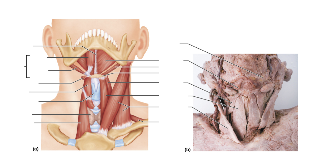
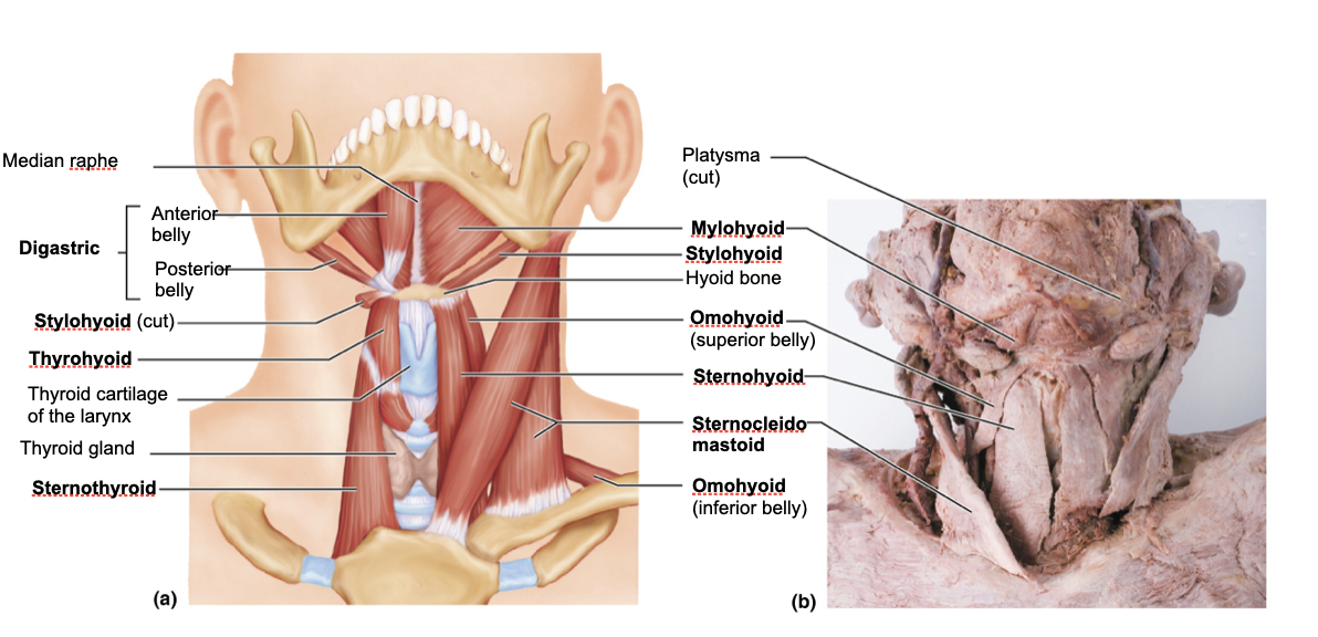
Suprahyoid muscles
Digastric
Stylohyoid
Mylohoid
Infrahyoid muscles
Sternohyoid
Sternothyroid
Omohhyoid
Thyrohoid
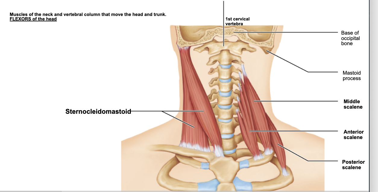
Sternocleidomastoid
Scalenes
Digastric
Comments - consists of two bellies united by an intermediate tendon, forming a V shape under the chin
Origin - lower margin of mandible (anterior belly) and mastoid process of temporal bone (posterior belly)
Insertion - by a connective tissue loop to hyoid bone
Action - open mouth and depress mandible
Nerve supply - mandibular branch of trigeminal nerve (cranial V) for anterior belly; facial nerve (cranial VII) for posterior belly
Stylohyoid
Comments - slender muscle below angle of jaw; parallels posteroid belly of digastric muscle
Origin - styloid process of temporal bone
Insertion - hyoid bone
Action - elevates and retracts hyoid
Nerve supply - facial nerve
Mylohoid
Comments - flat, triangular muscle just deep to digastric muscle; this muscle pair makes a sling that forms the floor of the anterior mouth
Origin - medial surface of mandible
Insertion - hyoid bone and median raphe
Action - elevates hyoid bone and floor of mouth
Nerve supply - mandibular branch of trigeminal nerve
Sternohyoid
Comments - most medial muscle of the neck: thin, superficial except inferiorly, where covered by sternocleidomastoid
Origin - manubrium and medial end of clavicle
Insertion - lower margin of hyoid bone
Action - depresses larynx and hyoid bone if mandible is fixed
Nerve supply - cervical spine nerves 1-3 through ansa cervicalis (slender nerve root in cervical plexus)
Sternothyroid
Comments - lateral and deep to sternohyoid
Origin - posterior surface of manubrium of sternum
Insertion - thyroid cartilage
Action - Depresses larynx and hyoid bone
Nerve supply - “as for sternohyoid” - cervical spine nerves 1-3 through ansa cervicalis (slender nerve root in cervical plexus)
Omohyoid
Comments - straplike muscle with two bellies united by an intermediate tendon; lateral to sternohyoid
Origin - superior surface of scapula
Insertion - hyoid bone, lower border
Action - depresses and retracts hyoid bone
Nerve supply - “as for sternohyoid” - cervical spine nerves 1-3 through ansa cervicalis (slender nerve root in cervical plexus)
Thyrohyoid
Comments - appears as a superior continuation of sternothyroid muscle
Origin - thyroid cartilage
Insertion - hyoid bone
Action - depresses hyoid bone or elevates larynx if hyoid is fixed
Nerve supply - first cervical nerve via hypoglossal nerve
Sternocleidomastoid
Comments - two-headed muscle located deep to platysma on anterolateral surface of neck; indicate limits of anterior and posterior triangles of neck
Origin - manubrium of sternum and medial portion of clavicle
Insertion - mastoid process of temporal bone and superior nuchal line of occipital bone
Action - flexion of neck, rotate head toward shoulder on opposite side
Scalenes - anterior, middle, and posterior
Comments - located more on lateral than anterior neck; deep to platysma and sternocleidomastoid
Origin - transverse process of cervical vertebrae
Insertion - anterolaterally on ribs 1-2
Action - elevate ribs (aid in inspiration)

Muscles of the neck and vertebral column: head movements and trunk extension
Iliocostalis
Longissimus
Spinalis
Semispinalis
Quadratus lumborum
Splenius
Comments - broad two-part superficial muscle extending from upper thoracic vertebrae to skull; capitis portion known as “bandage muscle” because it covers and holds down deeper neck muscles
Origin - ligamentum nuchae, spinous processes of vertebrae C7-T6
Insertion - mastoid process of temporal bone and occiptial bone (capitis); transverse processes of C2-C4 vertebrae (cervicis)
Action - extend or hyperextend head; prime mover
Errector spinae
Prime mover of back extension/trunk extension
Full length of back
Lifting a load or moving suddenly from a bent over position can injure muscles
Includes iliocostalis, longissimus, and spinalus
Iliocostalis
Comments - most lateral muscle group of erector spinae muscles; extend from pelvis to neck
Origin - iliac crests; inferior 6 ribs (thoracis); ribs 3-6 (cervicis)
Insertion - angles of ribs
rib to rib
Action - extend the vertebral column, laterally flex the vertebral column
Nerve supply - spinal nerves (dorsal rami)
Longissimus
Comments - intermediate 3-part muscle group of erector spinae; extend by many muscle slips from lumbar region to skull; mainly pass between transverse processes of vertebrae
Origin - transverse process
Insertion - transverse process
Action - extend vertebral column; laterally flex the vertebral column; extend and rotate the head
Nerve supply - spinal nerves (dorsal rami)
Spinalis
Comments - most medial muscle column of erector spinae; cervicis usually rudimentary and poorly defined
Origin - spinous process
Insertion - spinous process
Action - extends vertebral column
Nerve supply - spinal nerves (dorsal rami)
Semispinalis
Comments - composite muscle forming part of deep layer of intrinsic back muscles; extends from thoracic region to head
Origin - transverse process
Insertion - spinous process
Action - extends vertebral column and head; synergistically with opposite side
Nerve supply - spinal nerves (dorsal rami)
Quadratus lumborum
Comments - fleshy muscle forming part of posterior adominal wall
Origin - iliac crest and lumbar fascia
Insertion - transverse processes
Action - laterally flexes vertebral column
Nerve supply - T12 and upper lumbar spinal nerves (ventral rami)


Superficial Muscles of the Anterior and Posterior Thorax: Movements of the Scapula and Arm
Extrinsic shoulder muscles
Anterior muscles
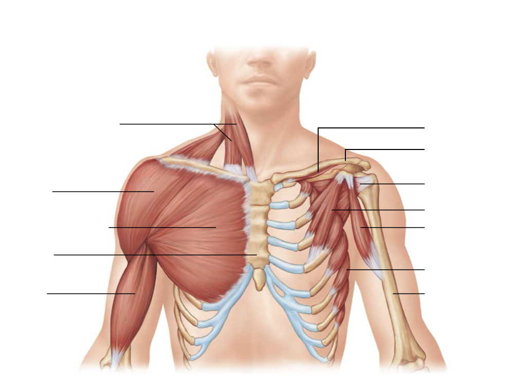
Pectoralis major (arm moving)
Pectoralis minor (move scapula)
Seratus anterior (move scapula)
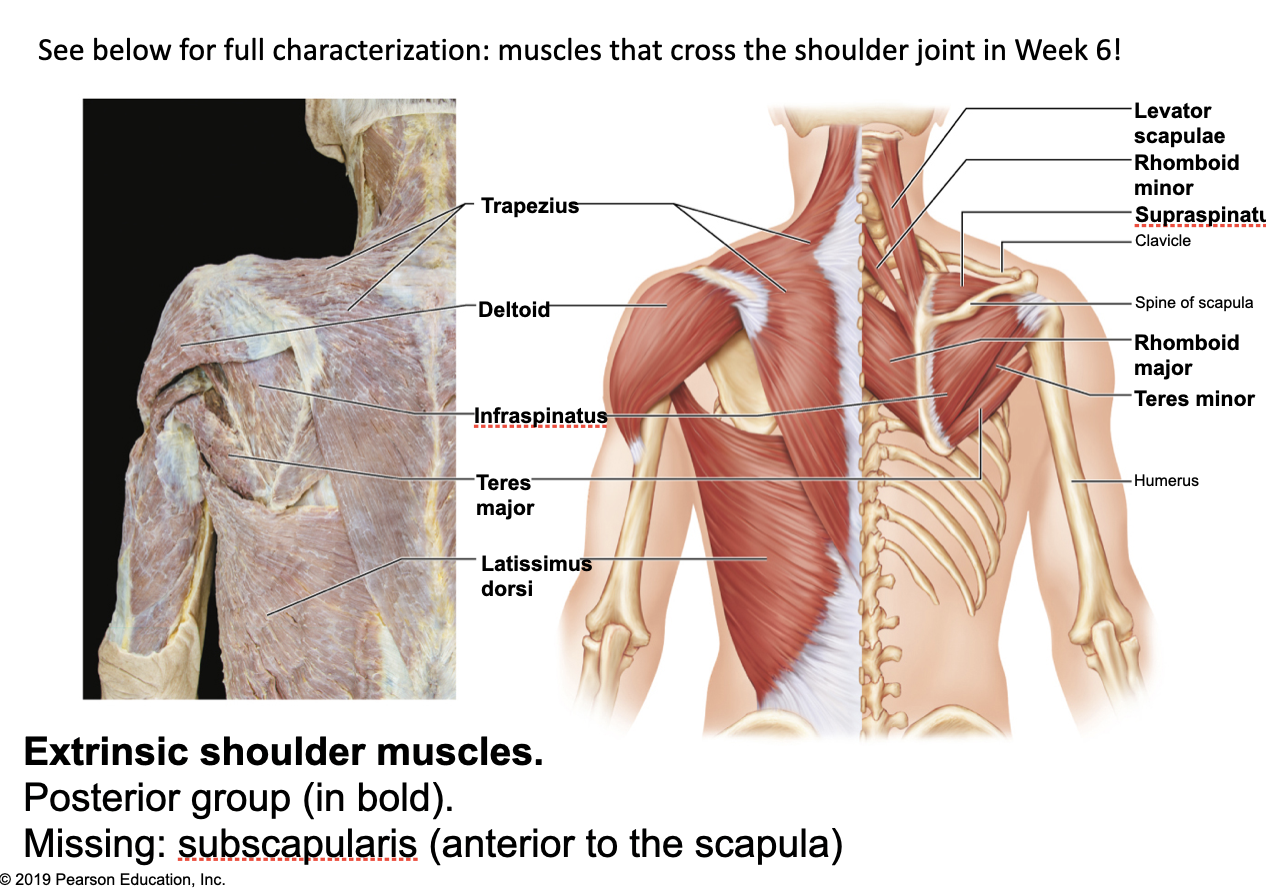
Posterior muscles
Latissimus dorsi (arm moving)
Trapezius
Levator scapulae (move scapula)
Rhomboids (move scapula)
Pectoralis minor
Comments - flat, thin muscle directly beneath and obscured by pectoralis major
Origin - ribs 3-5 (or 2-4)
Insertion - coracoid process
Action - with ribs fixed, pulls scapula forward and downward
Nerve supply - medial and lateral pectoral nerves
Serratus anterior
Comments - fan shaped muscle; lies deep to scapula, deep and inferior to pectoral muscles on lateral rib cage; forms medial wall of axilla; origins have serrated (sawtooth) appearance; paralysis results in “winging” of vertebral border of scapula away from chest wall, making arm elevation impossible
Origin - ribs
Insertion - medial border of scapula
Action - rotates scapula, prime mover to protract
Nerve supply - long thoracic nerve

Trapezius
Comments - most superficial muscle of posterior thorax; flat and triangular, upper fibers run inferiorly to scapula; middle fibers run horizontally to scapula; lower fibers run superiorly to scapula
Origin - occipital bone, ligamentum nuchae, spinous process, all thoracic vertebrae
Insertion - acromion, spine of scapula, lateral third of clavicle
Action - stabilizes, elevates, retracts, and rotates scapula
Nerve supply - acccessory nerve (cranial XI); C3 and C4
Levator scapulae
Comments - located at back and side of neck, deep to trapezius; thick straplike muscle
Origin - transverse processes
Insertion - medial border scapula
Action - stabilizes, elevates, retracts, and rotates scapula
Nerve supply - cervical spinal nerves and dorsal scapular nerve (C3-C8)
Rhomboids
Comments - two roughly diamond-shaped muscles lying deep to trapezius and inferior to levator scapulae; rhomboid minor is the more superior muscle
Origin - spinous process
Insertion - medial border of scapula
Action - retract (adduct); rotate downward
Nerve supply - dorsal and scapular nerve (C4 and C5)

Deep muscles of the thorax: breathing
External intercostals
Internal intercostals
Diaphragm
External intercostals
Comments - 11 pairs lie between ribs; fibers run obliquely (down and forward) from each rib to rib below; in lower intercostal spaces, fibers are continuous with external oblique muscle, forming part of abdominal wall
Origin - inferior border of rib above
Insertion - superior border of rib below
Action - pull ribs toward one another to elevate rib cage
Nerve supply - intercostal nerves

Internal intercostals
Comments - 11 pairs lie between ribs; fibers run deep to and at right angles to those of external intercostals (ie run downward and posteriorly); lower internal intercostal muscles are continuous with fibers of internal oblique muscle of abdominal wall
Origin - superior border of rib below
Insertion - inferior border of rib above
Action - pull ribs together and depress rib care
Nerve supply - intercostal nerves
Diaphragm
Comments - broad muscle pierced by the aorta, inferior vena cava, and esophagus; forms floor of thoracic cavity; dome shaped in relaxed state; fibers converge from margins of thoracic cage toward a boomerang-shaped central tendon
Origin - surface of rib cage and sternum
Insertion - lumbar vertebrae, central tendon
Action - prime mover of inspiration; flattens on contraction

Muscles of the Abdominal Wall: Trunk Movements and Compression of Abdominal Viscera
Rectus abdominis
External obliques
Internal obliques
Transversus abdominis
Rectus abdominis
Comments - medial superficial muscle pair; extend from pubis to rib cage; ensheathed by aponeurases of lateral muscles; segmented by three tendinous intersections
Origin - pubic crest and symphysis (not underlined)
Insertion - xiphoid process and costal cartilages of ribs
Action - flex lumbar region of vertebral column
External oblique
Comments - largest and most superficial of the three lateral muscles; fibers run downward and medially
Origin - lower eight ribs
Insertion - linea alba, pubic crest
Action - flex vertebral column and compress abdominal wall; trunk rotation and lateral flexion
Internal oblique
Comments - most fibers run upward and medially; however, the muscle fans so it is inferior fibers run downward and medially
Origin - lumbar fascia, iliac crest, and inguinal ligament
Insertion - linea alba, pubic crest
Action - “as for external oblique” - flex vertebral column and compress abdominal wall; trunk rotation and lateral flexion
Transverse abdominis
Comments - deepest (innermost) muscle of abdominal wall; fibers run horizontally
Origin - inguinal ligament, lumbar fascia, cartilages
Insertion - linea alba, pubic crest
Action - compresses abdominal contents


Muscles of the Pelvic Floor and Perineum: Support of Abdominopelvic Organs
Pelvic floor and perineum: three muscle layers that support the abdominopelvic organs (from inside out, superior to inferior):
Pelvic diaphragm (deep) - levator ani, coccygeus
urogenital diaphragm (NOW: PERINEAL MEMBRANE)- deep transverse perineal muscle
superficial perineal space (covered by skin) - ischiocavernosus, bulbospongiosus (SAME as bulbocavernosus!), superficial transverse perineal
Levitator ani
Comments - Broad, thin, three-part muscle (pubococcygeus, puborectalis, and iliococcygeus); its fibers extend inferomedially, forming a muscular “sling” around prostate (male) or vagina (female), urethra, and anorectal junction before meeting in the median plane
Origin - extensive linear origin inside pelvis from pubis to ischial spine
Insertion - inner surface of coccyx, levator ani of opposite side, and (in part) into the structures that penetrate it
Action - supports and maintains position of pelvic organs
Coccygeus, now ischiococcygeuus
Comments - Small triangular muscle lying posterior to levator ani; forms posterior part of pelvic diaphragm
Origin - spine of ischium
Insertion - sacrum and coccyx
Action - supports pelvic organs