Exam 2
 Call Kai
 Learn
 Practice Test
 Spaced Repetition
 Match
 Flashcards
 Knowt Play
1/43

There's no tags or description
Looks like no tags are added yet.
44 Terms
In what direction does phase encoding occur?
Perpendicular to frequency encoding direction; L-R on axial, R-L on coronal, Top to bottom on sagittal
In what direction does frequency encoding occur in?
Perpendicular to phase encoding; R-L on axial; Z axis on sagittal coronal
What methods can be used to compensate for phase encoding artifacts?
Increase FOV; add SAT bands; swap frequency and phase direction
What are three methods to reduce/eliminate cross talk?
add slice gap; adjust slice excitation order; add slice concatenation
Which cross talk elimination method does not reduce scan time?
Increasing slice gap
What causes alliancing artifacts in frequency direction?
Frequencies in echo aren’t sampled at the proper rate; frequency outside of FOV; and outside signal is encoded and included into FOV
What is the software option that compensates for frequency aliasing artifacts?
Frequency filter
What is the suggested temperature for the MRI scan room?
18-22 C
What is the optimal humidity range in MRI?
30-50% RH
What activity references to proactive and reactive?
Proactive - improve CONTINUOUSLY over time
Reactive - improve ONLY after a event
What is a quality control check that is performed on a phantom that is performed for something other than image quality?
Central frequency and transmitter gain
How often does a physicist check the coils?
Once a year
If there are air bubbles in a phantom (other than level bubbles), is it okay to proceed with the quality check?
No
What is the weekly allowed central frequency drift from the ACR menu?
1.5 ppm per week
What is “Table ok” checking for?
Accuracy of laser localizer
Are all shimming effects performed during installation only?
No
What material is used for RF shielding and magnetic shielding?
Copper, aluminum and steel
What value must the fringe field be restricted to inside the room?
5 gauss
What artifact can be made by a leak in RF shielding? How do you eliminate it?
Zipper artifact, can be eliminated by checking any RF leak such as door opened
What type of magnetic shielding is located inside the scanner?
Active shielding
What are two differences between MRI and CT contrast? (other than contrast agent?
CT contrast is in higher volume of contrast fluid and at a higher rate of injection
Screening MRI devices should locate and identify what type of objects?
Ferromagnetic objects
What are 3 types of hardware operation electronic devices for magnetic detectors?
Beat-frequency oscillator (BFO); pulse induction (PI); VLF
Describe a situation where a screening device can be helpful?
Can be helpful for patients with limited mobility
What are two cons of having screening devices?
Alarm fatigue and false safety
What are two type of lines that MRI techs are allowed to remove?
IV line and midline
What is the speed and volume for studies using power injector? (dynamic studies)
22 G can inject up to 3 ml/sec; 24 G at 1.5-2 ml/sec
What are some types of artifacts?
Motion artifact, physical artifacts, sampling artifacts, susceptibility artifacts, technical error, sampling artifacts, and hardware artifacts
Phase aliasing artifact
Shows: Anatomy overlaid onto anatomy inside FOV in direction of phase encoding (L→R)
Cause: Anatomy outside of FOV; outside signal is encoded and included into FOV; Extra signal is potentially at the same phase value as anatomy being scanned
Fix: Increase FOV (but lowers resolution); add sat bands (but increases SAR); add no phase wrap; swap phase and frequency direction
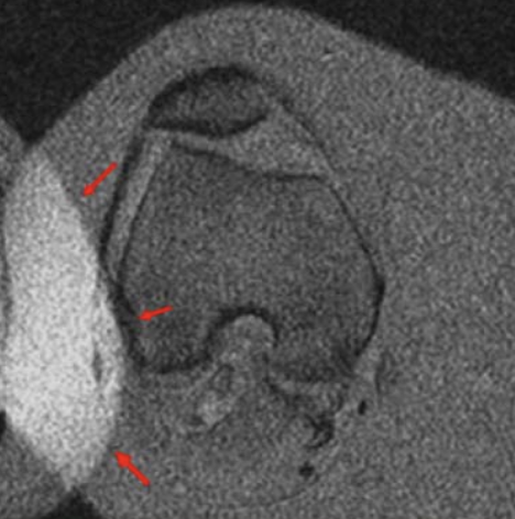
Frequency aliasing artifact
Shows: Anatomy overlaid onto anatomy inside FOV in direction of frequency encoding (top to bottom)
Cause: Frequencies in echo aren’t sampled at the proper rate; frequency outside of FOV; and outside signal is encoded and included into FOV
Fix: use frequency filter

Cross talk
Shows: Slices next to each other will have different tissue contrast
Cause: RF profile has protons in adjacent slices are already excited which = low/none signal
Fix: add slice gap; adjust slice excitation order; add slice concatenation

Slice overlap/cross slice
Shows: dark area with no signal
Cause: multi slice regions that are overlapping and saturated protons can’t produce signal
Fix: try position FOV or separation into two sequences (AX T1 Upper; AX T1 Lower)
Truncation artifact (Gibbs artifact)
Shows Low bright/dark lines though high intensity area
Cause: Under sampling of data so not enough k-space filled
Fix: Larger matrix size

Zipper artifact
Shows: Mini zipping lines across picture
Cause: Outside RF enters the room; defect in room RF shielding;
Fix: Make sure scan room door is shut; must find leak so call service engineer if not found

Spike artifact (k-space artifact)
Shows: Parallel bands
Cause: wrong electrical discharge gets encoded into k-space
Fix: Repeat scan; can’t be corrected
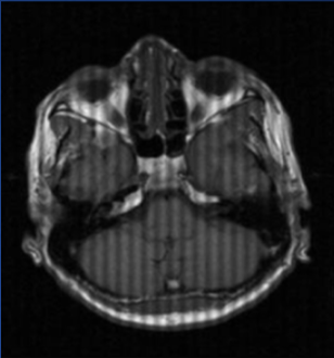
Propeller artifact (k-space artifact)
Cause: Using a propeller to fill k-space but anatomy is too large for FOV
Fix: Repeat scan and increase FOV
Moire artifact
Show: Shown only on gradient echo pulse sequences. Black and white banding near edge of FOV
Cause: wrap on one part of the body to the other; caused by field inhomogeneities
Fix: Use spin echo sequences; make sure body is not touching bore
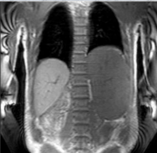
Shading artifact
Show: Shading around anatomy
Cause: Uneven uniform excitation; coil coupling; field inhomogeneities; coil selection
Fix: Select right size coil for anatomy; prevent anatomy from touching coil; make sure pre-scan calculation is performed by system

Quadrature coil artifact
Show: 180 degree ghosting
Cause: Signals from quadrature channels are summed incorrectly
Fix: Have coil serviced or repaired
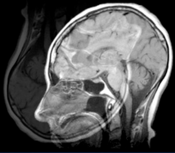
Geometric distortion artifact
Show: Image distortion and squishing
Cause: Improper gradient/gradient amplifier function which creates poor linear variation
Fix: Call service

Would more encoding be done to compensate a phase or frequency aliasing artifact? What direction?
Yes for phase encoding aliasing artifact so phase direction
ACR phantom; what value in mm is it measured in?
Sagittal is 148 mm and A/P and R/L is 190 mm with a ± 2mm allowed
ACR middle is shown to
measure whether laser/isocenter match
The arrows show in ACR
Outer edges show to measure diameter from A/P and L/R (in grid/slice 5/axial); arrows in sagittal localizer show SI length