ASLP 3025 Exam #3
 Call Kai
 Learn
 Practice Test
 Spaced Repetition
 Match
 Flashcards
 Knowt Play
1/135

There's no tags or description
Looks like no tags are added yet.
136 Terms
Which bone has projections which comprise the middle and superior nasal conchae?
Ethmoid
Which cranial/facial bone is unpaired?
Occipital
Which bone provides the alveolar ridge?
Mandible
Which bone comprises the posterior 1/4 of the hard palate?
Palatine
Which bone houses the lower dental arch?
Mandible
Which bone comprises part of the nasal septum?
Vomer
Mobile articulators
- Tongue
- Mandible
- Soft palate (velum)
- Lips
- Cheeks (buccae)
- Fauces
- Pharynx
Tongue
- Largest mobile articulator
- Occupies the floor of the mouth
- Superior surface - dorsum
- Anterior most portion - apex
- Median sulcus - divides the tongue into left and right
Soft palate (velum)
- Used to differentiate nasal sounds from non-nasal sounds

Lips
- Moved to produce different speech sounds
- Four layers of tissue
- Vermillion: red part of the lips
- Vermillion border: rim of paler skin around lips
- Philtrum: groove in the midline of the upper lip
Cheeks (buccae)
- Play a role in the changes of resonance of the oral cavity
- Continuous with the lips
Fauces

Pharynx
- Sounds resonate through the pharyngeal cavity
- Production of pharyngeal consonants in other languages
Immobile articulators
- Alveolar ridge
- Hard palate
- Teeth
Alveolar ridge

Hard palate

Cranial & facial bones
- Skull
- Mandible
- Maxillae
- Nasal bones
- Palatine bone
- Nasal conchae
- Vomer
- Zygomatic
- Lacrimal
Skull
- Provides protection of the brain
- Point of attachment for speech, mastication and swallowing muscles
- Composed of 22 bones: 8 paired, 6 unpaired
Mandible
- Second largest articulator
- Comprises the lower jaw
- Provides the lower dental arch
Landmarks:
- Symphysis menti
- Mental foramen
- Body
- Angle
- Ramus
- Condylar process
- Mandibular foramen

Maxillae
- Left and right portion comprise the upper jaw
- Provides the hard palate, alveolar ridge and upper dental arch
Landmarks:
- Infraorbital foramen
- Zygomatic, orbital and frontal processes
- Articulates with 9 bones: ethmoid, frontal, inferior nasal conchae, palatine, vomer, zygomatic

Nasal bones
- Paired bones which comprise the superior nasal surface
- Articulates with 3 bones: frontal, ethmoid and maxilla

Palatine bones
- Pair of "L" shaped bones
- Comprise the posterior 1/4 of the hard palate
- Soft palate attaches to the posterior border of the horizontal processes
- Articulates with 5 bones: ethmoid, frontal, inferior nasal conchae, maxilla and vomer

Inferior nasal conchae
- Makes up the lowermost part of the lateral wall of the nasal cavity
- Articulate with maxilla, palatine, and ethmoid bones

Vomer
- Unpaired bone
- Comprises the lower half of the nasal septum
- Articulates with the maxilla, palatine, ethmoid, and sphenoid bones

Zygomatic bones
- Form the cheek bones
- Articulates with maxillae, frontal bone, and temporal bone

Lacrimal bones
- Smallest bones of the facial skeleton
- Close to the lacrimal glands
- Articulate with the ethmoid, frontal, inferior nasal conchae, and maxilla
Bones of the cranial skeleton
- Ethmoid
- Sphenoid
- Frontal
- Parietal
- Occipital
- Temporal
Ethmoid
- Present in crania, nasal and orbital spaces
Landmarks
- Crista galli
- Perpendicular plate
- Cribriform plate
- Orbital plate
Sphenoid
Landmarks
- Corpus
- Hypophyseal fossa (sella turcica)
- Greater and lesser wings
- Lateral and medial pterygoid plates

Frontal
- Makes up the bony forehead, anterior cranial case, supraorbital region and the nasal cavity
- Overlies the frontal lobe of the cerebrum
- Articulates with 12 bones: ethmoid, sphenoid, lacrimals, maxilla, nasals, parietals and zygomatics
Parietal
- Overlies the parietal lobes of the cerebrum
- Forms the middle portion of the brain case
- Paired bones are united at the sagittal suture
- United with the frontal bone at the coronal suture
- United with occipital bone at the lambdoidal suture
- United with temporal bone at the squamosal suture

Occipital
- Overlies the occipital lobe of the cerebrum
- Makes up the posterior braincase
- Articulates with the temporal, parietal and sphenoid bones
Which of the following is an intrinsic lingual muscles?
Superior longitudinal
Which of the following is an extrinsic lingual muscle?
Genioglossus
Which of the following muscles is a mandible elevator?
Medial pterygoid
Which of the following muscles protrudes the mandible?
Lateral pterygoid
Which of the following muscles is a soft palate depressor?
Palatoglossus
Buccal dental surface
Space between the lips and teeth, and cheeks and teeth

Class I Malocclusion
Neutroclusion.
- First molar of the mandibular arch is one-half tooth advanced of the maxillary molar
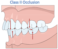
Class II Malocclusion
Mandibular molars are retracted at least one tooth behind the first maxillary molars
Class III Malocclusion
First mandibular molar is advanced more than one tooth ahead of the first maxillary molar

Torsiversion
Rotated or twisted on the long axis

Labioversion
Tilted toward the lip

Linguaversion
Tilts toward the tongue

Buccoversion
Tilted towards the cheek
Distoversion
Tilted away from the midline of the dental arch

Mesioversion
Tilted toward the midline of the dental arch
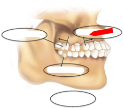
Infraversion
Inadequate eruption relative to the other teeth in the arch

Supraversion
Excessive eruption relative to the majority of teeth in the arch

Orbicularis Oris
- Constricts the oral opening
- Upper and lower act like a "drawstring" to pull the lips together and form a labial seal
- Interacts with other facial muscles to produce facial expressions

Risorius
Retracts lips at the corners

Buccinator
- Moves foo to the grinding surfaces of molars
- Constricts oropharynx

Levator Labii Superioris muscle
Elevates the upper lip with zygomatic minor and levator labii superioris alaeque nasi

Zygomatic Minor muscle
Elevates the upper lip with levator labii superioris and levator labii superioris alaeque nasi

Levator Labii Superioris Alaeque Nasi
Elevates the upper lip with levator labii superioris and zygomatic minor

Levator Anguli Oris
Draws the corner of the mouth up and medially

Zygomatic Major
Elevates and retracts angle of mouth
Depressor Labii Inferioris
Dilates orifice by pulling lips down and out

Depressor Anguli Oris
- Depressed the corners of the mouth
- Helps compress the upper lip against lower lip
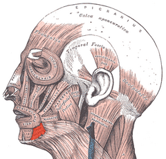
Mentalis
- Elevates and wrinkles the chin
- Pulls the lower lip out

Platysma
- Depresses the mandible
- Thin, flat and large muscle of the neck
- Covers the majority of the anterior and lateral surfaces of the neck

Superior Longitudinal muscle of the tongue
- Elevates the tongue
- Assists in retraction
- Deviates the tip

Inferior Longitudinal muscle of the tongue
- Pulls the tip down
- Assists in retraction
- Deviates the tongue

Transverse Longitudinal of the tongue
Involved with narrowing the tongue

Vertical muscle of the tongue
Pulls the tongue down to the floor
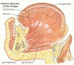
Genioglossus
- Anterior fibers retract
- Posterior fibers protrude
- Anterior and posterior depress

Hyoglossus
Pulls sides of tongue down

Styloglossus
Draws the tongue back and up

Palatoglossus
- Elevates back of tongue
- Depresses soft palate
- Comprises the anterior faucial pillar

Masseter
- Elevates the mandible
- Most superficial muscle of mastication
- Quadrilateral shaped muscle

Temporalis
- Elevates the mandible
- More rapid contraction than masseter

Medial Pterygoid
- Elevates the mandible
- Also known as internal pterygoid muscle
- Deep to the masseter
- Acts in conjuction with the masseter

Lateral Pterygoid
- Protrudes the mandible
- Also known as external pterygoid muscle
- Works in contrast of the mandibular elevators

Levator Veli Palatini
- Elevates and retracts posterior velum
- Makes up the bulk of the soft palate

Musculus Uvulae
Contraction shortens the soft palate, effectively bunching it up

Tensor Veli Palatini
- A major dilator of the Eustachian tube
- The only tensor of the soft palate
- Contraction stabilizes and flattens the soft palate

Palatopharyngeus
Assists in narrowing the pharyngeal cavity and lowering the soft palate

Superior Pharyngeal Constrictor
- Contraction pulls the pharyngeal wall forward and constricts the pharyngeal diameter, to assist with swallowing
- Forms the upper sleeve of pharyngeal wall

Middle Pharyngeal Constrictor
Narrows the diameter of the pharynx

Inferior Pharyngeal Constrictor
Comprised of the cricopharyngeus and thyropharyngeus

Cricopharyngeus
- Constricts superior orifice of esophagus
- Component of the inferior constrictor and forms the muscular orifice of the esophagus
- Important for swallowing

Thyropharyngeus
- Reduces the diameter of the lower pharynx
- Upper portion of inferior pharyngeal constrictor

Salpingopharyngeus
- Elevates the lateral pharyngeal wall
- Converges with palatopharyngeus

Stylopharyngeus
Assists in elevation and opening of the pharynx

Oral cavity
Extends from the oral opening to the faucial pillars
- Anterior limit: lips and teeth
- Lateral limit: cheeks and teeth
- Posterior limit: palatoglossal arch
- Superior limit: hard palate
- Inferior limit: tongue and floor of mouth
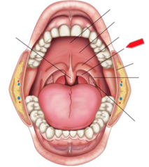
Anterior faucial pillars
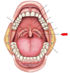
Posterior faucial pillars
Nasal cavity
- Produced by the maxillae, palatine, and nasal bones
- Divided by the nasal septum
- Nares to nasal conchae

Nasopharynx
Part of the pharyngeal cavity.
- Space above velum to nasal conchae

Oropharynx
Part of the pharyngeal cavity.
- Posterior to the fauces, velum to hyoid bone
- Letter B shown in picture

Laryngopharynx
Part of the pharyngeal cavity.
- Epiglottis to esophagus
- Letter C shown in picture

Individual teeth types functions
Incisors: 8; cutting
Canines: 4; tearing
Premolars: 8; tearing
Molars (including wisdom): 12; grinding
Deciduous teeth
Primary set of teeth, or baby teeth
Successional teeth
Teeth in the mature dental arch that replace corresponding teeth of the deciduous arch
Physiological elements of the lips
- Facial muscles act upon the lips to form a range of movements
- Facial muscles exert forces on the lips which determine the direction of lip movement
- Lower lip achieves a greater velocity and force than the upper lip. The extra force is due to the mentalis muscle
- Lower lip adapts to being closer to/further from the upper lip
- Lips are resistant to interference; they adjust and accommodate to physical restraints
Physiological elements of the mandible
- Carries the lips, tongue, and teeth to their targets
- Movement is slight during speech, compared to chewing
- Paralysis of the muscles of mastication can highly decrease speech intelligibility
- Mandibular elevation is more important than depression for articulation
Physiological elements of the tongue
- The most important articulator
- Involved in the production of the majority of the sounds in English
- Extrinsic muscles set the basic posture
- Intrinsic muscles manage the fine movements needed for articulation
Physiological elements of the velum
- Rapid and precise opening and closing of the velopharyngeal port must occur
- Velopharyngeal mechanism acts as a "regulator"
- Generally closed for non-nasal speech through contraction of levator veli palatini with help from musculus uvulae
- Production of high pressure consonants requires greater velopharyngeal effort
Mechanism of resonance
- The shape of the supraglottal vocal tract is constantly changing
- Pharyngeal constrictors and other supraglottal muscles change the shape of the pharynx
- Higher frequency sounds resonate best when the pharyngeal wall is highly tense
- Lower frequency sounds are better amplified by a larger more relaxed pharynx
- Mouth is capable of most variation of shape and size out of all resonators
- Moving structures of mouth-tongue, velum, cheeks, mandible, and lips - are more important in resonance than the fixed structures
- Tongue movements along with the lowering and closing of the mandible contributes to the formation of specific vowels
- Elevation and tensing of the velum is important for achieving velopharyngeal closure
Hypernasality
Excessive nasal cavity resonance during non-nasal vowels and non-nasal voiced consonants