Practical 2 Structures
 Call Kai
 Learn
Learn Practice Test
 Spaced Repetition
 Match
Match Flashcards
 Knowt Play
1/105

There's no tags or description
Looks like no tags are added yet.
Name | Mastery | Learn | Test | Matching | Spaced | Call with Kai | Chat |
|---|
No analytics yet
Send a link to your students to track their progress
106 Terms
Nasal Cavities

Nasal Aperture

Dorsal Nasal Concha

Ventral Nasal Concha

Ethmoidal conchae
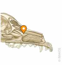
Nasal Septum

Dorsal nasal meatus

Middle nasal meatus

Ventral nasal meatus
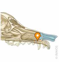
Common nasal meatus
From top to bottom of the nasal meatuses
Frontal sinus

Nasopharynx

Trachea

Basihyoid bone

Epiglottis
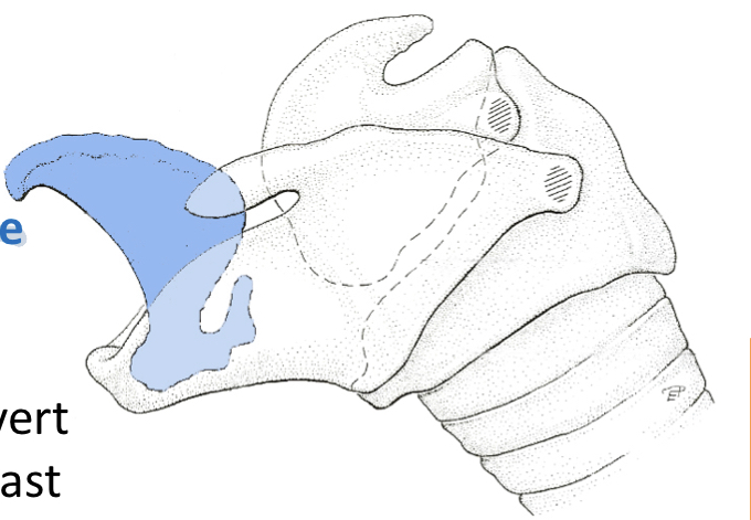
Thryoid Cartilage

Arytenoid cartilage

Cricoid cartilage

Vocal Folds

Glottis

Cricothyroid muscle
tense the vocal folds

cricoarytenoideus dorsalis muscle

Abduct the vocal folds
cricoarytenoideus lateralis muscle

adducts the vocal folds
thyroartyenoideus muscle
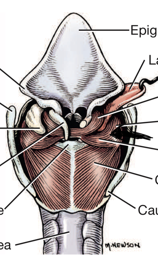
Relaxes the vocal folds
External Intercostal m

Internal Intercostal muscle

Costal Pleura
Section of parietal pleura that lines the inner surface of the chest wall
Diaphragmatic Pleura
Section of the parietal pleura that covers the upper surface of the diaphragm in the thoracic cavity
Pleural Cavity
Fluid filled space between the visceral pleura and parietal pleura
Mediastinal pleura
Central area of the chest cavity between the lungs and outlining the heart
Hilus of the lungs

Internal thoracic artery

Diaphragm

Cranial Lobe of Left Lobe

Caudal Lobe of Left Lung

Cranial Lobe of Right Lung

Caudal Lobe of Right Lung

Middle Lobe of Right lung

Accessory Lobe of Right Lung

Primary Bronchi

Carina
Where the lungs separate, and where they bifurcate
Frontal Bone

Cornual Process
ox

Ethmoid Bone

Infraorbital canal

Thyrohyoid Bone
C

Lingual Process

Choanae

Ethmoidal Meatuses
Horse and Ox
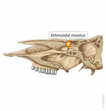
Ventral conchal sinus
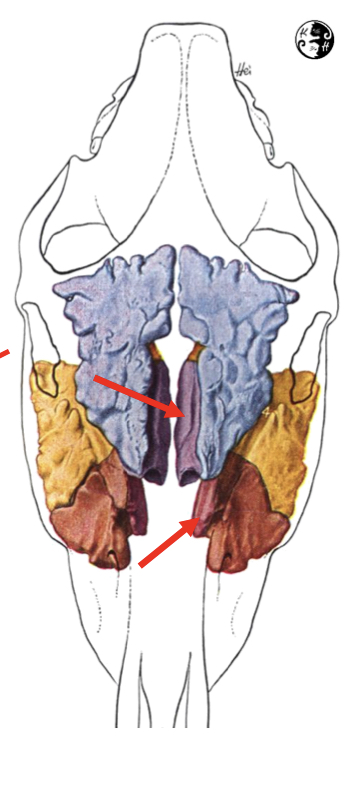
Deeper magenta- Horse
Dorsal Conchal Sinus

Purple- Horse
Cornual Diverticulum

Sinus in the horn-Ox
sphenopalatine sinus

Continuous with green(horses only)
Rostral and Caudal maxillary

Horses(ox have one)
Conchofrontal sinus

Horse
Frontomaxillary aperture

Horse
Palatine Sinus
ox

Thyroid Notch
Horse

Nasopharynx

Pharynx

Connects the nasopharynx to the trachea
Ox lung

Horse Lung

larger, externally smooth and very little CT septa. Fewer external fissurations(separations), so the lobes are not obvious. There are fewer lobes
Tracheal Bronchus
Ox- A right sided structure

Cranial Vena Cava

1
brachiocephalic vv
Dog

External jugular vein

Subclavian veins

4
Azygos Vein

5
Aorta

3
Ascending aorta
Bottom part of the aorta

Aortic arch
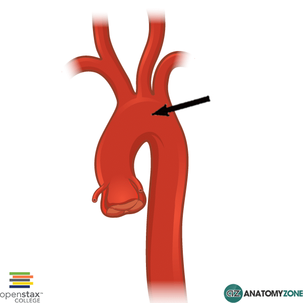
Descending aorta
Right side

Brachiocephalic trunk
Left trunk

Bicarotid trunk
B- Horse and ox

Common corotid artery
On both sides

Subclavian artery
On both sides

Phrenic Nerve

12
Sympathetic trunk

11
Cervicothoracic ganglion

6
Ansa Subclavia

4 and 4’- dog
vagosympathetic trunk

1
Middle cervical ganglion

3- dog
Vagus nerves
Cervical Nerve

Ventral Vagal Trunk
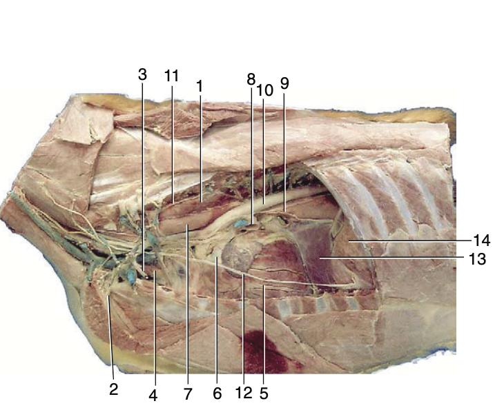
9
Pericardial Sac

Black Circle
Parietal pericardium

Green
Fibrous pericardium

Yellow
Pleural Cupula

Red- extension of the parietal pleura through thoracic inlet
Pulmonary Ligament

Blue- Double layer of pleura that reflects off the mediastinum and onto the caudal loeb of the lungs, caudal to the hilus
Costodiaphragmatic recess

Green- potential space at the junction of the diaphragmatic and costal pleura, lungs don’t enter this space during normal breathing
Pleural Reflection

Reflection of the costal pleura onto the diaphragm, Sternum to 11th costochondral junction to dorsal part of the 13th rib. Caudal extent of the thoracic cavity
Costodiaphragmatic Recess

Green Line
Basal Border of the lung

Blue- 6th costochondral junction to middle of the 8th rib to dorsal end of the 11th intercostal space
Frontal Sinus

Green-Dog
Maxillary Recess Sinus
Yellow-Dog

Ventral Conchal Sinus

Horse- Communicates with rostral maxillary sinus
Nasomaxillary Apeture

Horse- Opening between maxillary sinuses and middle meatus of the nasal cavity. All sinuses drain into a maxillary sinus then into middle meatus
Auricles
Ear-like appendages
Atria

Receive veins, they’re thin walled and have less forceful contractions. Interatrial septum separates RA from LA
Interventricular Septum(IVS)

Separates Ventricles