IB BIO C3.1 Integration of Body Systems ☑️
 Call Kai
 Learn
 Practice Test
 Spaced Repetition
 Match
 Flashcards
 Knowt Play
1/36

There's no tags or description
Looks like no tags are added yet.
37 Terms
(C3.1.1) Define system integration.
groups of specialized cells (tissues) → groups of specilized tissues (organs) → groups of organs (body systems) - specialized for nutrition, respiration, exrection, reproduction, coordiantion → organism
* coordination is needed for component parts of a system to collectively perform an overall function.
(C3.1.2) Define tissue, organ and organ systems.
- hierarchy of organisation (animal / plant) -
1. cell: smooth muscle cell // guard cell
2. tissue: muscular wall // stoma
3. organ: bladder // leaf
4. organ system: nervous, respiratory, circulatory, digestive, excretory, muscular, skeletal, integumentary, reproductive, endocrine // vascular
5. organism: white-tailed deer // magnolia tree
(C3.1.2) Define emergent property.
the whole is greater than the sum of the parts;
arise due to integration of subsystems
(C3.1.2) State an example of an emergent property for each level of biological organization within a multicellular organism.
Cheetah: integration of nervous, skeletal and muscular system
- large heart and lungs
- flexible spine that acts as a spring while running and increases stride length
- lean body with long legs
- longer and heavier hind limb bones enable longer strides
- long muscular tail acts as a stabiliser while running at high speeds
(C3.1.3) State the two primary mechanisms by which animals integrate organ systems.
hormonal (endocrine system) and nervous signalling:
- autonomic nervous system (ANS): communication without conscious knowledge e.g. regulation of heart rate, blood glucose level
- adrenal (endocrine) glands secrete hormones into bloodstream to help communication between organs (target tissue)
e.g. release of epinephrine from adrenal glands:
- sensory organs transmit info to nervous system that indicate epinephrine is needed as part of response
- ANS send impulses to adrenal glands to release epinephrine
- increased heart rate, increased blood flow to muscles to prepare body for immediate increased activity
(C3.1.3) Compare the type of signal, transmission of signal, effector response, speed and duration of response between hormonal and nervous signals.
similarities:
- both used for communication between cells/tissues/organs
- hormones and neurotransmitters are both chemicals (bind to receptors)
- both can stimulate / inhibit processes in target cells
- both can work over long distances
- both under control of the brain
- both use negative feedback
differences: (hormonal VS nervous)
- type: chemical messenger VS electrical nerve impulses
- duration: long term VS short term
- response speed: slower VS faster
- transmit through: bloodstream VS neurons
- carried throughout body VS carried to specific cell
- wide range of tissues affected VS only muscles / glands receive signals
- control involuntary VS control both
- examples: epinephrine, insulin VS dopamine, acetylcholine
(C3.1.4) State the function of the brain.
central information integration organs:
- regulate and monitors uncounscious body processes e.g. blood pressure, heart rate, breathing
- process information combined from several inputs, and respond by controlling balance, muscle coordination and most voluntary movements
- speech, emotions, problem solving
- learning and memory
(C3.1.4) List the receptors which send information to the brains.
receptors (conscious level):
1. photoreceptors: located within retina of eyes for visual information, travels to occipital lobe
2. chemoreceptors: located within tongue for tasting, travels to parietal lobe
3. thermoreceptors: located in skin to provide information on temp changes
4. mechanorecptors: located in inner ear, sensitive to sound vibrations, travels to temporal lobe
receptors (subconscious level):
1. osmoreceptors: located in carotid arteries and hypohtalamus of brain, sense solute and water content of blood
2. baroreceptors: located in carotid arteries and aorta, sense blood pressure based on how much a blood vessel is stretched by internal pressure
3. proprioceptors: located in muscles and joints, provide brain with sense of balance and coordination
(C3.1.4) List the three main areas in the brain.
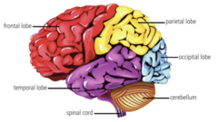
1. cerebrum: responsible for conscious activities
- divided into right and left cerebral hemispheres
- each hemisphere consists of 4 lobes: frontal (learning and memory activities mainly in), temporal, parietal, occipital (順時針 Flower POT)
2. cerebellum: coordinates voluntary movements, controls balance and equilibrium
3. brainstem: responsible for subconscious functions /associated with ANS
- medulla oblongata in brainstem regulates breathing and heart rate
(C3.1.5) List organs of the central nervous system.
spinal cord (內灰外白) + brain (內白外灰)
- SC contains unconscious reflexes assocated with balance and skeletal muscles (= can only coordinate unconscious processes)
- 31 pairs of spinal nerves bring sensory information into CNS from body, allowing motor information to be sent out
(C3.1.5) Outline the two main tissues types of the central nervous system.
in spinal cord (內灰外白):
- white matter (composed of axons of neurons, carries neural impulses to and from brain)
- grey matter (contains neurons and synapses)
when sensory info enters grey matter of spinal cord, motor info is immediately sent back out, this pathway of impulse = reflex arc (involuntary)
(C3.1.6) Outline the function of sensory neurons sensory receptors and sensory neurons.
- receptor: a modified neuron capable of transduction
- transduction: conversion of a physical stimulus into an electrical signal called action potential that is carried along a neuron
- receptors are specialized to transduce one type of physical stimulus e.g. photoreceptors transduce light, thermoreceptors transduce heat / lack of heat, baroreceptors transduce pressure
- sensory neurons carry action potential from receptors to CNS
(C3.1.7) Outline the function of motor neurons.
- cerebrum uses sensory info to make decisions concerning movements
- motor cortex in cerebrum: sends action potential, located in the most posterior portion of frontal lobe of cerebrum
- motor neurons: carry action potential from CNS to muscles (effectors), form synapses called motor end plate / neuromuscular junctions with muscule fibres
- when AP arrived at motor end plate,, acetylcholine is released (NT that initiates contraction of sacromere, leading to shortening of the muscle and movement of a bone
(C3.1.8) Define nerve and neuron, give examples of neurons.
neuron: cell of nervous system
- sensory, motor, interneuron
nerve: group of myelinated or unmyelinated neurons
- contain either or both sensory and motor neurons
- myelinated: Schwann cells wrapped around axon, intervening areas: nodes of Ranvier
- action potential of myelinated axon can skip from one NOR to next → faster transmission speed
(C3.1.9) Outline the input, processing and output of the pain reflex arc, including the role of receptors, sensory neurons, interneurons, motor neurons and effectors.
pain reflex arc: involuntary
1. nocireceptor located in skin of finger initates afferent (sensory) action potential
2. action potential travel through hand and join a spinal nerve
3. afferent neuron synapses with a short interneuron loacted entirely within grey matter of spinal cord
4. afferent neuron synapses with a motor neuron
5. action potential goes to arm muscle (effector)
6. hand withdrawal
significance: limit damage to body tissue by generating a quick reaction involving 3 neurons only
withdrawal action occurs faster than pain sensation:
- needs to travel to cerebrum to be integrated by many neural syanpses before a sensation is felt
(C3.1.10) State the function of the cerebellum.
coordinating skeletal muscle contraction and balance
1. motor cortex initiates muscle contraction
2. cerebellum receives feedback impulses
3. cerebellum send out impulses to coordinate movement
4. smooth and balanced muscular activity, coordinated movements
(C3.1.11) Define circadian rhythm.
pattern or physiology based on a 24-hour cycle e.g. wake and sleep cycle
- diurnal, nocturnal
(C3.1.11) State how circadian rhythm is controlled.
controlled by secretion of melatonin by pineal gland (located between cerebrum and brainstem) in preparation sleep - higher levels during night for diurnal animals
(C3.1.12) Outline the mechanism of action of epinephrine as a signaling molecule.
1. epinephrine is secreted by adrenal glands in preparation for vigorous exercise / fight or flight response (muscle contraction)
2. epinephrine is hydrophilic, cannot pass through plasma membrane
3. epinephrine binds to binding site on GPCR protein → GPCR change shape → G protein release GDP (replaced by GTP) → activate G protein - alpha subunit dissociates from beta and gamma → alpha activate adenylyl cyclase → AC converts ATP to cyclic AMP (cAMP)
4. cAMP is a secondary messenger: triggers cascade of reactions → change in metabolism
- cAMP binds to / activates kinase A (PKA)
- activation of PKA further continue the signalling cascade, including multiple phosphorylation
one of the final cellular responses for epinephrine: breakdown of glycogen → more glucose available for energy during fight-or-flight response
(C3.1.12) Outline the effects of epinephrine on the body.
- contraction of skeletal muscles
- accelerated heart rate with higher stroke volume / blood pressure
- vessel dilation: increased blood supply to muscles
- increased blood sugar levels by stimulating glycogen conversion to glucose in liver
- pupil dilation
- increased ventilation rate + dilation of air passages - more air received by lungs to supply O2 and CO2
(C3.1.13) Outline the role of the hypothalamus as a link between nervous and endocrine systems.
hypothalamus links nervous system to endocrine system via the pituitary gland:
- hypothalamus contains specialized areas called nuclei
1. operates one or more control systems using info from various sources
2. some have sensors for blood temp, blood glucose conc, osmolarity, conc of hormones
3. many receives signals from sense organs directly or indirectly via cerebral hemispheres / medulla oblongata / hippocampus / amygdala
2 parts of pituitary gland: anterior lobe and posterior lobe - both secrete hormones into blood capillaries
- anterior: make and store its own hormones
e.g. HGH (human growth hormone), TSH (thyroid-stimulating hormone), FSH (follicle-stimulating hormone), prolactin, LH (luteinizing hormone)
- posterior: stores hormones made by hypothalamus
e.g. ADH (antidiuretic hormone), oxytocin
(C3.1.13) List body processes that are monitored by the hypothalamus.
1. osmoregulation:
- osmoreceptors in hypothalamus monitor solute concentration of blood
- this input influences the amount of ADH produced by neurosecretory cells in hypothalamus
- axons of neurosecretory cells transport the ADH to pituitary gland, where it is secreted into blood capillaries
2. puberty:
- hypothalamus stimulates puberty by secreting GnRH (a hormone that stimulates secretion of LH and FSH by pituitary gland)
- LH and FSH stimulates secretion of testosterone in males and oestradiol and progesterone in females, leading to changes associated with puberty

(C3.1.14) Described the structures and functions of nervous tissue that can regulate heart rate, including the role of the medulla oblongata, sympathetic nerve, vagus nerve, baroreceptors and chemoreceptors.
myogenic heart rate can be adjusted by neural and endocrine feedback mechanisms:
1. cardiovascular centre receives sensory inputs from:
(a) baroreceptors in walls of aorta and carotid arteries → allows control of blood pressure by negative feedback (low bp, ↑ heart rate)
(b) chemoreceptors in aorta and carotid arteries → allows control of blood O2 conc and pH by negative feedback (low oxygen conc + low pH, ↑ heart rate)
2. pacemaker / sinoatrial node receives signals from cardiovascular centre in medulla oblongata via:
- sympathetic nerve: causes the pacemaker to ↑ heart rate
- vagus nerve: decrease heart rate
3. SA node sets heart rate by initiating each beat
4. SA node also increases heart rate in respond to epinephrine in blood:
- amygdala sends distress signals to hypothalamus
- hypothalamus sends signals directly via nerve fibres to cells in adrenal glands that secrete epinephrine

(C3.1.15) Outline the feedback loop that regulates the ventilation rate, including the role of chemoreceptors, brainstem, diaphragm and intercostal muscles.
ventilation rate: number of times air is inhaled and exhaled per minute, regulated by respiratory center in brainstem
1. chemoreceptors in the respiratory centre of the medulla oblongata + walls of aorta and carotid arteries detect a fall in blood pH resulting from a rise in [CO2] in blood;
2. respiratory center sends more nerve impulses to the external intercostal muscles and diaphragm muscle, causing them to contract more frequently and more forcefully, expanding the lungs
3. increased rate and depth of breathing, CO2 can be removed from the body at a faster rate
(C3.1.16) Outline the role of the central and enteric nervous systems in movement of material into, through and out of the gut.
into: swallowing (CNS, voluntary)
- striated muscle in tongue is under voluntary control by brain
- to initiate swallowing, tongue pushes food to the back of the mouth cavity
- food stimulates touch receptors in the pharynx
- signals from receptors pass to brainstem, which stimulates muscle contractions that push food into oesophagus
through: peristalsis (ENS, involuntary)
- peristalsis: the wave of contraction and relaxation in the wall of the gut that moves food from mouth, stomach, intestines to the anus
- circular (內) and longitudinal (外) muscles relax in front of a bolus, contract behind to shorten and make the lumen narrower, pushing the food along the gut
out: defecation (involuntary → voluntary)
defecation: removal of faeces from rectum via anus
- anus contains a ring of smooth muscle (sphincter)
- wall of rectum contains layers of circular and longitudinal smooth muscle
(C3.1.17) Contrast positive and negative tropism.
tropism: differential growth to directional stimuli
positive tropism: growth towards the stimulus
negative tropism: growth away from stimulus
(C3.1.17) Outline phototropism and gravitropism in roots and stems.
shoot: positively phototropism (grow towards light) + negatively gravitropic (grow upwards in darkness)
roots: positively gravitropic (grow downward to absorb water and minerals + anchor the plant, same direction as gravity)
(C3.1.18) Outline the cause and consequence of positive phototropism in a plant shoot.
cause: shoot tip detects it is not growing towards brightest light
consequence: differential growth
benefit: increases the amount of light absorbed by a shoot's leaves for use in photosynthesis
(C3.1.19) Define phytohormone.
plant hormones which are signalling chemicals that control growth, development and response to stimuli in plants
(C3.1.20) State two roles of the hormone auxin.
1. maintain concentration gradients of phytohormones
2. promotes stem growth and causes differential growth response of phototropism
(C3.1.19) List examples of chemicals that function as phytohormones.
1. promote or inhibit growth by affecting rates of cell division + cell enlargement
auxin: cell elongation; produced by shoot tips and are transported to roots
cytokinin: cell differentiation (increase rate of cell division)
gibberellin: stem elongation and seed germination
2. promote or inhibit development e.g. produce side shoot
ethelene: promotes fruit ripening
(growth)
3. response to stimuli e.g. communication using electrical signals
jasmonic acid: triggers secretion of enzymes to digest the fly caught by Venus flytrap plant
(C3.1.20) Describe the mechanism of movement of auxin into and between plant cells.
1. auxin enter cells by passive diffusion if its carboxyl group is uncharged
2. trapped inside because cytoplasm is slightly alkaline and carboxyl group of auxin dissociates by losing a proton
3. plant cells produce auxin efflux carriers to pump auxin with negative charge (COO-) across the plasma membrane into the cellulose cell wall (slightly acidic due to presence of H+ ions which loosens connections between cellulose fibres)
4. auxin reverts to uncharged state, diffuse into adjacent cell (from lighter side to darker side)
5. plants cell coordinate the production of auxin efflux carriers on the same side, so auxin can be actively transported until there is a high concentration
(C3.1.21) Explain how auxin concentrations allow for phototropism.
(C3.1.21) Describe the mechanism of action of auxin in the phototropic response, including the role of H+ ions and cellulose crosslinks.
1. auxin promotes synthesis of proton pumps and their insertion into plasma membrane
2. proton pumps transport H+ ions from inside of cell/protoplast to cell wall outside/apoplast, acidifying the apoplast
3. cellulose microfibrils are inelastic, main structural component of plant cell wall
4. microfibrils are crosslinked by other carbohydrates e.g. pectin
5. decrease in pH weaken the links, allowing the wall to extend
6. conc gradient of auxin cause gradients of apoplastic pH and differences in cell growth in phototropism
(C3.1.22) Outline the source and transport of auxin and cytokinin in plants.
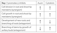
auxin: cell elongation; produced in shoot tips, transported in phloem down stems and into roots; inhibits growth of axillary buds
cytokinin: cell differentiation; produced in root tips, transported in xylem up roots and into stems
(C3.1.22) Explain how root and shoot growth are regulated by the interaction of auxin and cytokinin.
多auxin多cytokinin:
- cell devision + cell growth in root and shoot tip meristems
多auxin少cytokinin
- development of new roots + branching of roots
多cytokinin少auxin
- growth of axillary buds (新苗) to replace lost main shoot
- produce auxin: stimulates root growth, inhibits development of axillary buds
(C3.1.23) List changes that occur to a fruit as it ripens.
stimulated by ethylene in many plant species:
1. color of fruit changes from green
2. cell wall are partly digested, softening the flesh
3. acids and starch are converted to sugar, making the fruit palatable
4. volatile substances are synthesized to give the fruit a distinctive scent
(C3.1.23) Describe the positive feedback mechanism of fruit ripening.
1. ethylene stimulates ripening
2. ripening fruits release ethylene as a volatile vapour which diffuses to other fruits and initiates their ripening
3. rapid and synchronized ripening of fruits e.g. peach tree → increase attractiveness of plant with fruits to animals, encourage dispersal of seeds