PCB675 Week 6 Cellular Effects of Ionising Radiation
 Call Kai
 Learn
Learn Practice Test
 Spaced Repetition
 Match
Match Flashcards
 Knowt Play
1/50

There's no tags or description
Looks like no tags are added yet.
Name | Mastery | Learn | Test | Matching | Spaced | Call with Kai | Chat |
|---|
No analytics yet
Send a link to your students to track their progress
51 Terms
What is Radiobiology, define the 2 levels.
Study of radiation effect at cellular and molecular levels
Cellular level: the basic functional unit of human
Molecular level: such as water, protein, carbohydrates, and DNA
What happens when cell is damaged? (Hint: 3 outcomes)
Injured cells repair themselves through defence mechanisms, results in no residual damage
The cell dies and is replaced
Cell repairs itself improperly, results in a viable but modified cell.
How radiation affects cells? (3 Steps) 1. energy deposition 2. effect of energy deposition 3. chemical bond
Irradiation of biological material results in a random and uneven distribution of energy deposition in tissues and cells.
Much of the deposited energy causes excitation and ionisation.
Random distribution of chemical bond breakage is responsible for biological effects e.g. molecular change
Effects of ionising radiation at various levels
Molecular - DNA
Subcellular - Cell membranes and chromosomes
Cellular - inhibition of cell division
Tissue/Organ - Radiation sickness to gastrointestinal (GI), bone marrow (haematopoietic), and CNS
Whole body - death, cancer
Population - genetic
Biologic Factors - types of cell damage (examples)
Damage to nucleus and DNA
Damage to cell membrane (ion and nutrient leakage)
Damage to mitochondria in the cell’s cytoplasm (food supply of cell affected)
Damage to lysosomes (Causes release of enzyme which results in the cell digesting it’s own internal structures)
DNA damage examples
Base damages
Cross links between different DNA strands
Protein cross link
Intercalation
Point lesions
Single and double strand breaks
DNA damage outcome and reversibility
Can be reversible, less likely when more DNA damage is experienced
The outcome can be cell death, malignant disease, genetic damage
DNA damage - Double strand breakage
A break in both strands of DNA
While lesions (breaks or changes in chemical structure) can travel a few base pairs along the DNA 2 ionisation events are still required near each other, and that possibility is dependent on radiation type and dose.
number of single strand breaks and base damages per unit radiation is typically nearly 2 orders of magntude larger than the number of double strand breaks
number of double strand breaks correlates best with cell kill, therefore:
It is generally assumed that double strand breaks are the most significant lesion for cell kill
Direct and indirect effects of IR
Direct - ionising effect occurs on radiosensitive moelcule (DNA)
Indirect - Initial ionising event occurs on any other molecule, and energy is transferred to the DNA
What does Radiolysis of water result in the production of?
Free radicals
Radiolysis of water - principal damaging products after radiolysis of water are formation of…
OH* + OH* -> H2O2 (Hydrogen Peroxide Formation)
H* + O2 → HO2* (Hydroperoxyl formation)
Radiolysis of water - life time of a radical
The longer the life time of a radical the further it can travel which makes H2O2 (hydrogen peroxide) an important radiation product in radiobiology
Radiolysis of water - damaging effects of free radicals and relation with oxygen
Damaging effects of free radicals is enhanced by oxygen! (reduces the probability of the radicals converting back to simple water or hydrogen).
Indirect effect of IR - What happens if the free radical formation occur close to the DNA?
The radicals can diffuse to react with the DNA, causing radiation damage (indirect effect)
What effect is the most common to cause damage in low LET (x-rays, gamma, electrons) IR? how common?
Indirect effect, accounts or 2/3 of damage on low LET IR, leaving direct effect causing 1/3 damage .
Why is indirect effect so damaging?
Due to cells being made up of mostly water, the IR is most likely to reach water molecules which is where radiolysis occurs via the indirect effect.
Surviving fraction
expression of the magnitude of the effect of a given dose of radiation on cell reproductive capacity.
surviving fraction formula
S=Number of cells surviving a given dose/number of cells in the original unexposed sample. = N/No
Factors affecting biological radiosensitivity (4 physical)
Radiation dose
Dose rate, quantity of dose per unit of time
Type of radiation: LET and Relative Biologic effectiveness (RBE)
Protraction and fractionation
Factors affecting biological radiosensitivity (4 biological)
Chemical agents
Cell type
Phase of cell cycle, age
TIme between fractions / Recovery effect
Physical factor - Dose Rate explained
For the same total dose, higher dose rates are more effective than low dose rates due to the lowered potential for repair of radiation damage
Physical factors - Type of Radiation
High LET radiation such as neutrons and protons are more effective at cell killing than low LET radiation
Physical factors - Type of Radiation LET explained, Unit of LET, Diagnostic x-ray LET
LET is a measure of the rate at which energy is transferred from ionising radiation to soft tissue.
Unit - keV/µm
LET of diagnostic x-ray is approximately 3keV/µm
Physical factors - Type of Radiation (Relative Biologic Effectiveness)
RBE (relative biological effectiveness) is another method of measuring relative effectiveness of different types of radiation in producing damage - aka tissue weighting factor
RBE formula
RBE = Dose of standard radiation (250 keV x-rays) necessary to produce a given effect/Dose of another radiation source needed to produce the same biologic response
RBE vs LET for
Physical factors - Protraction and Fractionation explained with examples
Protraction: the dose is delivered continuously but at a lower dose rate, allowing time for cell repair and tissue recovery.
Example: 2Gy/min is lethal for a mouse, but it survives 10mGy/hr for a total of 600hrs.
Fractionation: the dose is delivered in a number of separate fractions over a long time. Cell repair and recovery occur between doses. This is used routinely in radiation oncology.
Example: a mouse survives a 2Gy/min dose rate in 12 equal fractions of 500mGy), each separated by 24hrs.
Surviving fraction - Single dose
Single dose: Initial “shoulder” on the curve indicating accumulation of sublethal damage, followed by almost exponential decrease in surviving fraction.
Surviving fraction - fractionated dose
repopulation and recovery occur between multiple doses – not necessarily at a constant rate. Surviving fraction in fractionated dose is higher than receiving a single dose of the same magnitude.
Biological factors - Sensitisers (oxygen)
Tissues/cells are more sensitive in the presence of oxygen (aerobic state) than in hypoxic (low oxygen)
Useful knowledge in Radiotherapy since tumors have areas of hypoxic cells which are difficult to kill with low LET radiation.
Biological factors - Sensitisers (oxygen) formula
The oxygen enhancement ratio is the ratio of doses which produce the same level of biological effect (e.g. level of survival) in hypoxic compared with oxygenated conditions:
OER = Dose necessary in hypoxic conditions/ Dose necessary in aerobic conditions
Biological factors - Sensitisers (oxygen) The OER is LET dependen… (3 things)
Highest for low LET radiation (max of 3)
OER of 2.5 for diagnostic radiation
Decreases to about 1 for high-LET radiation
Biological factors - Sensitisers (oxygen) 2 types of hypoxia as a result of tumour growth
acute hypoxia resulting from temporary closing/blockage of blood vessels
Chronic hypoxia is a result of long periods of closing/blockage of blood vessels, as some cells are too far from capillary for oxygen to diffuse
tumour shrinkage linking hypoxia and oxygenated cells
Fractionated dose kills some hypoxic cells but more oxygenated cells near the capillaries due to them being more sensitive.
Tumor shrinks towards the capillaries and previously hypoxic cells become better oxygenated.
Process is repeated and tumour shrinks
High LET radiation in relation to hypoxic and oxygenated cell survival
High LET radiation shows similar/equal damage between oxygenated and hypoxic tumour cells
Biological factors - Cell type
Radiosensitivity is greater for cells that:
High mitotic rate
Long mitotic future
Undifferentiated (haven’t formed into final cell type yet)
Biological factors - Phase of the cell cycle
Cells exposed in mitosis and the gap (G2) between preparatory stage for synthesis and mitosis are most sensitive
Less sensitive during late DNA synthesis (S), survival curves show broader shoulder = more chance to repair
Radiosensitivity with age
radiosensitivity decreases with age, which is why radiation safety for children and pregnant women is considered
Biological Factors - Time between fractions, Define the recovery effect of fractionated dose (aka time between fractions).
If radiation is insufficient to kill cell before next division, cell will recover from IR.
Biological factors - cell type (Target theory)
Cell death occurs after radiation exposure and when the “target molecule” is inactivated.
DNA is the principal radiation-sensitive molecule (target molecule)
Mathematical description of target theory (3)
Single target, Single Hit model
Multitarget, Single hit model
Linear-Quadratic model
What happens when radiation damage is inflicted on a molecule which is in abundance within a cell?
May not result in noticeable cell injury as similar molecules are still available to support the cell
What happens when radiation damage is inflicted on a molecule which is NOT in abundance within a cell?
Loss of function as no similar substitutes can carry on the function of the particular cell.
Single-Target, single hit model explained
Start with 100 cells and ascribe a random hit pattern: o After one hit, one of the 100 cells has been hit and killed. o At the second hit, there’s a 99% chance that it will hit a different cell to the first hit and a 1% chance that it will re-hit the first cell. Note: All hits per target in excess of one, represents wasted radiation dose, as the cell has already been killed by the 1st hit). o At the third hit, there’s a 98% chance that it will hit a new cell, and a 2% chance that it will hit a cell that’s already been hit (from hit 1, and hit 2). o As the number of hits increases, it will become more probable that a given cell will be hit twice or more.
Poisson distribution - single target single hit model
when number of hits = number of cells, 63% of cells will be hut at least once, and 37% survive
Calculation of surviving fraction formula

What does a high and low D37 value indicate?
High D37 = low radiosensitivity, low D37=high radiosensitivity
Multitarget, single hit model
The model assumes that within each cell there are multiple targets all of which must be hit once to cause reproductive cell death.
Multi target, single hit model formula
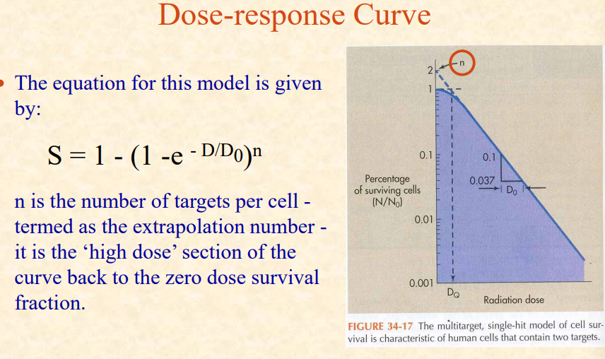

Multitarget, single hit model - explained (Dq, D0, n, shoulder)
At low doses (i.e. the first few hits), there is nearly 100% survival (there is a threshold).
As the D increases, fewer cells will survive (all targets have been hit).
A high D, all cells will be hit at least once on each of the targets and it starts to look like the single hit model - which accounts for the bend (shoulder) in the curve.
D0 (Mean Lethal Dose ) = The dose required to reduce the survival fraction to 37% , is the slope of the straight line portion of the graph.
DQ (Threshold Dose) = a measure of the width of the shoulder of the curve related to the capacity of the cell to recover from the sublethal damage.
Linear Quadratic Model
This is currently the preferred model and assumes that there are two components to cell killing:
One due to single event killing (e.g. double strand break caused by a single ionising particle). The probability of NO damage is e-αD
The other one due to two event killing (e.g. double strand break caused by two separate ionising particles). The probability of NO damage is e-βD 2
Hence: S = e-αD - β D 2 (where α and β are constants)
This relationship has an initial non zero slope which is in agreement with experiment but results in a continuously ‘bending’ curve which may not be the case.