X zone photo questions
 Call Kai
 Learn
 Practice Test
 Spaced Repetition
 Match
 Flashcards
 Knowt Play
1/84

There's no tags or description
Looks like no tags are added yet.
85 Terms
D. The sonographer has not carefully obtain the correct circumference view.
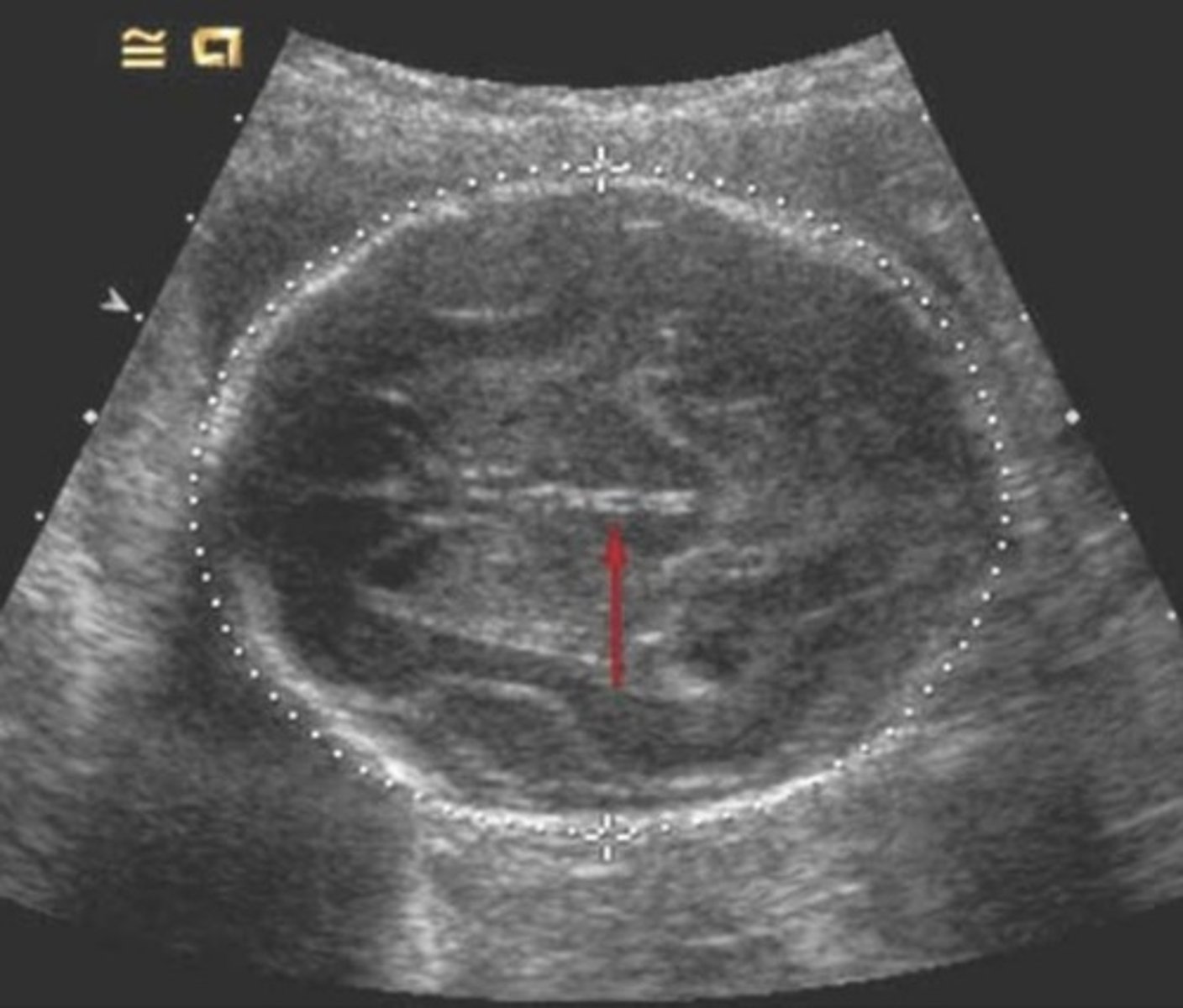
Which of the following statements about the image below is not accurate
A. Midline structures demonstrated in this view for measuring the head, circumference or normal.
B. The fetus is not younger than 20 weeks gestational age.
C. The calipers outlining the head circumference are accurately positioned.
D. The sonographer has not carefully obtain the correct circumference view.
D. Macrosomia.
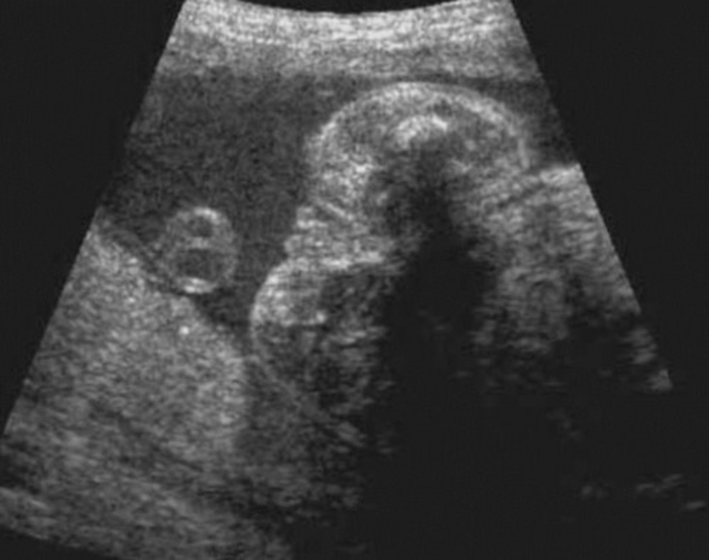
The finding in the image image below is associated with all of the following, except
A. Trisomy 18.
B. Unilateral renal agenesis.
C. VACTERL syndrome.
D. Macrosomia.
E. Fetal cardiac anomalies.
a. The measurement being obtained is appropriate when it includes this structure
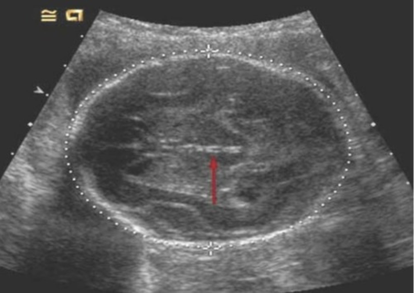
The red arrow in the image is pointing to a specific anatomic structure. Which of the following statements about that structure is true?
a. The measurement being obtained is appropriate when it includes this structure
b. The measurement being taken will be abnormal if this anatomic structure is demonstrated
c. This structure should not be seen if the measurement is accurately performed.
d. The fetus has an intracranial abnormality, and this measurement should not be included in estimating gestational age.
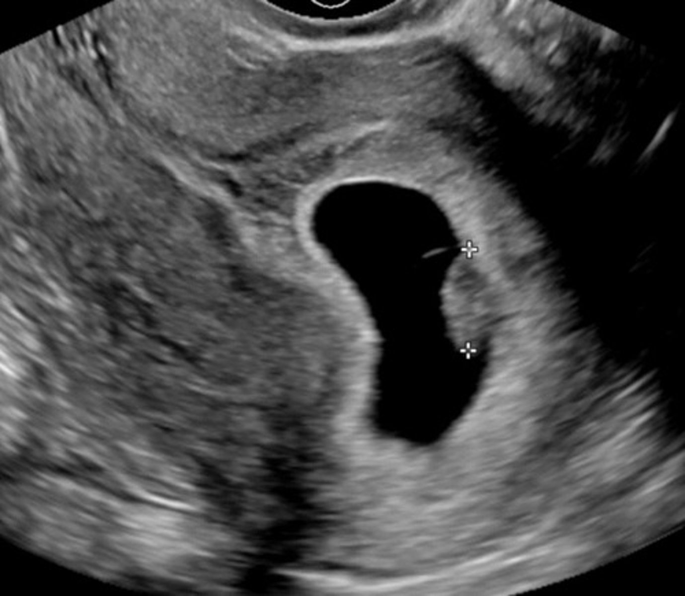
D. 7w 2d
The embryo in the image below measures 10.2mm CRL. What is the approximate gestational age of this pregnancy?
A. 6w 3d
B. 8w 2d
C. 5w 1d
D. 7w 2d
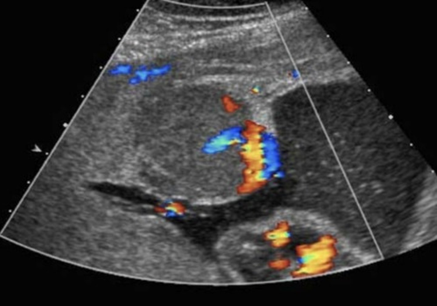
E. Chorioangioma
The presence of color flow in the structure below helps confirm the diagnosis of
A. Placental lake
B. Placental abruption
C. Placental infarct
D. Velamentous sord insertion
E. Chorioangioma
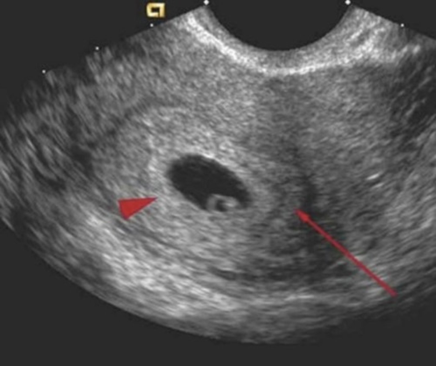
E. The decidua parietalis
In the image below, the long access is pointing to what specific structure
A. The decidua capsularis
B. The uterine cavity
C. The chorion frondosum
D. The chorionic membrane
E. The decidua parietalis
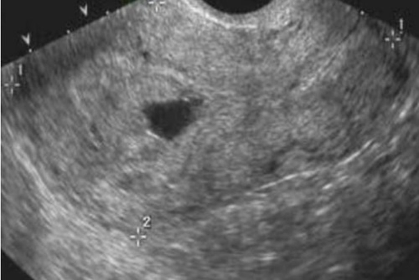
A. Incomplete abortion.
A patient presents with cramping and vaginal bleeding for two days, and an hCG level of 1600. An Indo vaginal image is shown below. What is the most likely diagnosis?
A. Incomplete abortion.
B. Ectopic pregnancy
C. Normal gestational, but low implantation
D. Anembryonic pregnancy
b. Decidua basalis
In the image below, the arrowhead is identifying
a. Decidua capsularis
b. Decidua basalis
c. Normal myometrium
d. Decidua parietalis
e. The uterine cornu
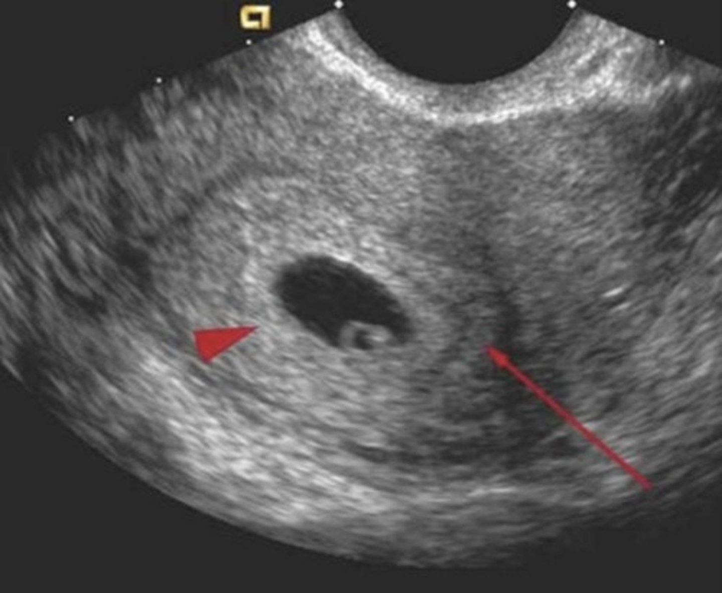
C.The distal femoral epiphysis ossification
The image below is a measurement of a fetal femur bone. What structure is indicated by the arrowhead?
b. The proxibul in
The proximal tibial bone
C.The distal femoral epiphysis ossification
d. The greater trochanter ossification
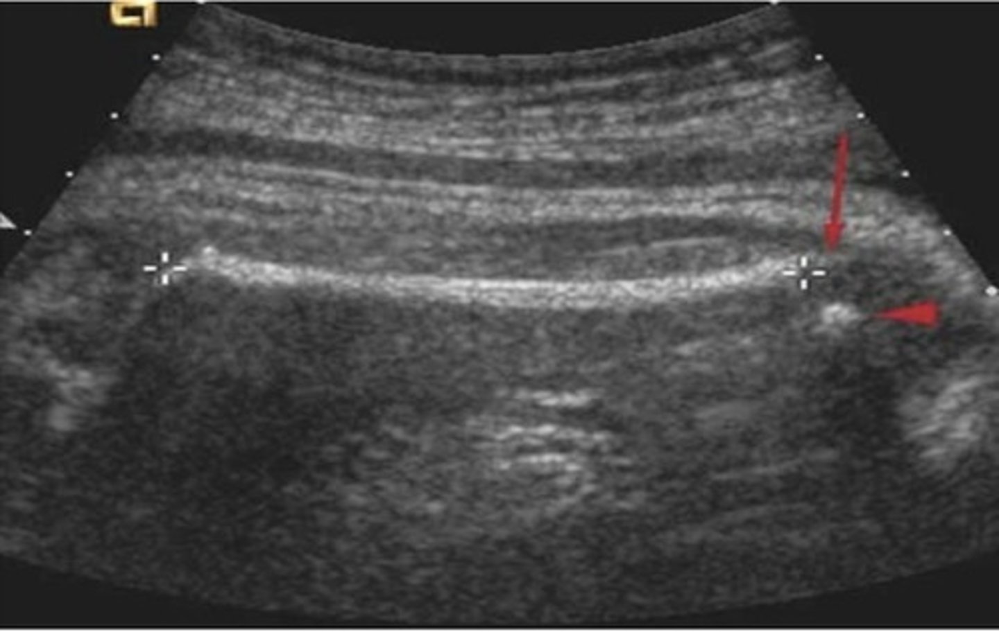
B. The rhombencephalon
In the image below, to what is the arrow pointing?
a. A Dandy-Walker malformation
B. The rhombencephalon
C. First trimester hydrocephalus
D. The cisterna magna
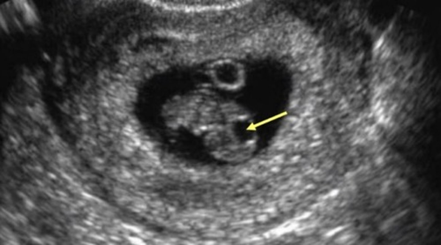
a. 33 weeks
The fetus in the image below is at least what gestational age?
a. 33 weeks
b. 30 weeks
c. 38 weeks
d. 35 weeks
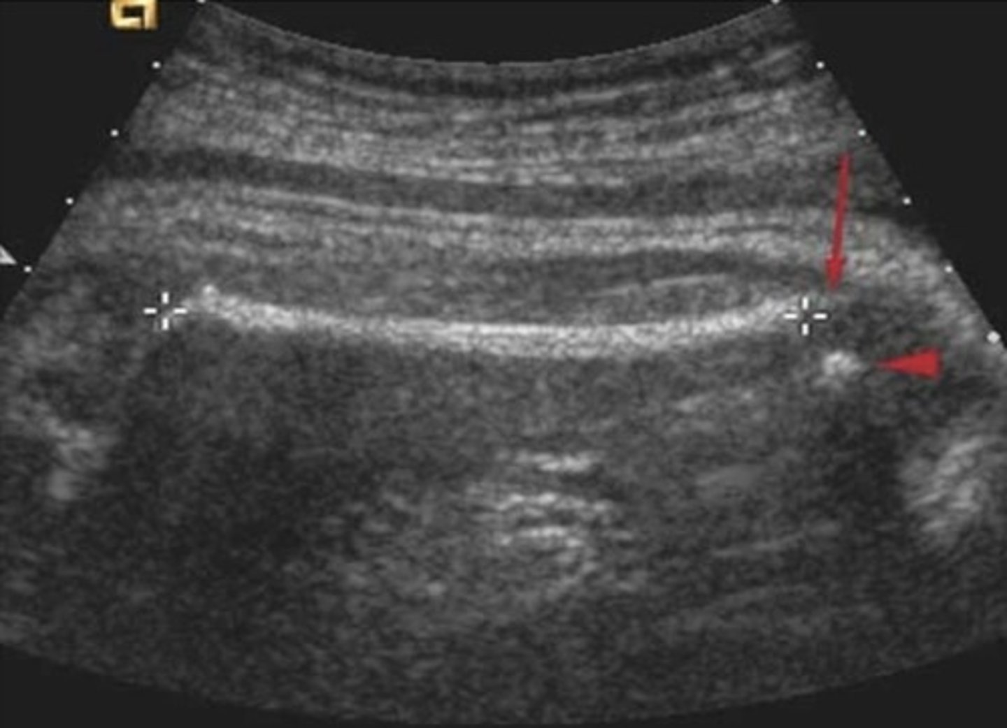
D. Placental infarct
The finding in the image below may be associated with all of the following except
A. Polyhydramnios
B. Fetal hydrops
C. Increased MS AFP
D. Placental infarct
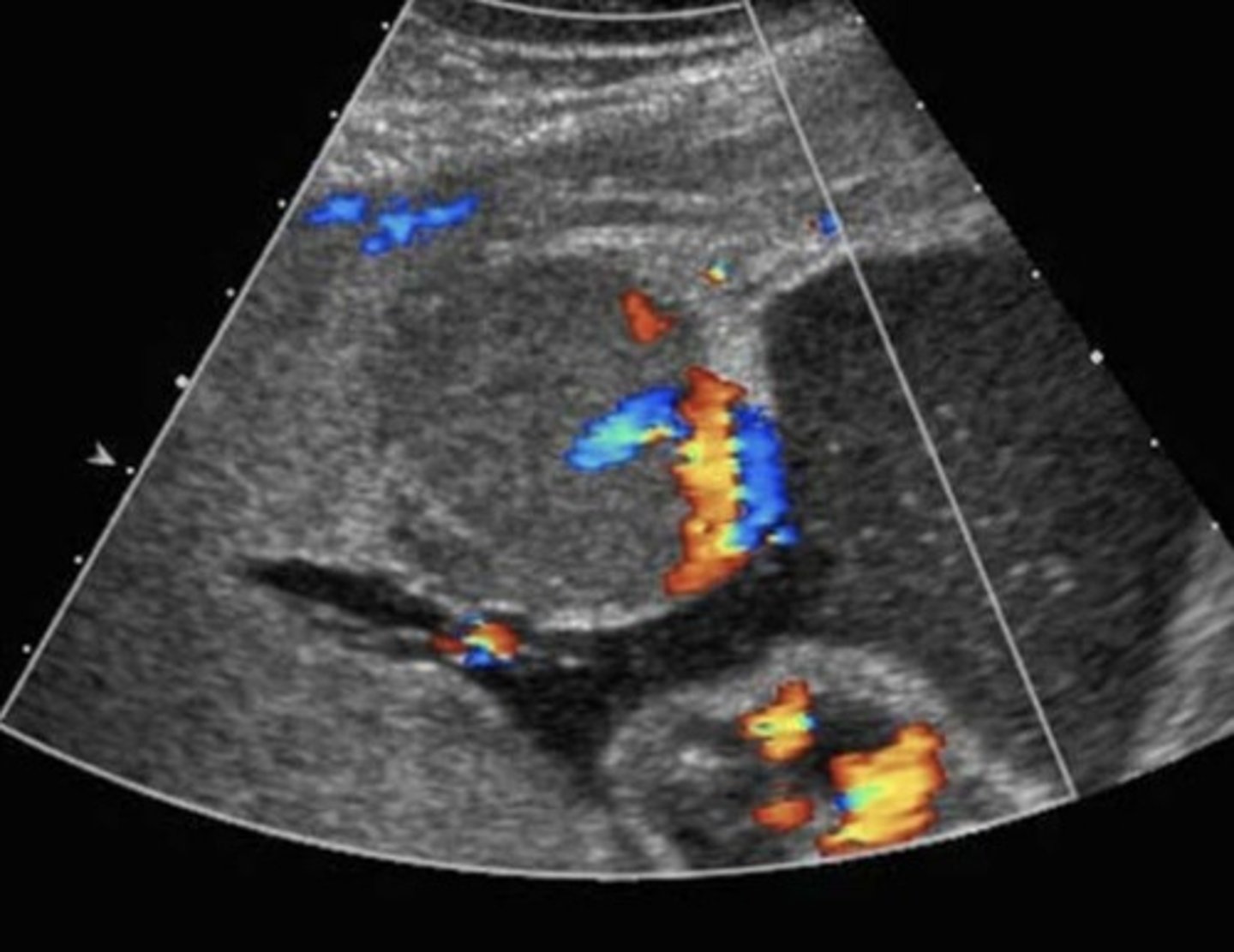
c. The arrow indicates the reflective edge of the distal femoral epiphysis
In the image below, the arrow is located just distal to a "+", which the sonographer has placed measurement. Which of the following statements is true?
a.The femur length measurement is underestimated
b. The femur length measurement is overestimated
c. The arrow indicates the reflective edge of the distal femoral epiphysis
d. The sonographer has misplaced the caliper, and it should be moved to the location of the arrow
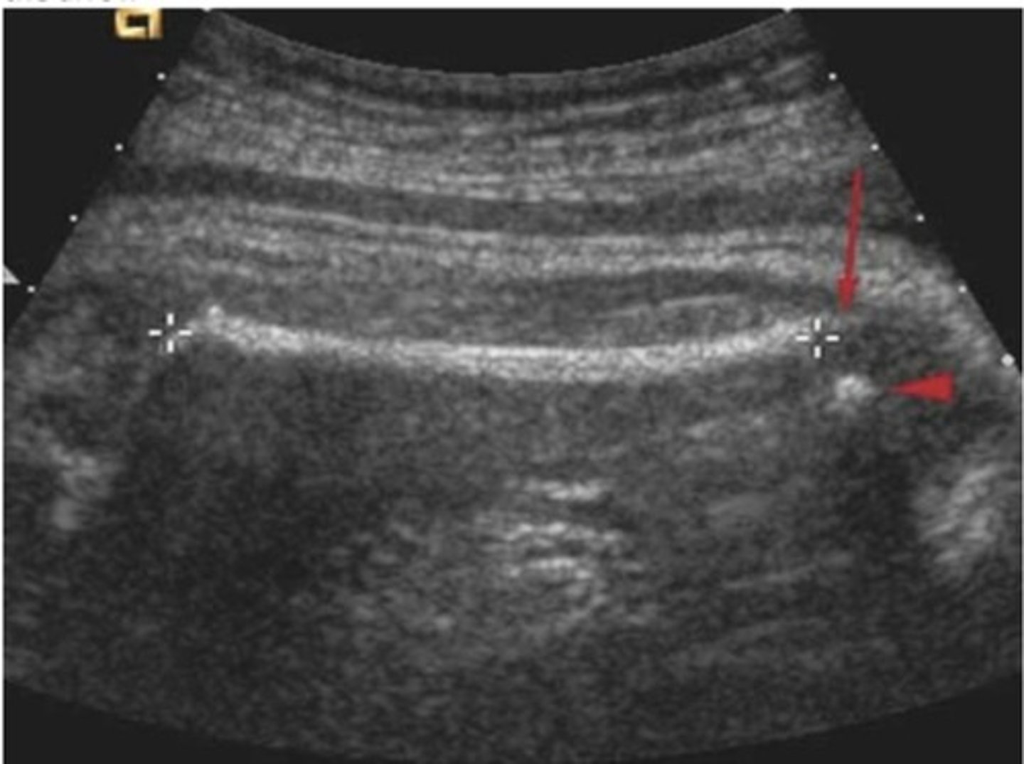
d. Abnormally elevated hCG
A patient with the following sonographic finding may have all of the listed clinical symptoms except
a. History of pelvic inflammatory disease
b.Vaginal spotting
c. Slower than expected rise in hCG levels
d. Abnormally elevated hCG
e. Right lower quadrant pain
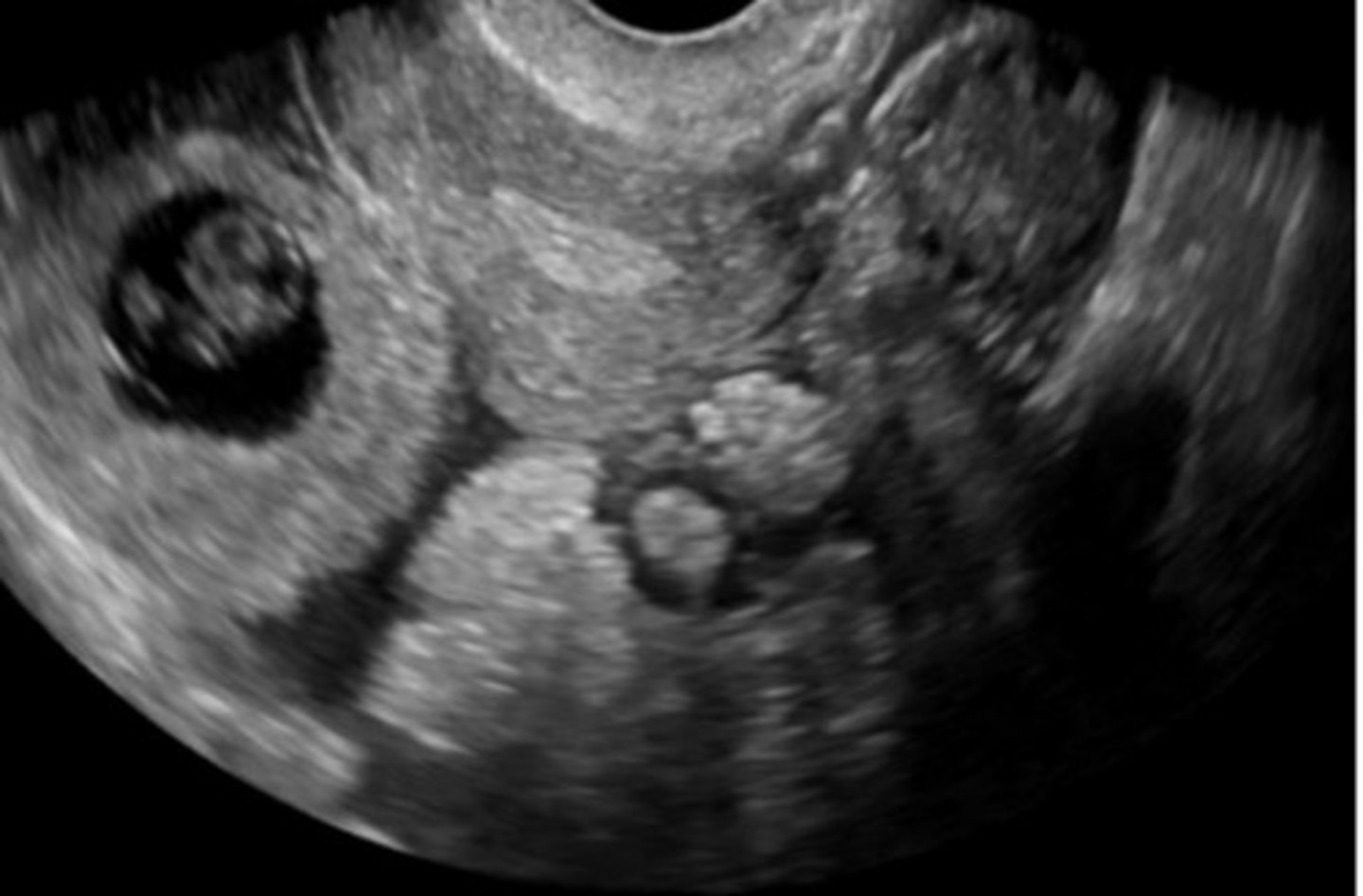
d. Failure of Mullerian duct fusion
The abnormality in the image below is most consistent with:
a. Mullerian and Wolffian duct agenesis
b. A urogenital sinus defect
c. Failure of Mullerian duct formation
d. Failure of Mullerian duct fusion
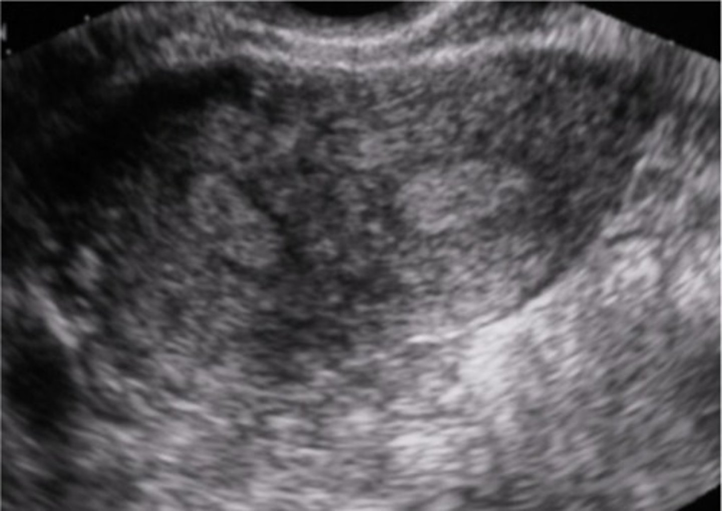
b. 4mm
The image below shows an endometrial measurement of 3cm. What dimension is the upper limits of normal for a postmenopausal woman who is experiencing vaginal bleeding?
a. 8mm
b. 4mm
c. 10mm
d. 20mm
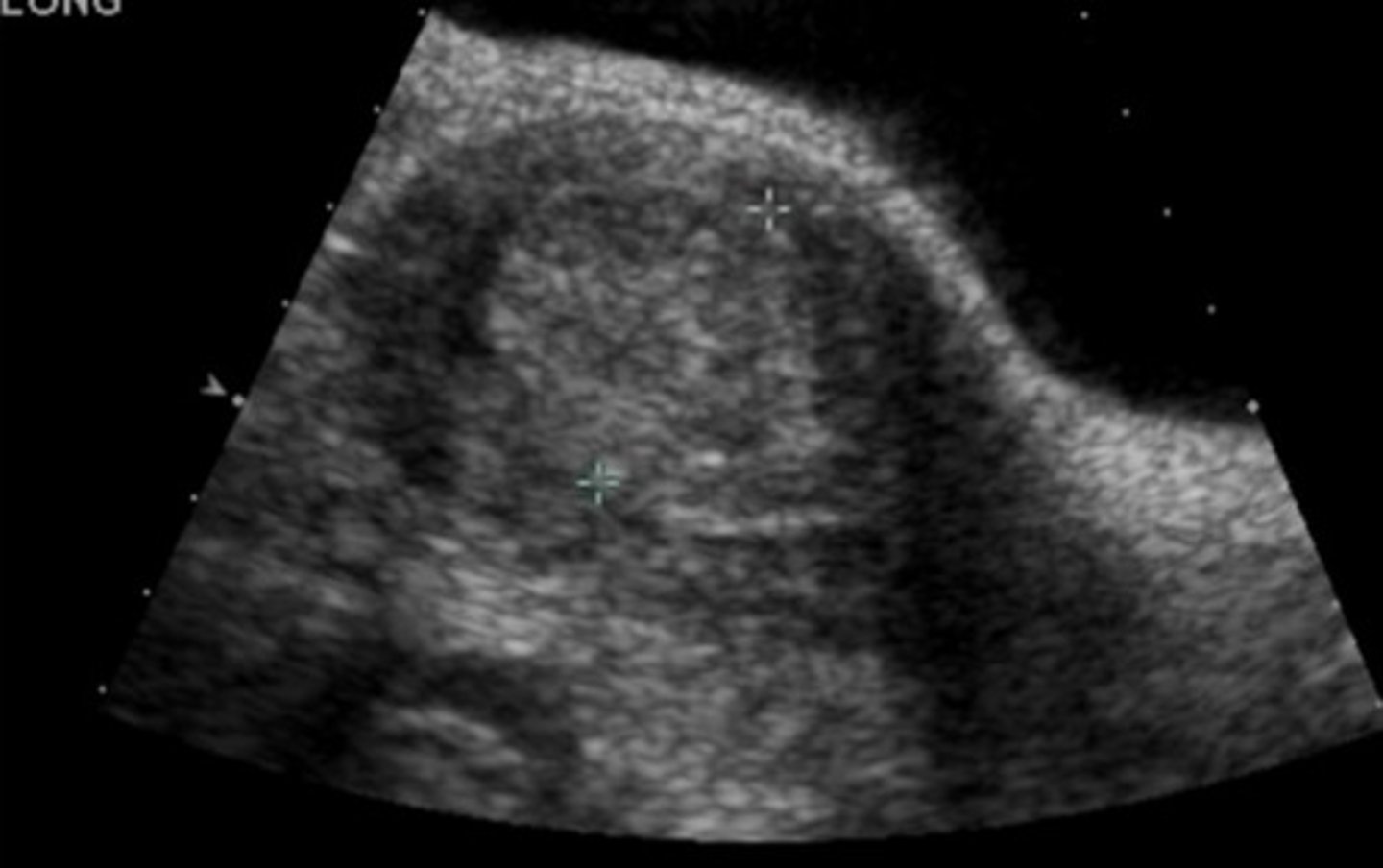
c. Fever and leukocytosis
The mass seen adjacent to the uterus in the image below maybe associated with all of the following symptoms except:
a. Dysmenorrhea
b. Chronic pain
c. Fever and leukocytosis
d. Infertility
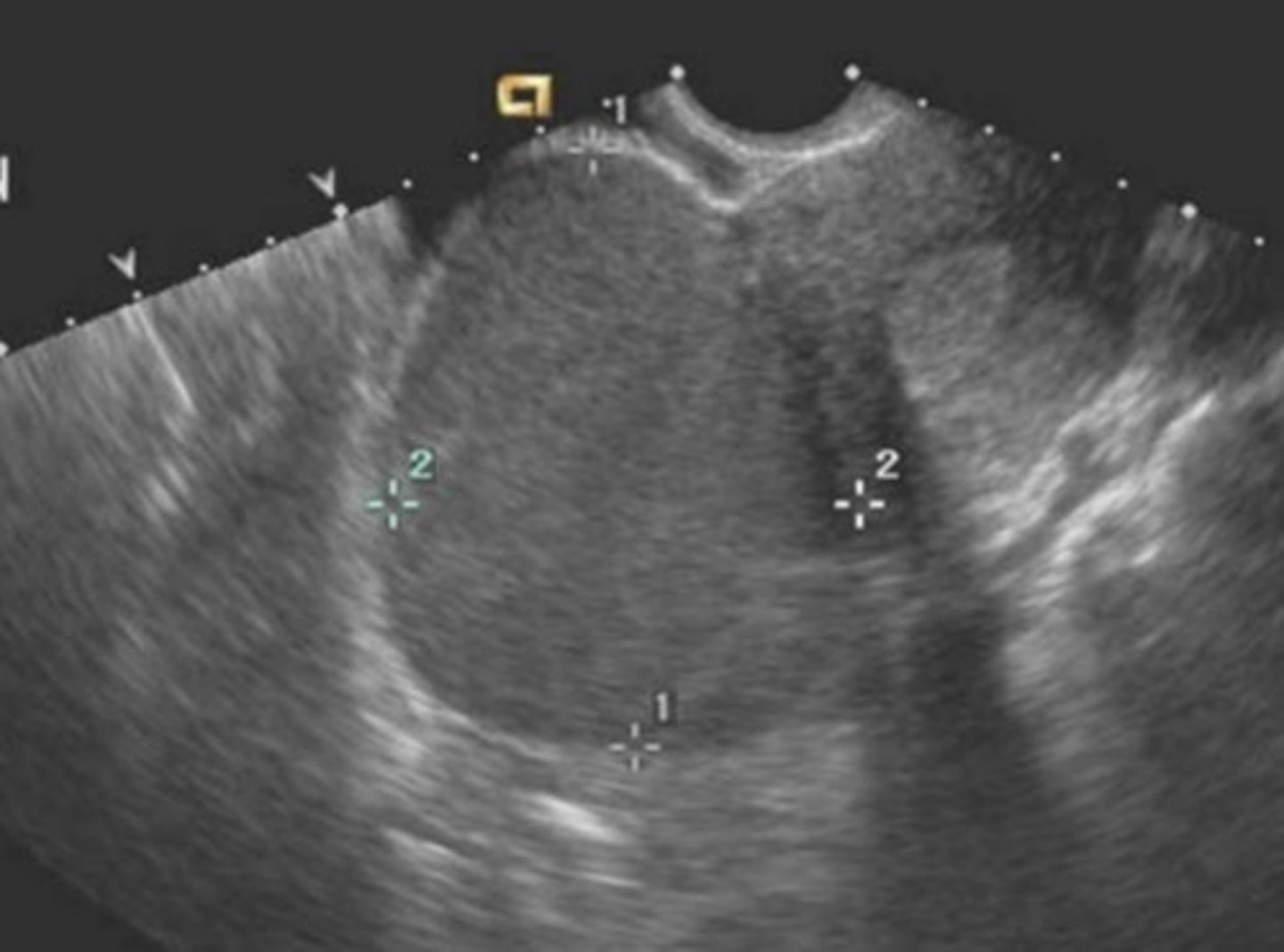
b. A hemorrhagic ovarian cyst
A patient with the following sonographic finding in the adnexa most likely has
a. A krukenberg tumor
b. A hemorrhagic ovarian cyst
c. A dysgerminoma
d. An ovarian teratoma
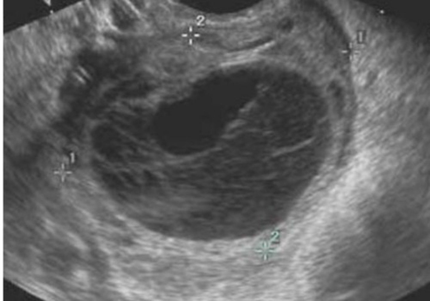
b. Metastatic ovarian neoplasms
The patient with the following sonographic findings most likely has a diagnosis of:
a. Bilateral ovarian torsion
b. Metastatic ovarian neoplasms
c. Stage I PID
d. Bilateral ovarian teratomas
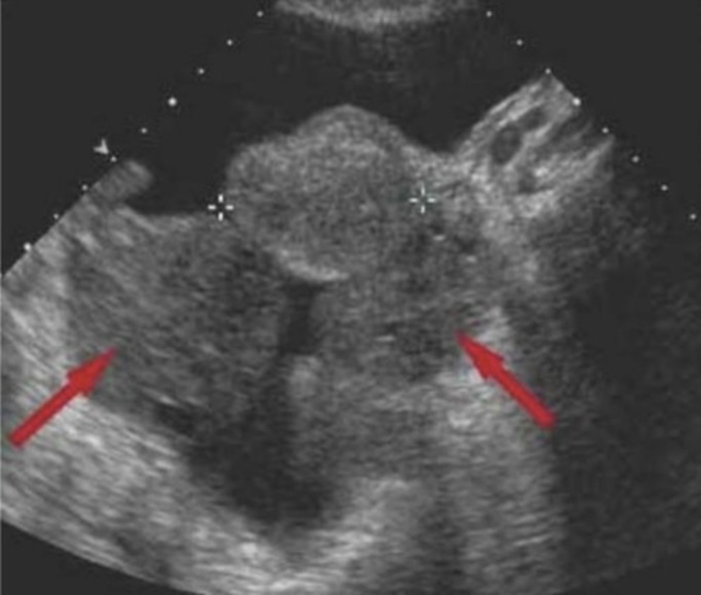
b. A submucosal myoma
The red arrowhead in the image below most likely indicates
a. A focal endometrial carcinoma
b. A submucosal myoma
C. An intramural myoma
d. An endometrial polyp
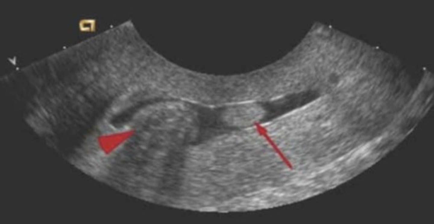
b. Adenomyosis
A 42-year-old patient presents with dysmenorrhea and irregular vaginal bleeding. What is the most likely diagnosis?
a. Endometrial carcinoma
b. Adenomyosis
c. Endometrial hyperplasia
d. Multiple uterine myomas
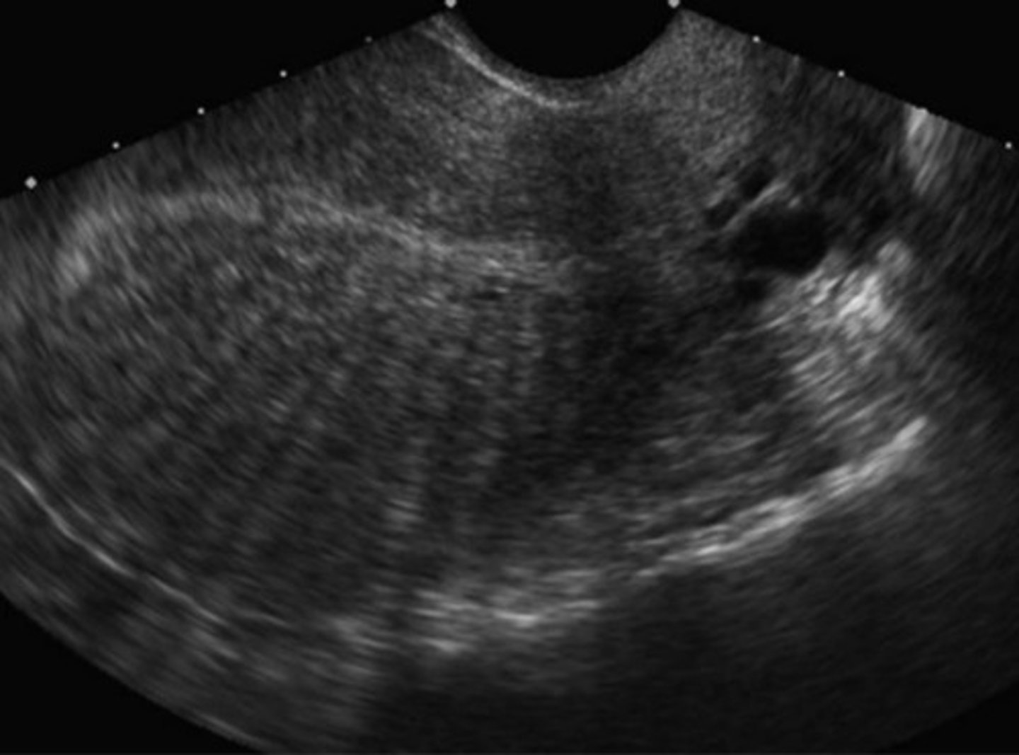
d. Retroflexed
The uterine position in the image below is best described as
a. Anteverted
b. Anteflexed
c. Retrovered
d. Retroflexed
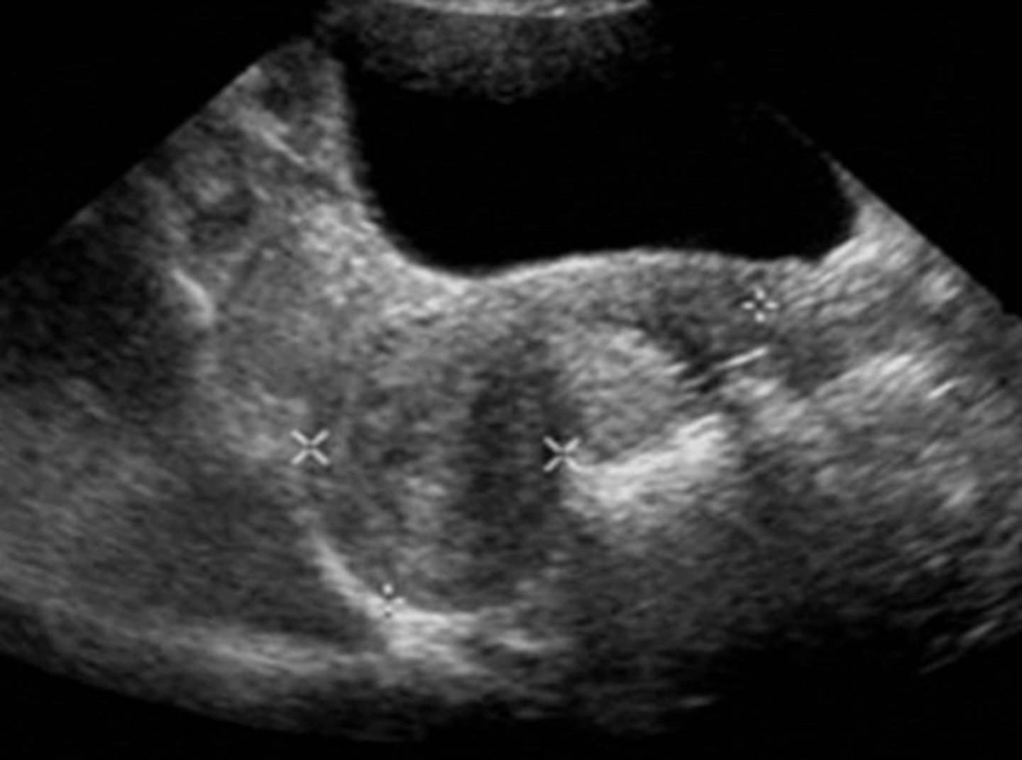
a. A Gartner's duct cyst
The cystic structure located posterior to the urinary bladder in the image below is most consistent with
a. A Gartner's duct cyst
b. Free fluid in the posterior cul-de-sac
c. Fluid in the vaginal fornices
d. A Nabothian cyst
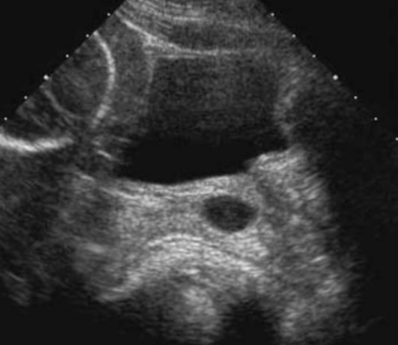
a. Endometrial carcinoma
A healthy postmenopausal woman on unopposed estrogen presents with vaginal spotting and a feeling of fullness in the lower abdomen. Based on the sonographic image below, what is the most likely diagnosis?
a. Endometrial carcinoma
b. Hydrometra
C. Myomatous uterus
d. Endometrial polyp
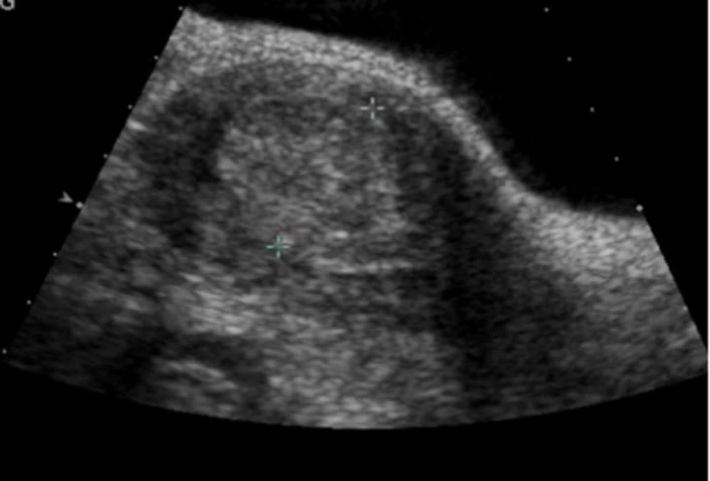
a. Muscinous cystadenoma
A patient presents to the emergency department complaining of a sudden onset of severe left lower quadrant pain. Sonographic imaging of the left ovary is shown below. Based on this image and the patient's symptoms, which of the following is least likely to be the diagnosis?
a. Muscinous cystadenoma
b. Ovarian torsion
С. Hemorrhagic ovarian cyst
d. Tubo-ovarian abscess
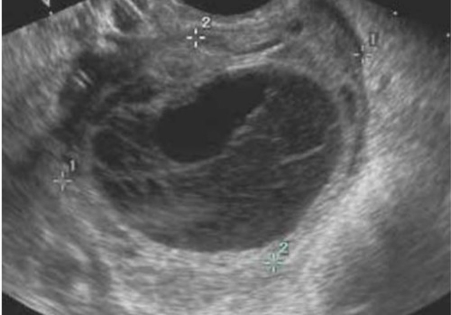
c. Sudden onset of mid-cycle pain
A patient is referred for evaluation of the pelvis, and the following image is obtained in the left adnexa. Which of the following clinical symptoms is least likely in this patient?
a. History of chlamydia exposure
b. Elevated white cell count and fever
c. Sudden onset of mid-cycle pain
d. Vaginal discharge
e. Pain with intercourse
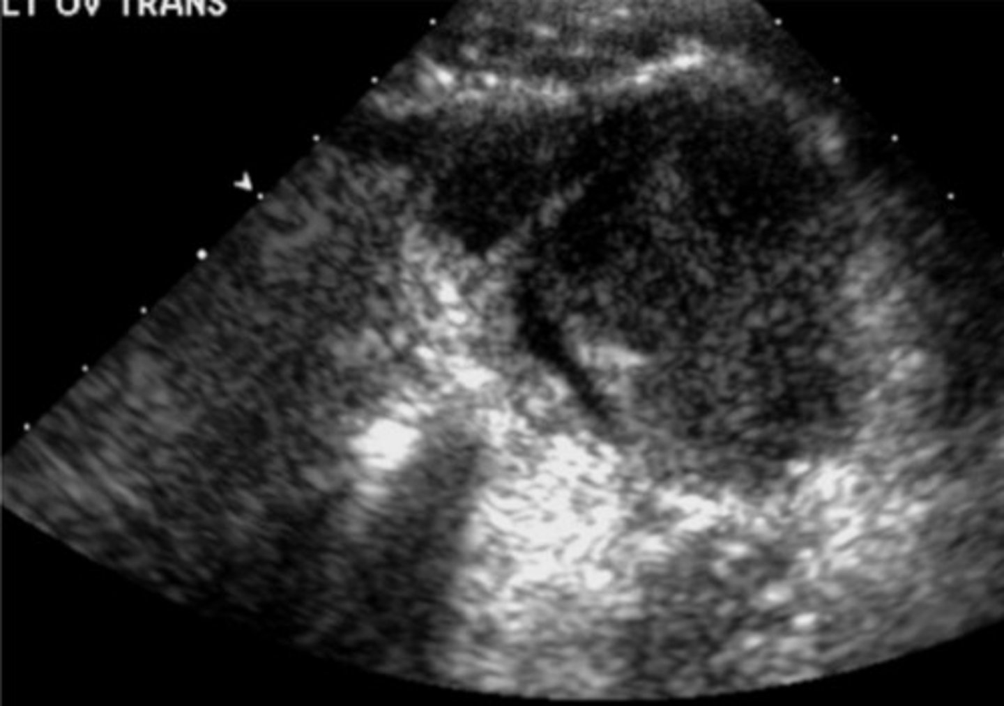
d. An endometrial polyp
The image demonstrates which endometrial pathology?
a. An intramural myoma
b. A submucosal myoma
c. A focal endometrial carcinoma
d. An endometrial polyp
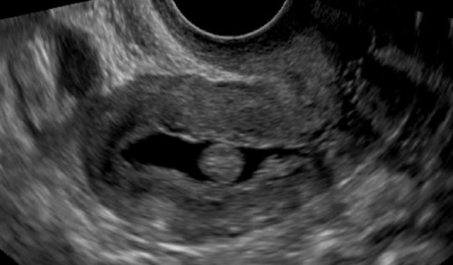
c. Dysmenorrhea and irregular bleeding
The sonographic features of the image below suggest diffuse involvement of the posterior myometrium. What clinical symptoms may be associated with this finding?
a. Acute pain and hemorrhaging
b. Purulent vaginal discharge
c. Dysmenorrhea and irregular bleeding
d. Fever and elevated white cell count
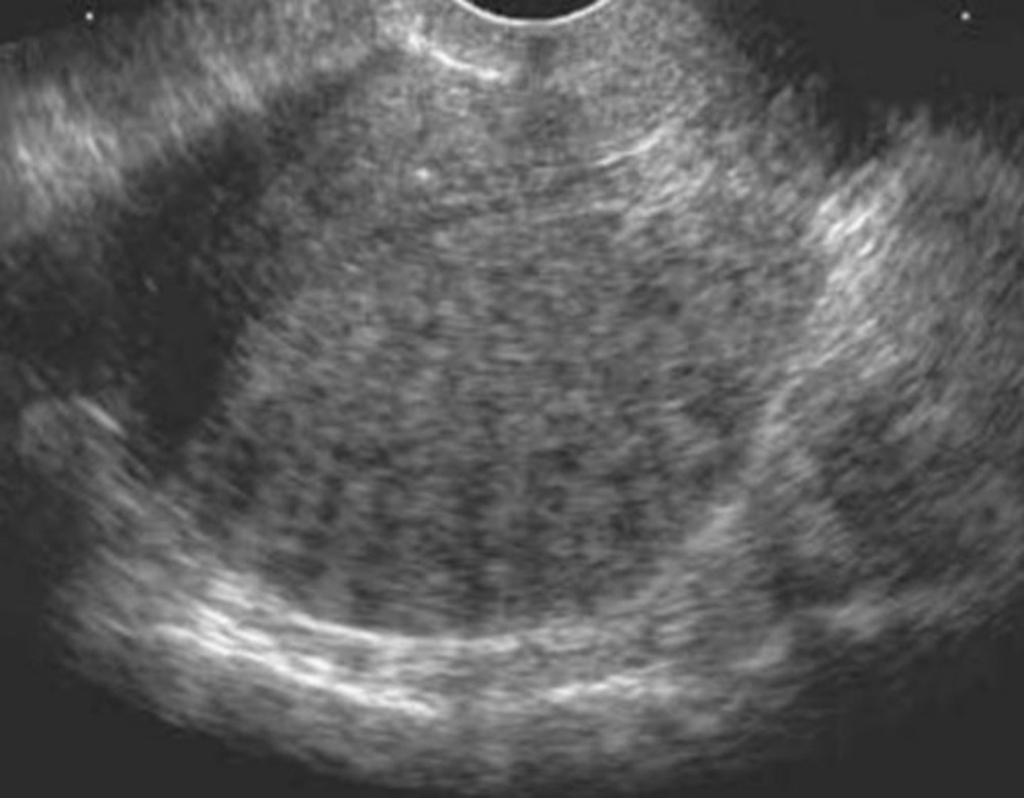
c. Adenomyosis
A 42-year-old patient presents with dysmenorrhea and irregular vaginal bleeding. What is the most likely diagnosis?
a. Endometrial carcinoma
b. Multiple uterine myomas
c. Adenomyosis
d. Endometrial hyperplasia
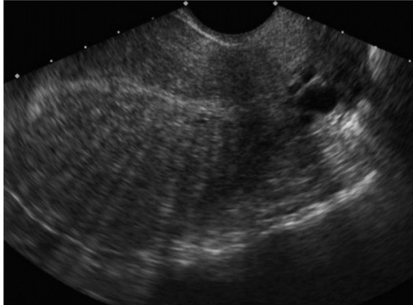
d. 4mm
The image below shows an endometrial measurement of 3cm. What dimension is the upper limits of normal for a postmenopausal woman who is experiencing vaginal bleeding?
a. 20mm
b. 10mm
c. 8mm
d. 4mm
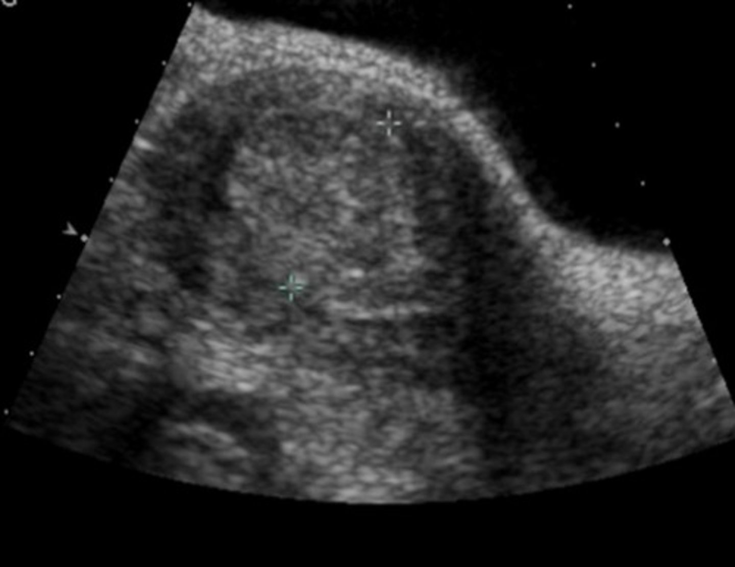
a. A leiomyoma
Saline infusion sonohysterography is helpful in the following image to delineate what abnormality?
a. A leiomyoma
b. Endometrial hyperplasia
c. Endometrial irregularities
d. An endometrial polyp
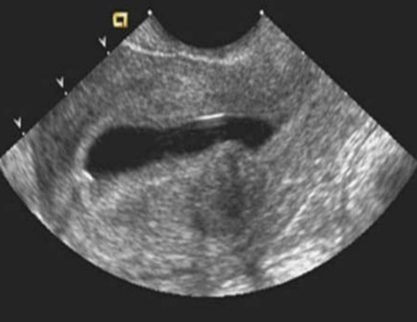
a. Endometritis
The sonographic appearance of the endometrium in the image below may be due to all of the following except
a. Endometritis
b. Endometrial polyp
c. Focal endometrial carcinoma
d. Focal endometrial hyperplasia
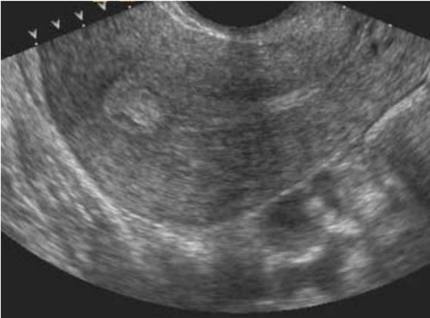
d. Failure of the mesonephric duct to disappear
The anechoic structure posterior to the bladder in the image below is most likely a result of
a. An obstructed cervical gland
b. Ruptured membranes
c. Recent vaginal instrumentation
d. Failure of the mesonephric duct to disappear
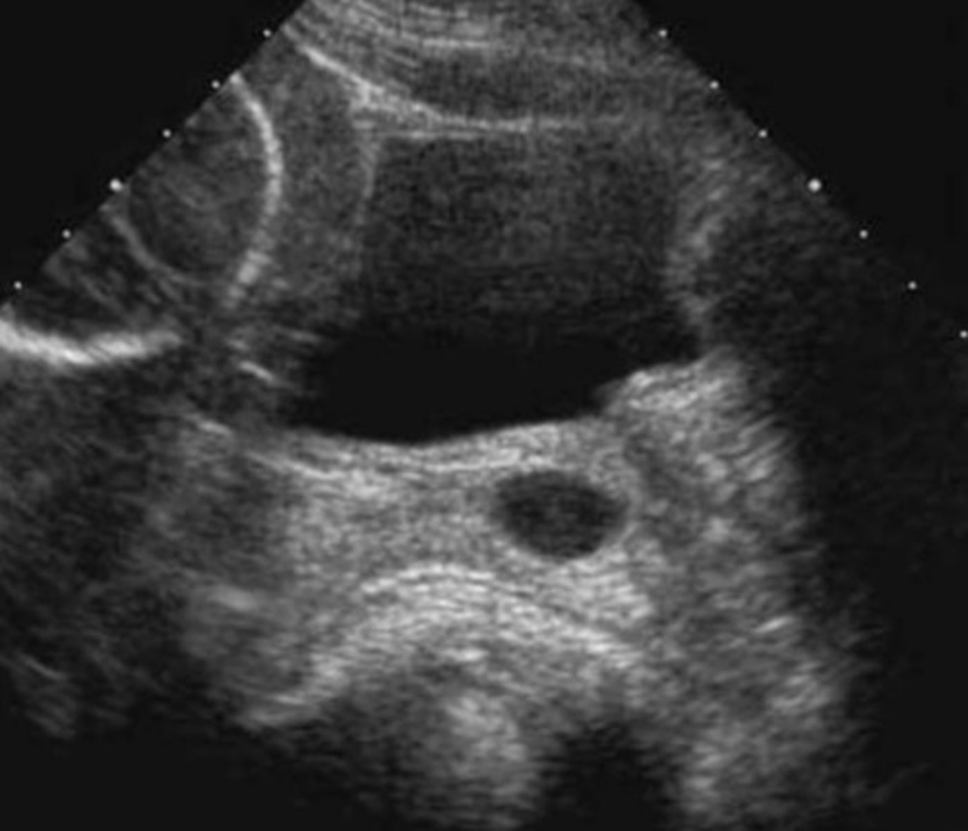
c. The fetal spleen
Immediately posterior to the fetal stomach in the image below is a slightly hypoechoic structure. What is this structure?
a. Fluid in the fetal abdomen
b. Fetal lung parenchyma
c. The fetal spleen
d. The fetal liver
e. The placenta
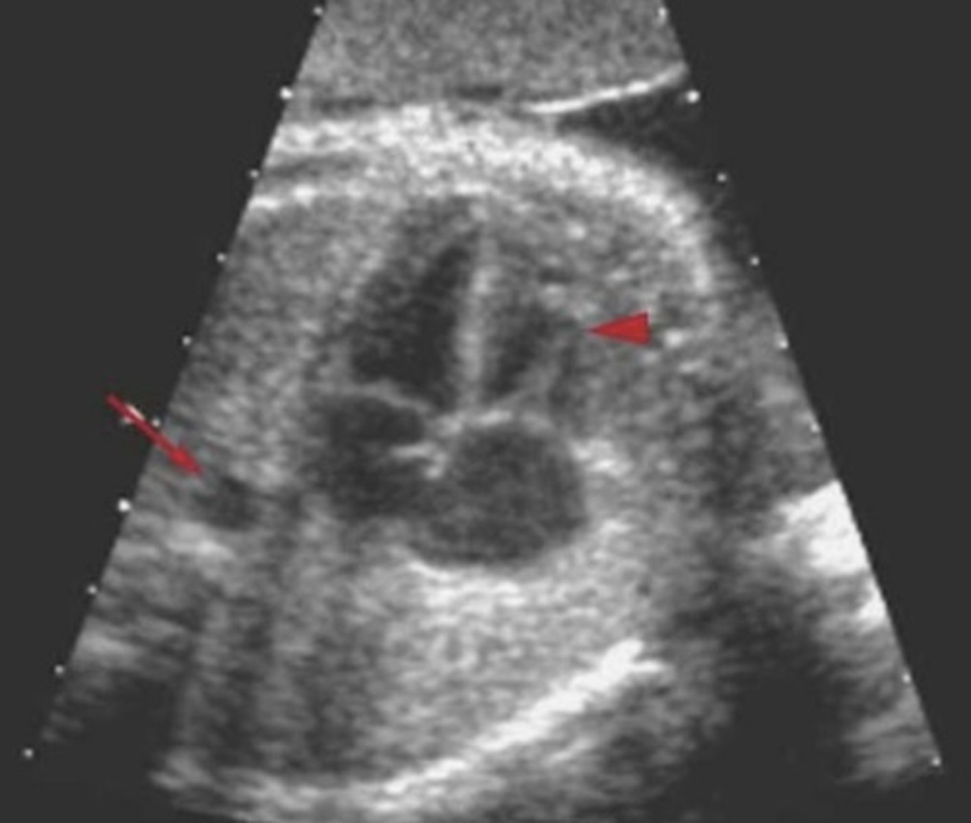
d. Right ventricle
In the image below, the arrowhead points to what heart chamber?
a. Right atrium
b. Left ventricle
c. Left atrium
d. Right ventricle
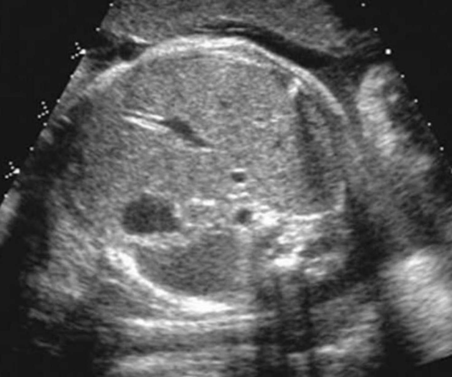
d. Fetal gallbladder
To what is the arrow in the image below pointing?
a. Fluid in the fetal intestinal tract
b. Fetal urinary bladder
c. Fetal stomach
d. Fetal gallbladder
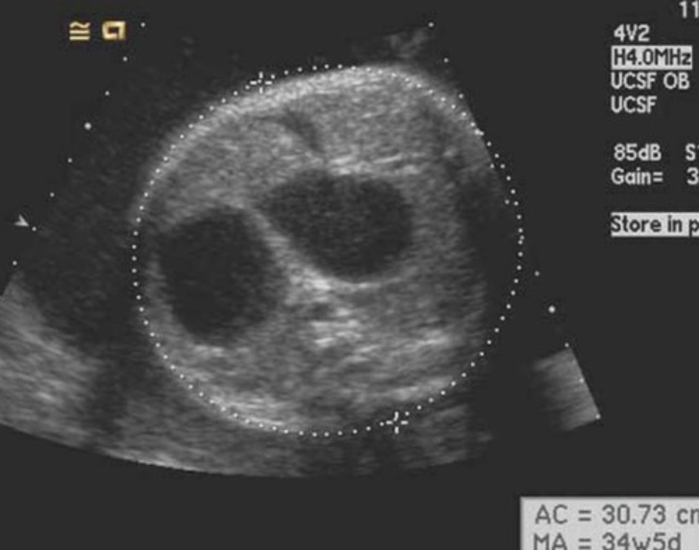
d. Elevated MSAFP level
Which maternal clinical finding is most likely to be associated with the image below?
a. Rh incompatibility
b. Small for dates
c. Deceased MSAFP level
d. Elevated MSAFP level
e. Maternal hypertension
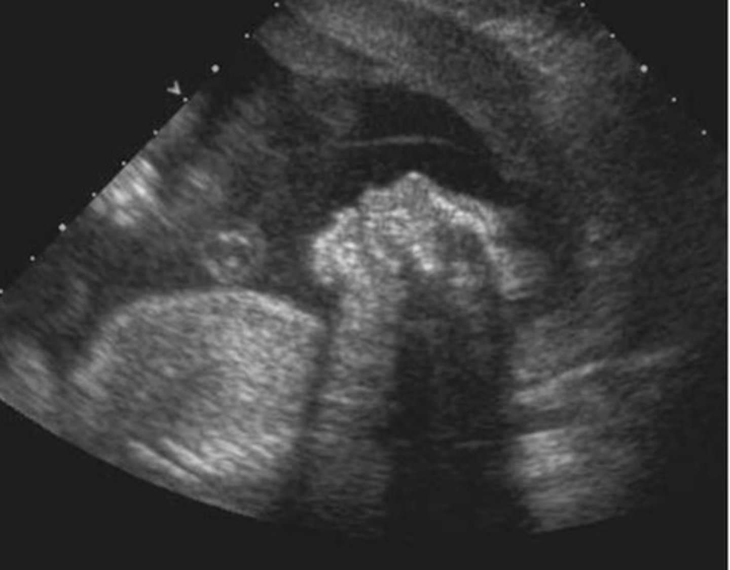
c. Esophageal atresia
All of the following may be associated with the sonographic findings in the image below except
a. Polyhydramnios
b. Cardia anomalies
c. Esophageal atresia
d. Choledochal cyst
e. Down syndrome
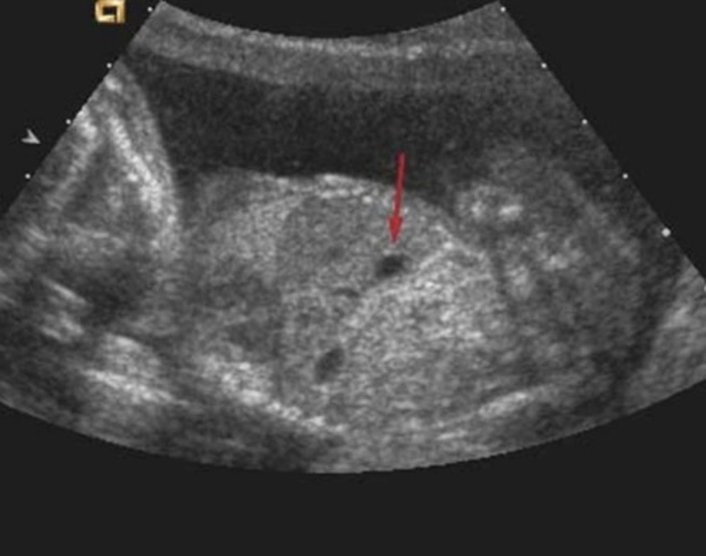
d. The fluid-filled fetal stomach
In the image below, the red arrow was pointing to
a. A pulmonary sequestration
b. The fetal spleen
c. A congenital diaphragmatic hernia
d. The fluid-filled fetal stomach
e. A hypochoic lung mass
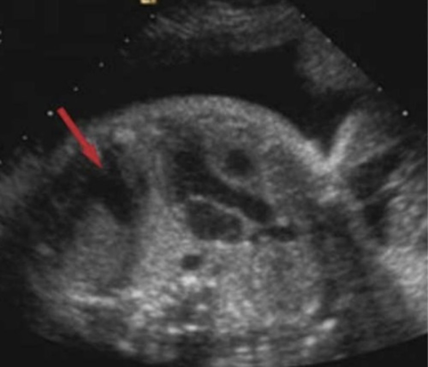
b. The left iliac crest
If the image below is a transverse view through the gravid uterus, and the fetus is in a breech presentation, which bone is indicated by the red arrow
a. The right femur
b. The left iliac crest
c. The right iliac bone
d. The left femur
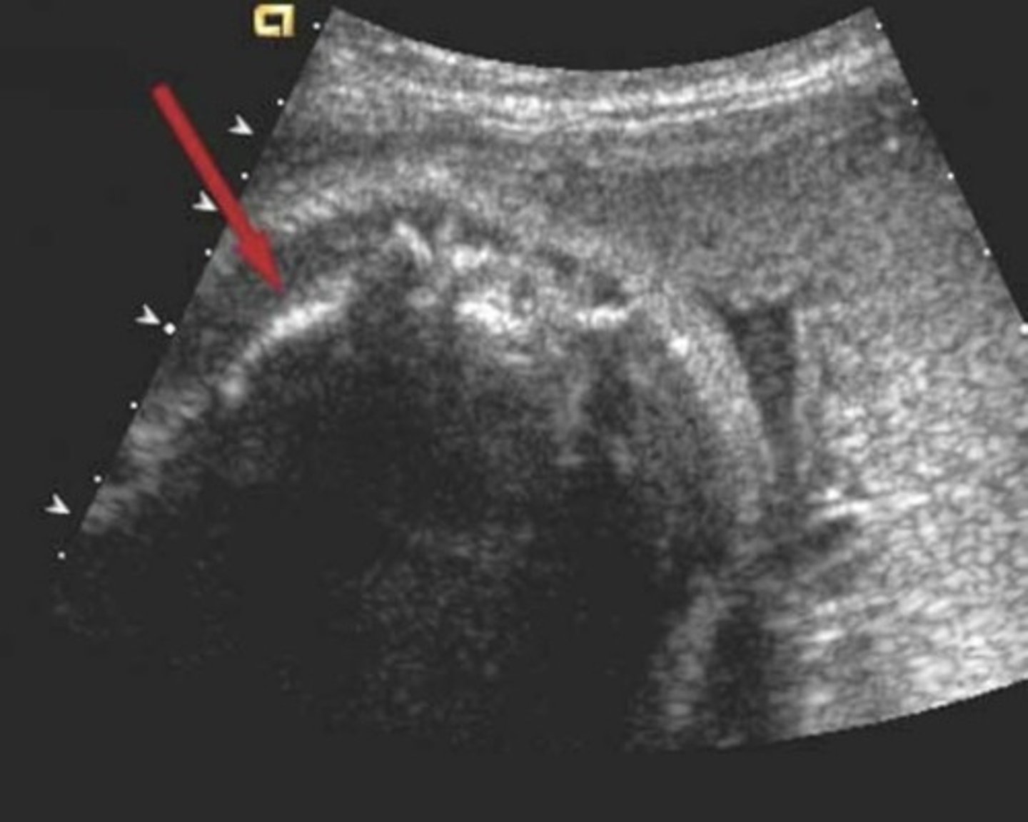
d. Trisomy 13
The abnormaility in the image below is most typically a finding with which chromosomal aberration?
a. Trisomy 18
b. Trisomy 21
c. Turner's syndrome
d. Trisomy 13
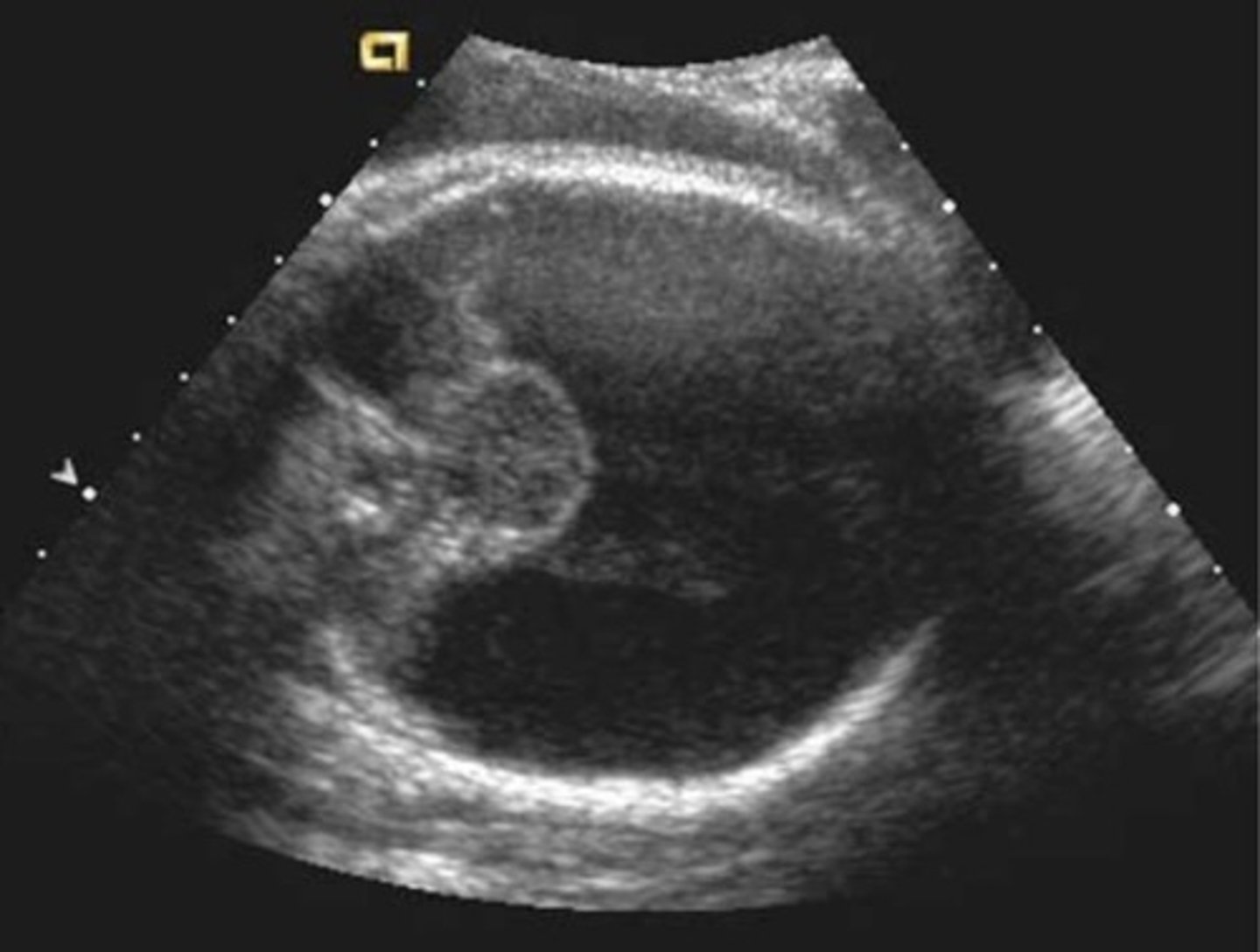
d. Limb amputations
The fetus in the image below may have any of the following except
a. An abnormal karyotype
b. Hydrops fetalis
C. Diffuse lymphangiectasia
d. Limb amputations
e. Turner's syndrome
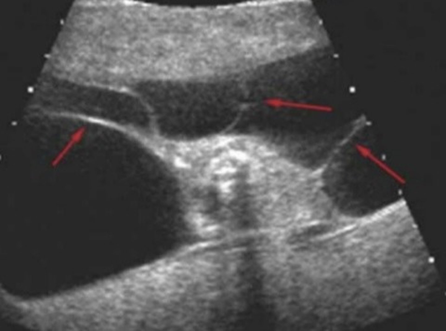
a. The parietal bones
The image below is a measurement of the fetal cranium. What cranial bones are immediately adjacent to the two calipers?
a. The parietal bones
b.The frontal bone
c. The occipital bone
d. The sphenoid bone
e. The temporal bones
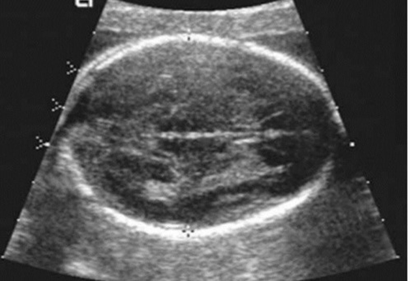
a. Holoprosencephaly
The fetal profile in the image below is most likely associated with which of the following?
a. Holoprosencephaly
b. Encephalocele
c. Spina bifida
d. Hydranencephaly
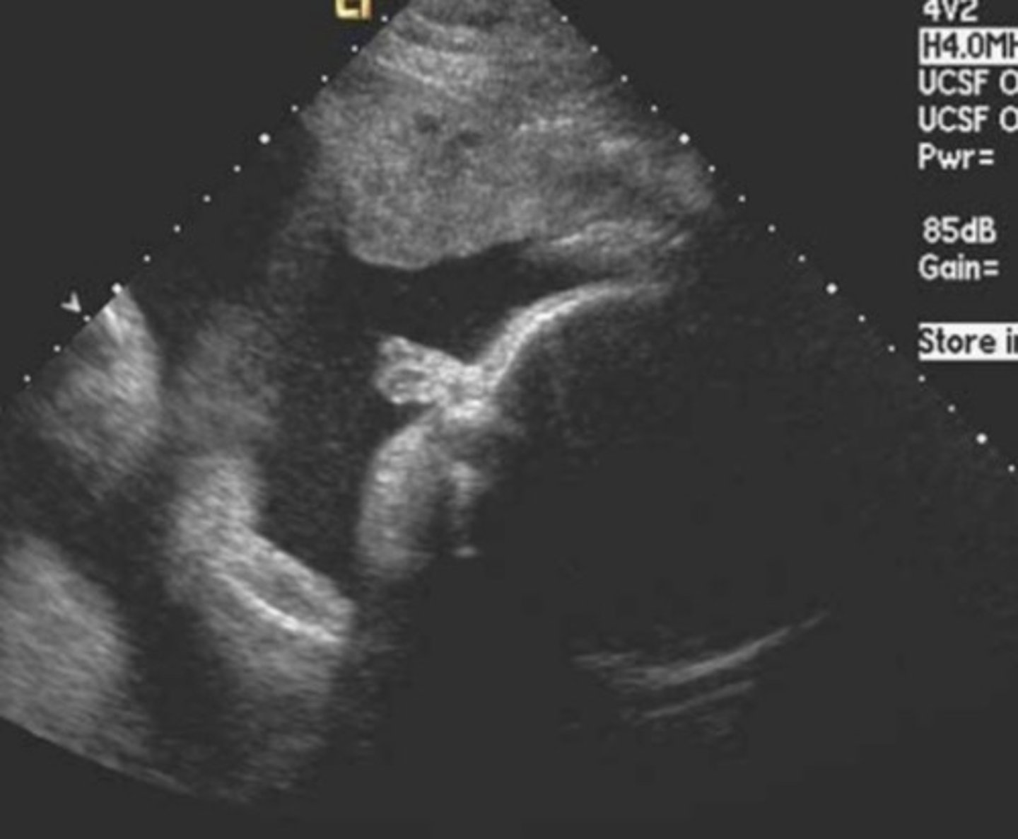
b. Approximate gestational age
If calipers were placed to measure the structure indicated by the red arrow heads in the image below, what can be determined by this fetus
a. Microcephaly
b. Approximate gestational age
c. The presence of a spina bifida
d. The presence of hydrocephalus
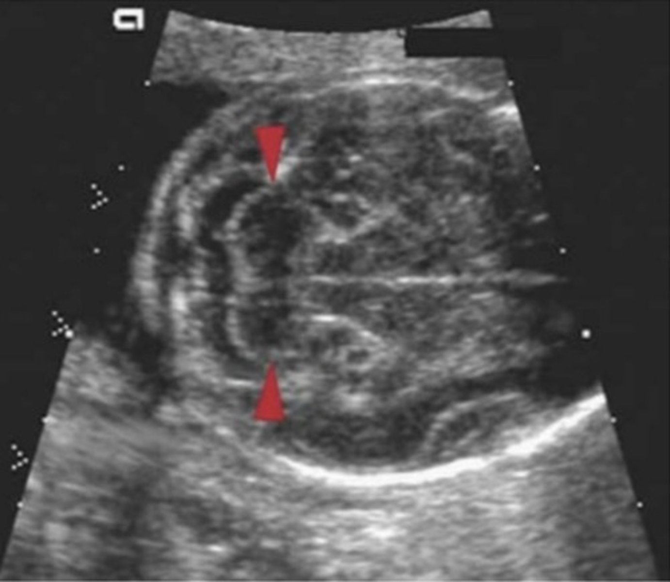
d. The fetal lung parenchyma is normal echogenicity
Regarding the image below, which of the following statements is least likely true?
a. The fetal lung parenchyma is replaced by cysts
b. There is mass effect on the right hemidiaphragm
c. The fetus has developed ascites because of the existing abnormality
d. The fetal lung parenchyma is normal echogenicity
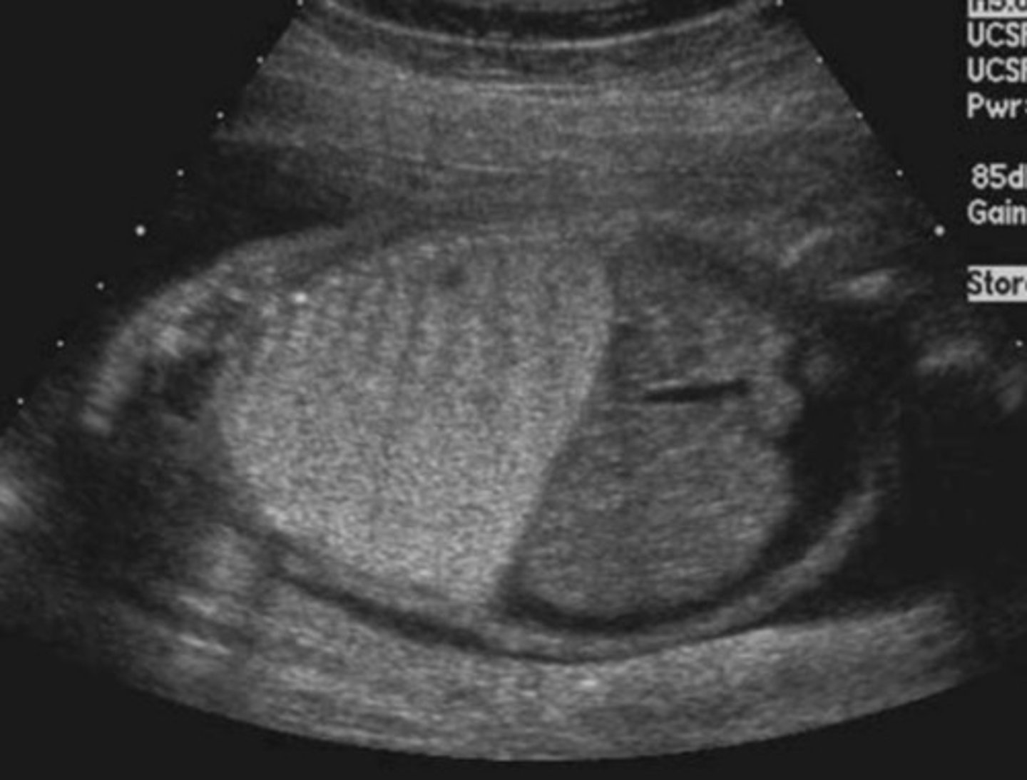
c. Septations of a cystic hygroma
The red arrows in the image below are pointing to...?
a. A large myelomeningocele
b. Amniotic bands
c. Septations of a cystic hygroma
d. Fetal anasarca
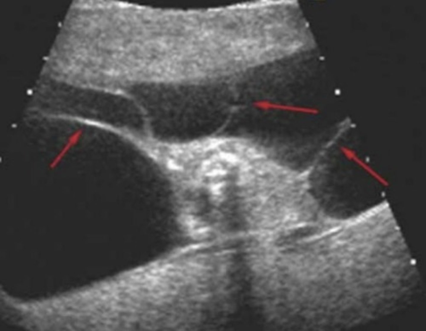
b. A fluid filled duodenum
The midline anechoic structure in the image below represents
a. A congenital diaphragmatic hernia
b. A fluid filled duodenum
c. A multicystic dysplastic kidney
d. A fetal gallbladder
e. The fetal stomach
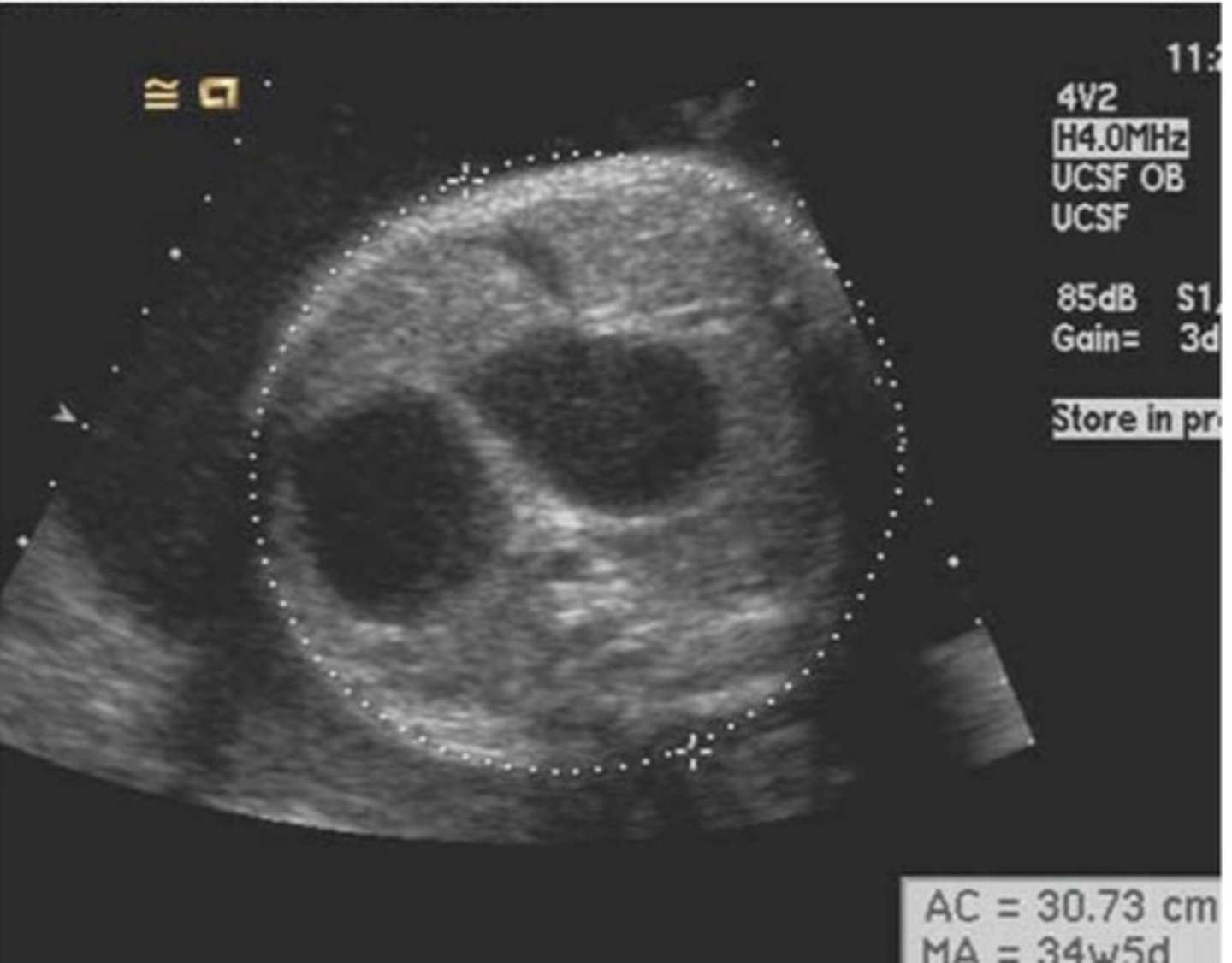
A. Uteropelvic junction obstruction
What is your best diagnosis for the image below?
A. Uteropelvic junction obstruction
B. Unilateral renal agenesis
C. Multicystic dysplastic kidney disease
D. Mild hydronephrosis
E. Intestinal atresia
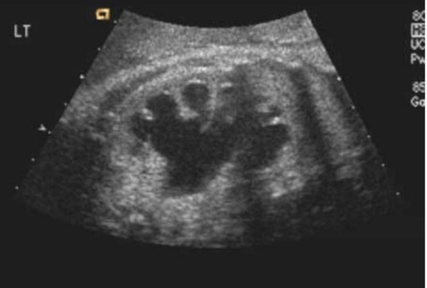
d. Meconium peritonitis
All of the following may be associated with the findings in the sonographic image below except
a. Pulmonary hypoplasia
b. Posterior urethral valve syndrome
c. Ectopic ureterocele
d. Meconium peritonitis
e. Oligohydramnios
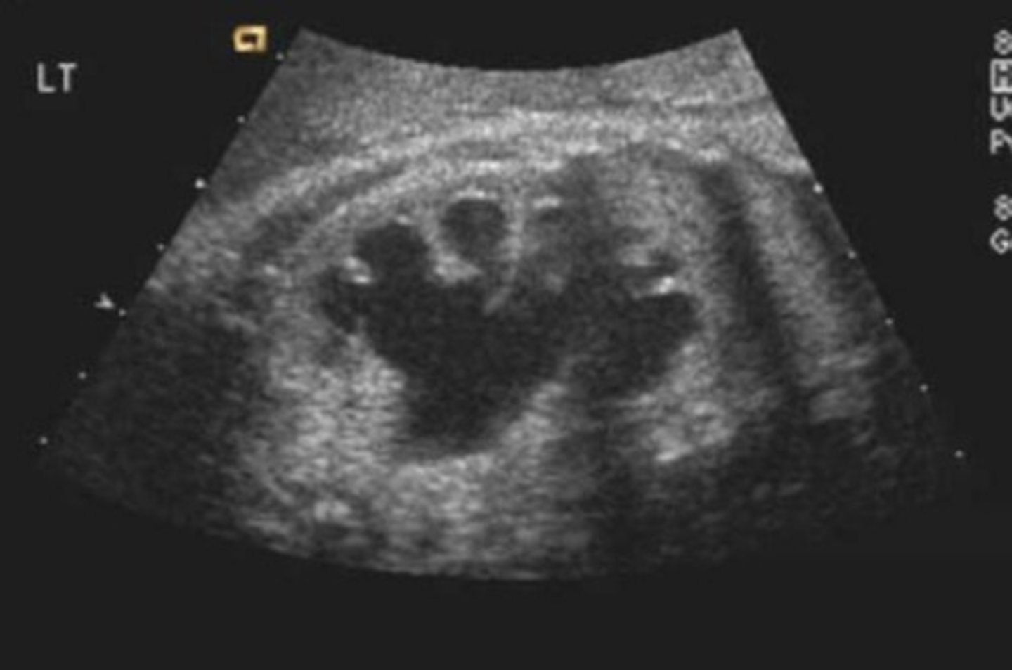
b. Pulmonary hypoplasia
Which of the following is least likely associated with the fetus in the image below?
a. Ventriculomegaly
b. Pulmonary hypoplasia
c. Elevated MSAFP level
d. Chiari Il malformation
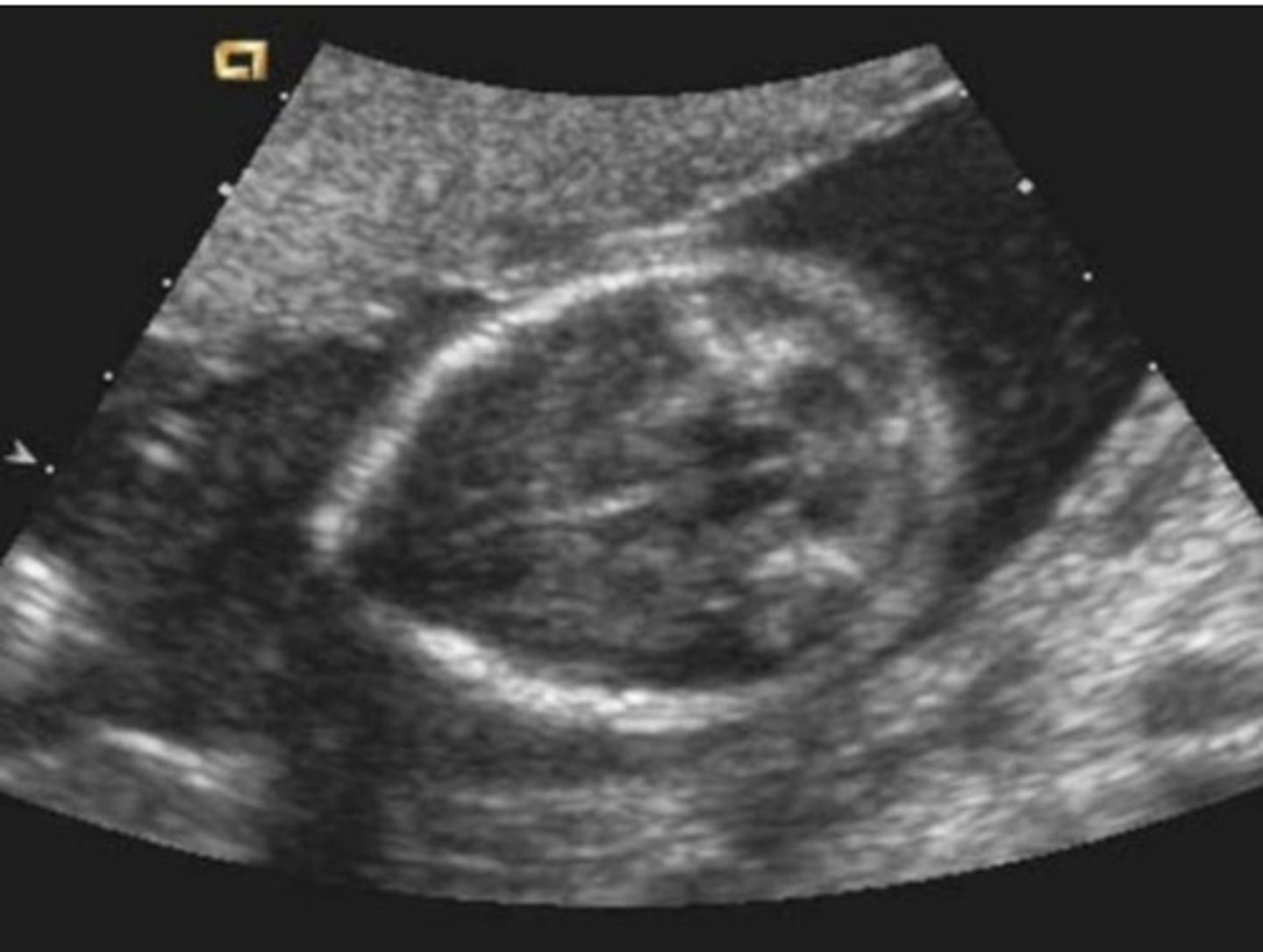
a. Breech
The image below is a longitudinal view through the gravid uterus. What is the fetal position?
a. Breech
b.Transverse, with head to the maternal left
c. Cephalic
d. Transverse, with the head to the maternal right
e. Vertex
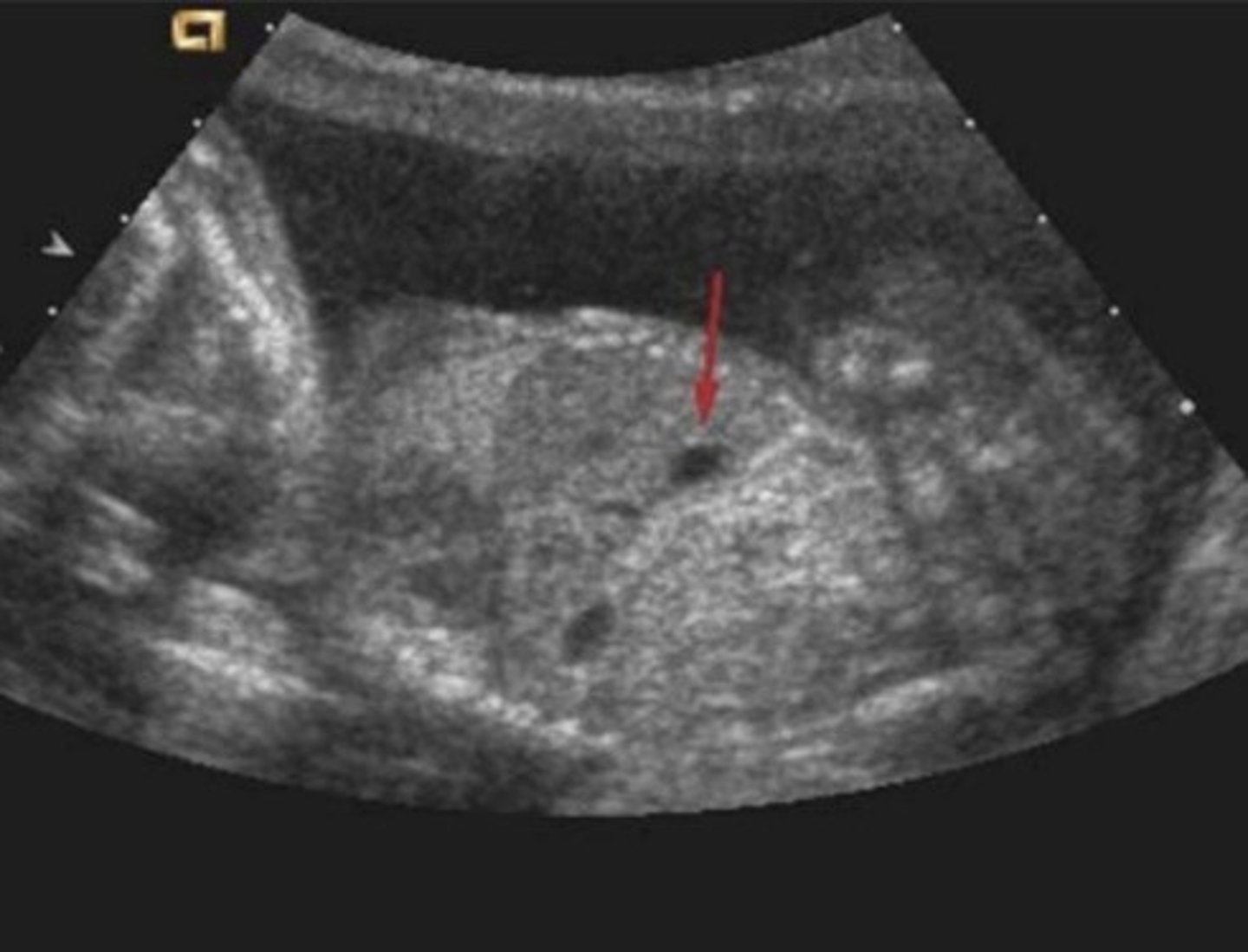
a. Bilateral renal agenesis
In the image below, what is the least likely diagnosis?
a. Bilateral renal agenesis
b. Bilateral renal obstruction
c. Bilateral Multicystic dysplastic kidney
d. Unilateral megaloureter
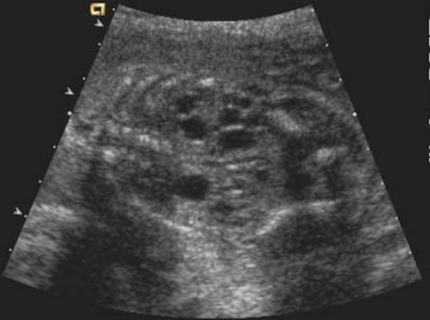
d. Spina bifida
The cause of the abnormality in the image below most likely is
a. Idiopathic
b. A chromosomal abnormality
c. Hydranencephaly
d. Spina bifida
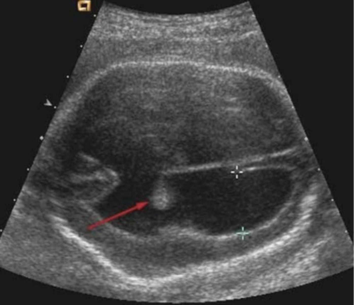
D. Myelomeningocele
The sonographic findings in the image below are most likely associated with
A. Encephalocele
B. Urinary obstruction
C. Thanatophoric dysplasia
D. Myelomeningocele
E. Facial Anomalies
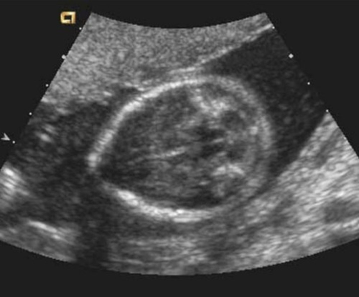
d. Muscle
The arrowhead in the image below is pointing to...?
a. Spinal cranial
b. Subcutaneous fat
c. Rusperitoneal fuid
d. Muscle
e. Ascites
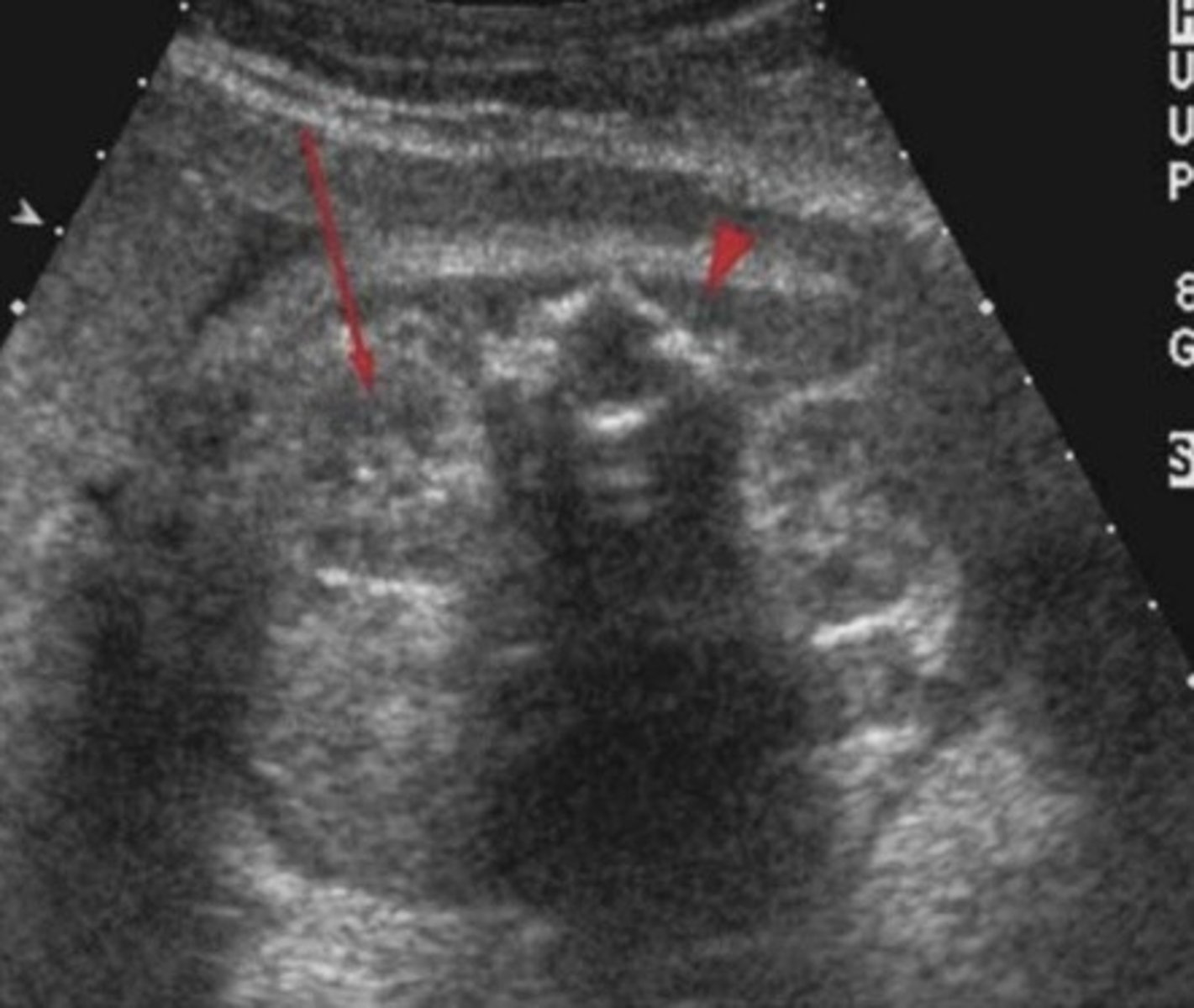
b. An abnormal kidney
All of the following are likely to be associated with the findings in the image below except
a. Elevated AFAFP
b. An abnormal kidney
c. Elevated MSAFP
d. Bowel edema as gestation advances
e. An abdominal wall muscular defect
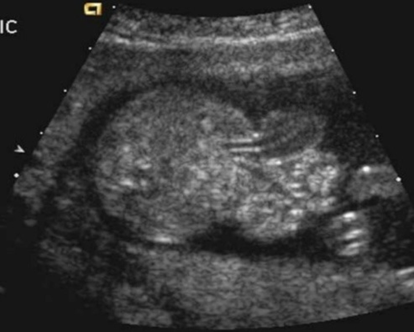
d. Hypoplastic left heart syndrome
The abnormality in the image below is most likely
a. Congenital diaphragmatic hernia
b. Pulmonary sequestration
c. Transposition of the great arteries
d. Hypoplastic left heart syndrome
e. CCAM type III
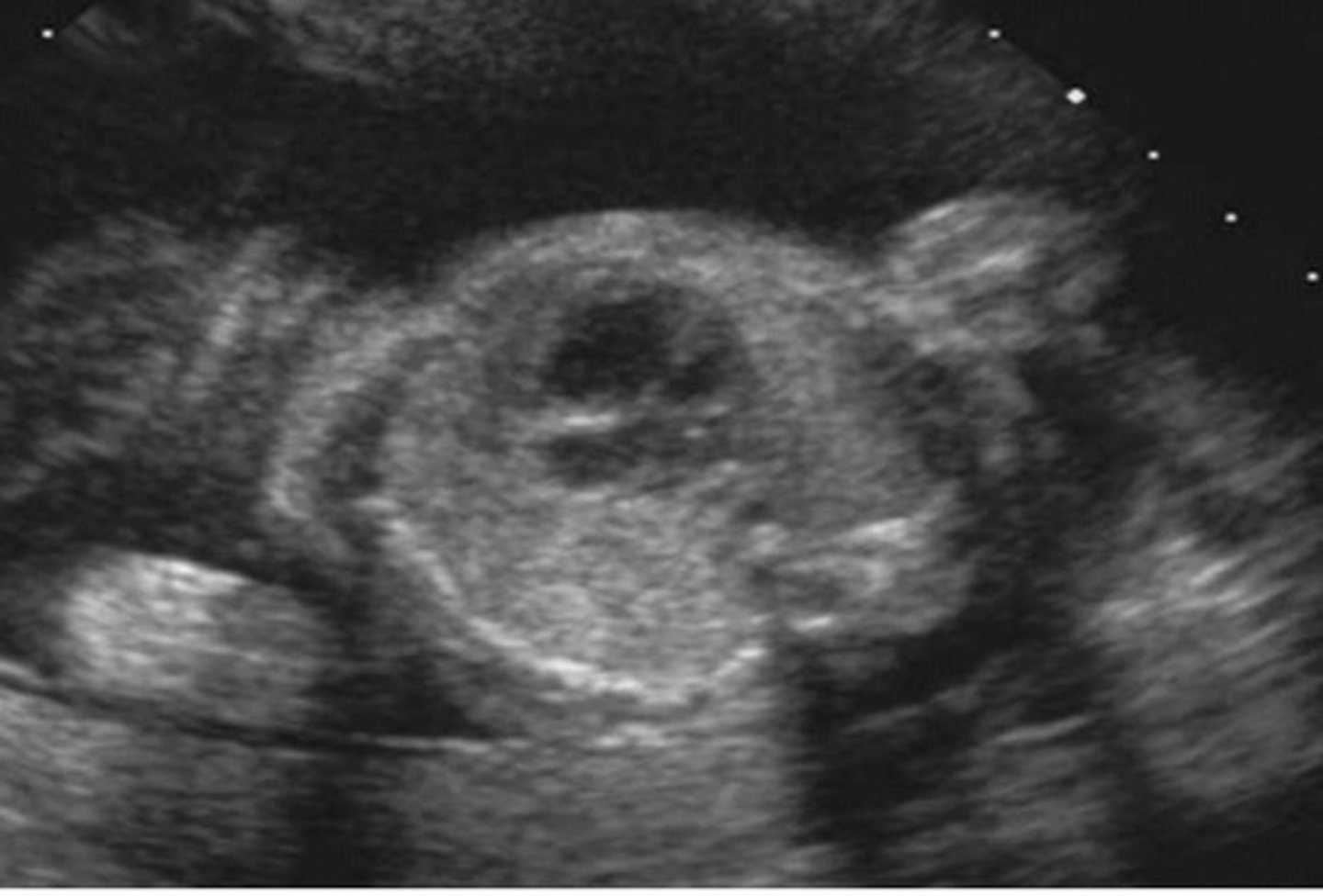
b. Dangling choroid plexus
The red arrow in the image below is pointing to...?
a. The thalamus
b. Dangling choroid plexus
c. Intraventricular hemorrhage
d. The third ventricle
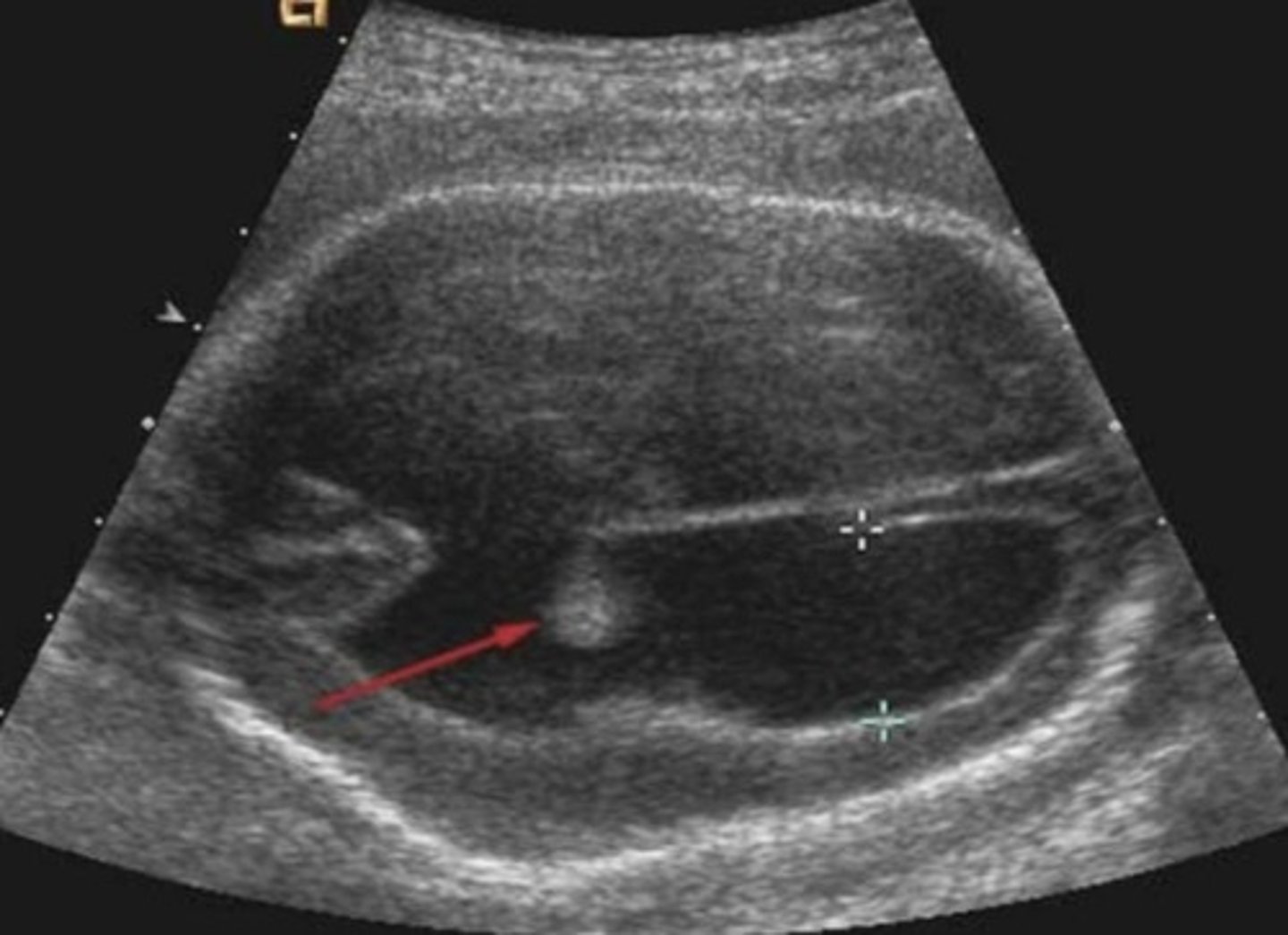
b. Anencephaly
The sonographic finding in the image below may be associated with all of the following except
a. Dandy-Walker syndrome
b. Anencephaly
c. Down syndrome
d. Encephalocele
e. Myelomeningocele
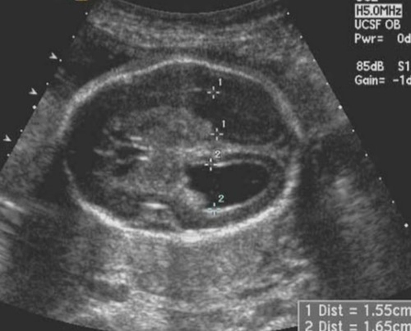
b. The iliopsoas muscle
Which structure is seen immediately posterior to the ovary in the image below?
a. The uterus
b. The iliopsoas muscle
c. Bowel
d. The iliac bone
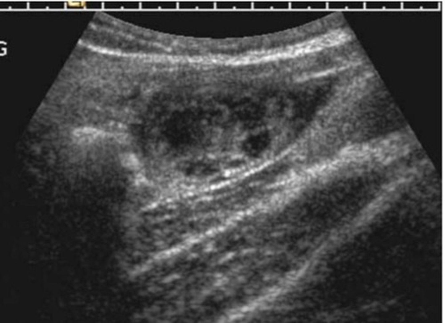
d. An enlarged myomatous uterus
The uterine dimensions in the image below are most consistent with:
a. A normal postmenopausal uterus
b. A normal nulliparous uterus
c. A multiparous uterus d.
d. An enlarged myomatous uterus
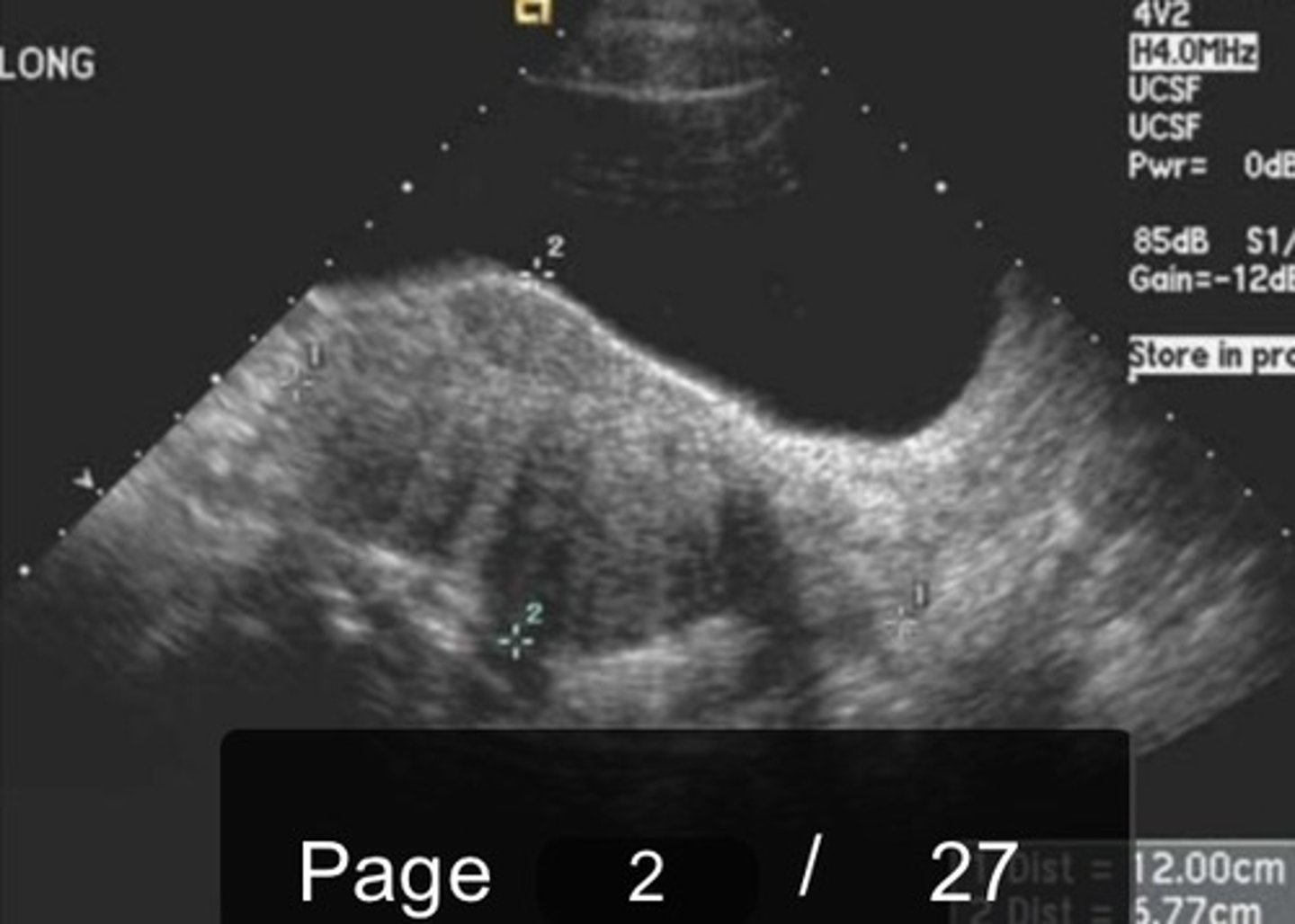
a. Sacral bones
The red arrows in the image below point to what reflective structure(s)?
a. Sacral bones
b. Calcified pelvic vessels
c. Gas in the rectum
d. A pelvic mass
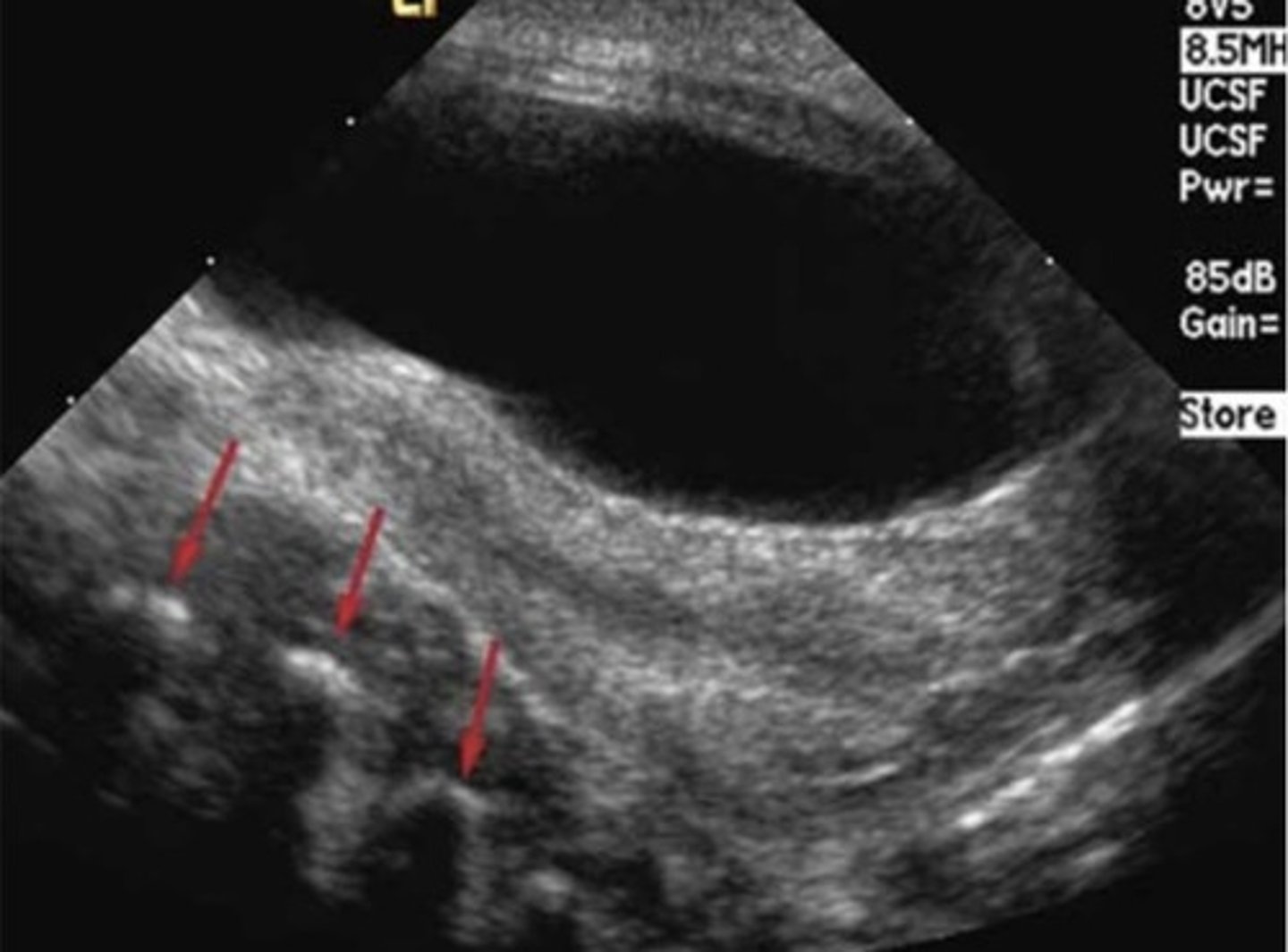
a. Ovarian hyperstimulation syndrome
The size and appearance of the ovary in the image below may be consistent with all of the following scenarios except
a. Ovarian hyperstimulation syndrome
b. Normal postmenopausal ovary
c. Early follicular ovarian phase
d. Inadequate response to ovulation induction drugs
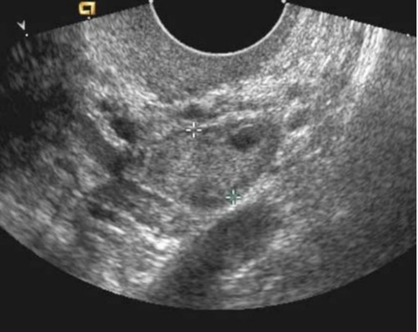
d. Uterine arcuate vessels
In the image below, the arrow points to a region of decreased echogenicity in the uterus. This most likely represents:
a. Myometrial Cysts
b. Uterine Spiral Vessels
c. Perimetrium
d. Uterine arcuate vessels
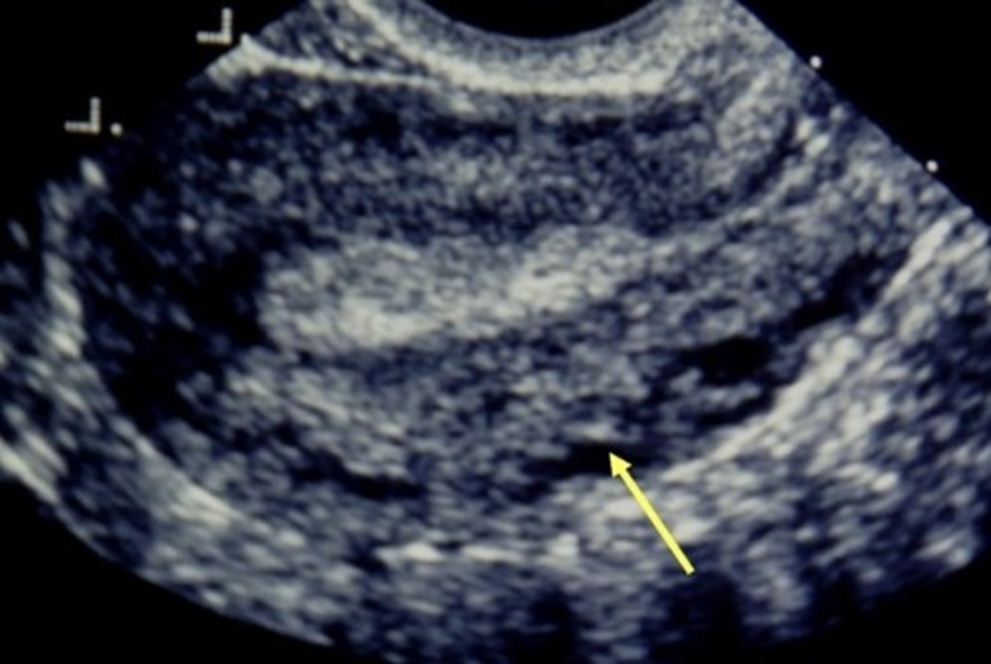
b. Posterior acoustic enhancement
What sonographic characteristic in the image below is most suggestive of fluid in the uterus?
a. Dilatation of the endometrial cavity
b. Posterior acoustic enhancement
c. Marked attenuation of sound
d. Posterior acoustic shadowing
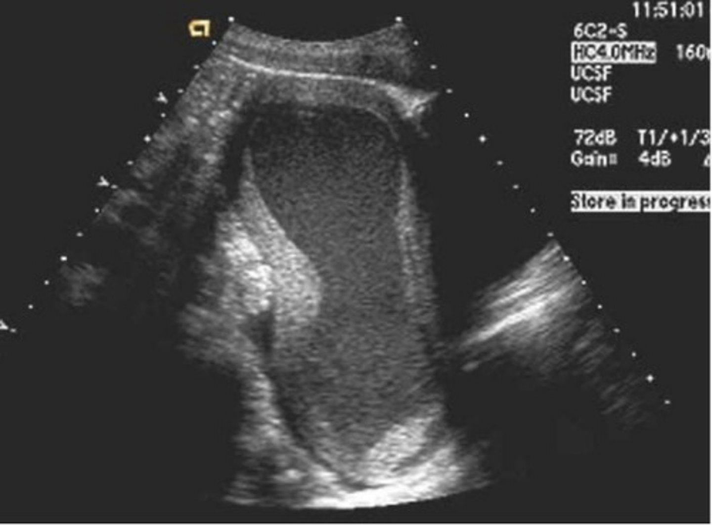
a. Proliferative
The uterine phase, according to the appearance of the endometrium, is most likely
a. Proliferative
b. Follicular
c. Late secretory
d. Menstrual
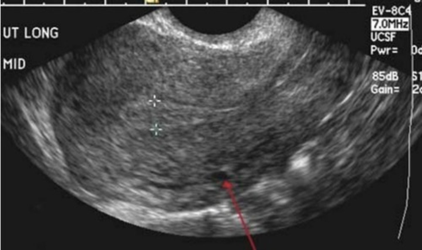
d. Menorrhagia
A postmenopausal uterus, as in the image below, is noted to have a small amount of intracavitary fluid. This may be associated with all of the following except:
a. Cervical stenosis
b. Postmenopausal bleeding
c. Hormone administration
d. Menorrhagia
e. Endometrial atrophy
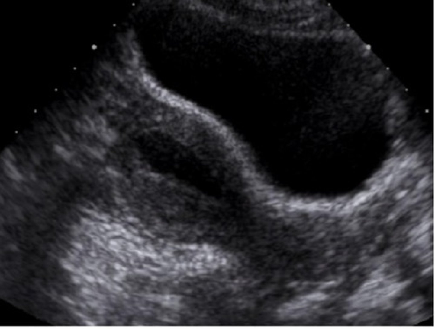
c. The string is visible and the upper vagina
All of the following are demonstrated shadowing correct IUD placement in the image below except
a. The IUD is not embedded in the myometrium
b. The reflective echo of the IUD is within the endometrial cavity
c. The string is visible and the upper vagina
d. The IUD is positioned in the fundus and mid corpus
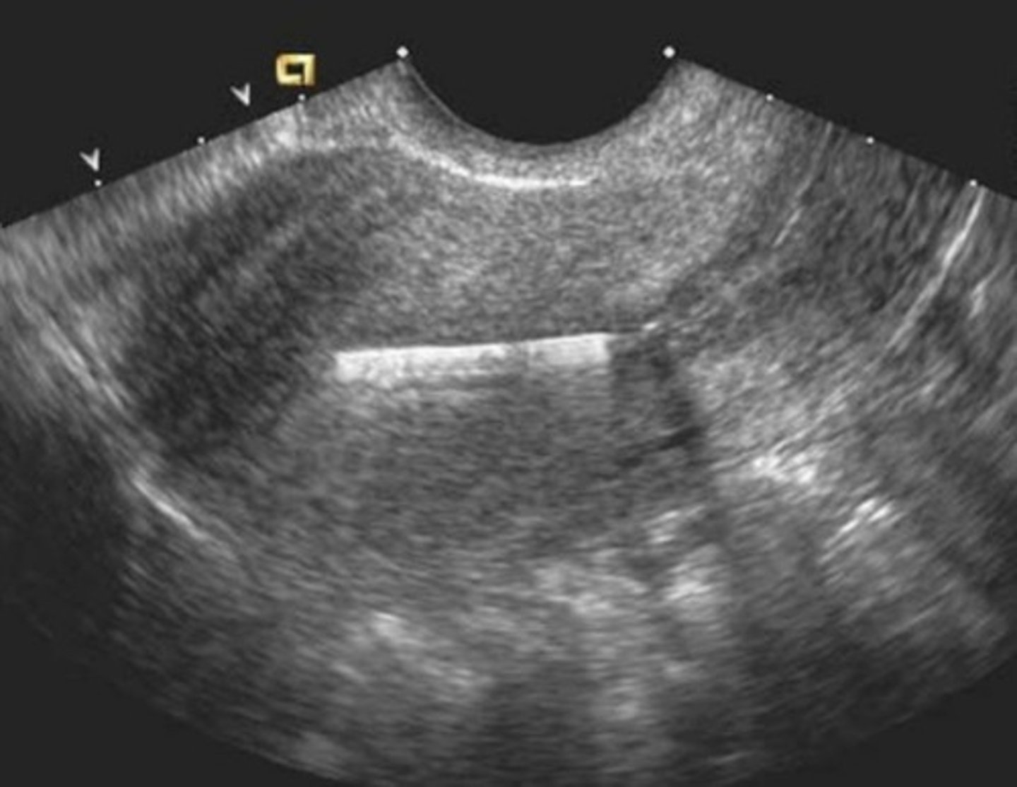
a. Ovulatory phase
Based on the finding in the image below, this patient is most likely in what part of her menstrual cycle?
a. Ovulatory phase
b. Menstrual phase
c. Luteal phase
d. Late secretory phase
e. Follicular phase
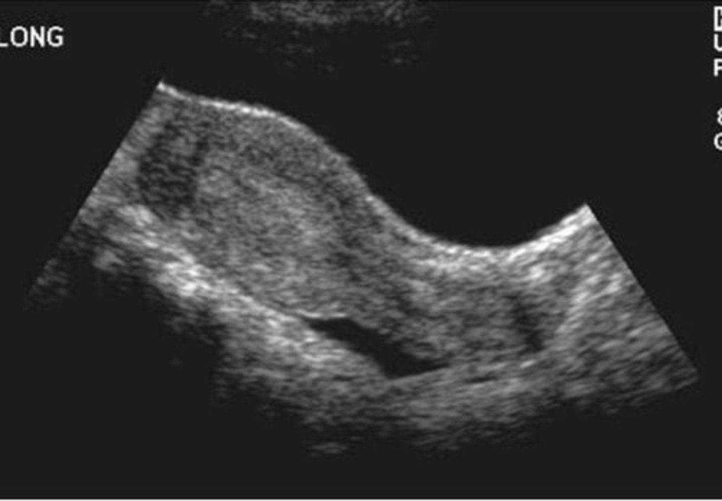
b. Levator ani
The arrows in the image below indicate which muscle/muscle group?
a. Piriformis
b. Levator ani
c. Obterator internus
d. Iliopsoas
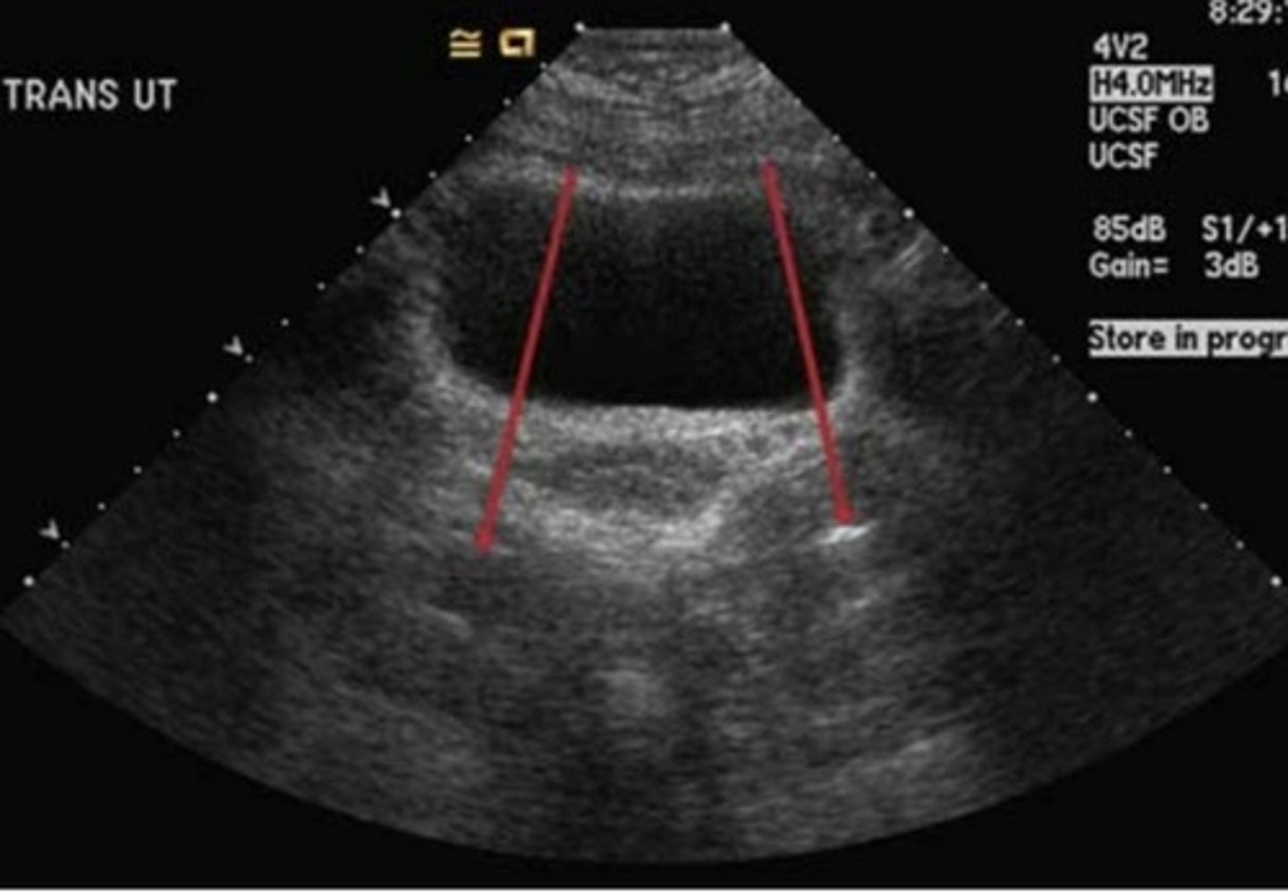
a. A neonate
The uterus in the image below is typical in appearance and size for what age?
a. A neonate
b. Ages 4-7 years
c. A nulliparous female
d. Ages 7 years to puberty
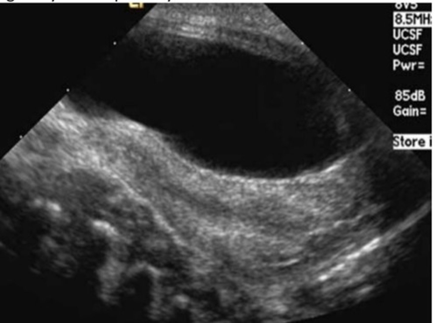
a. Retroflexed
The position of the uterus in the image below is most accurately described with which terminology?
a. Retroflexed
b. Anteflexed
C. Anteverted
d. Retroverted
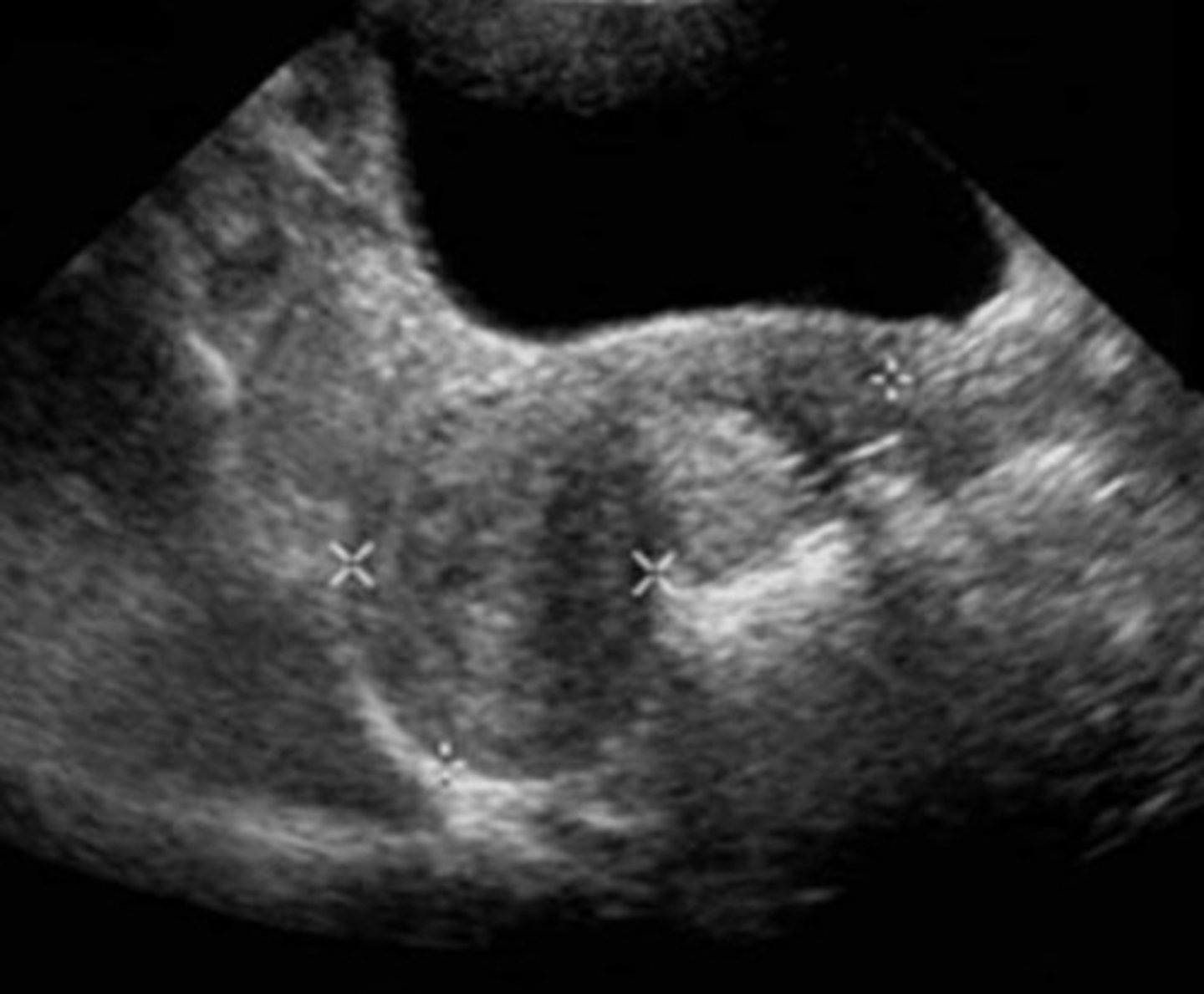
d. Vesicouterine space
The red arrow points to a specific peritoneal space. All of the following are names for that space except:
a. Posterior cul-de-sac
b. Pouch of Douglas
c. Rectouterine space
d. Vesicouterine space
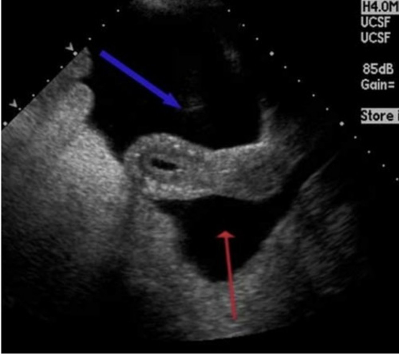
c. A nulliparous female who is mid cycle
The appearance of the uterus is consistent with which scenario?
a. A multiparous female with pelvic inflammatory disease
b. A pediatric patient who is no older than 2 years of age
c. A nulliparous female who is mid cycle
d. A postmensatesale me vinie ndometrial carcinoma
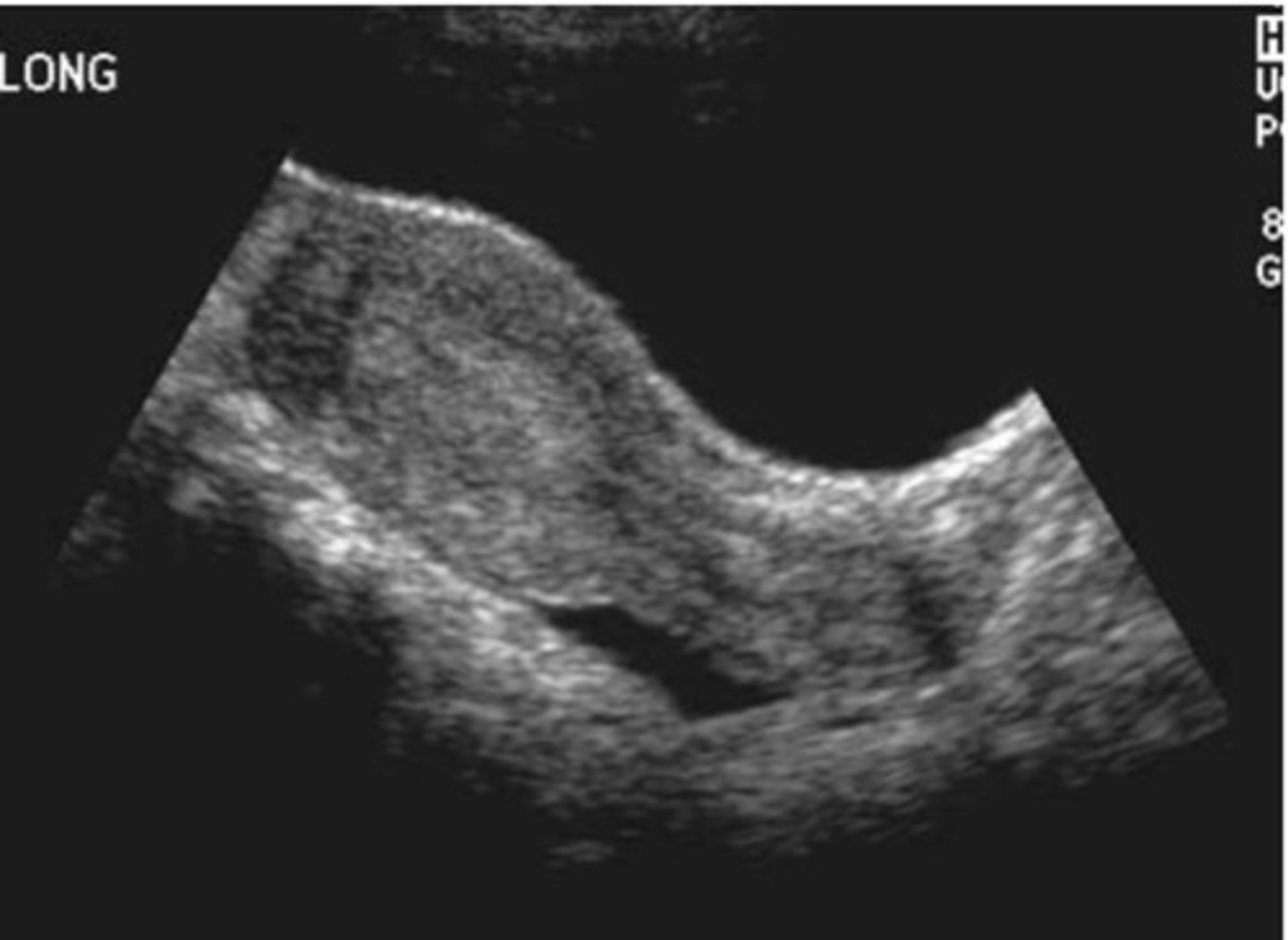
d. Incarcerated uterus
If the patient becomes pregnant, she is at risk of having:
a. Macrosomia
b. Multiple gestation
c. Ectopic pregnancy
d. Incarcerated uterus
e. Hydatidaform mole
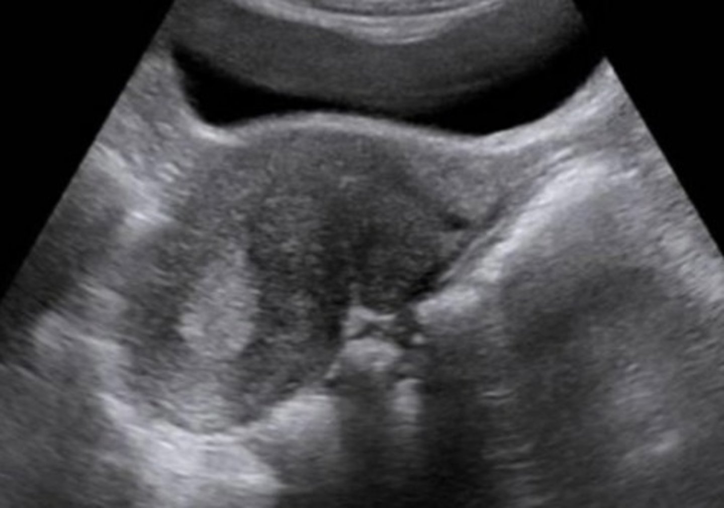
b. To visualize the broad ligaments and fallopian tubes
Ability to image the uterine fundus through an adequately full bladder is significant for all of the following reasons except:
a.To obtain an accurate uterine length measurement
b. To visualize the broad ligaments and fallopian tubes
c. To displace bowel from the anterior pelvic cavity as much as possible
d. To identify superiorly located masses
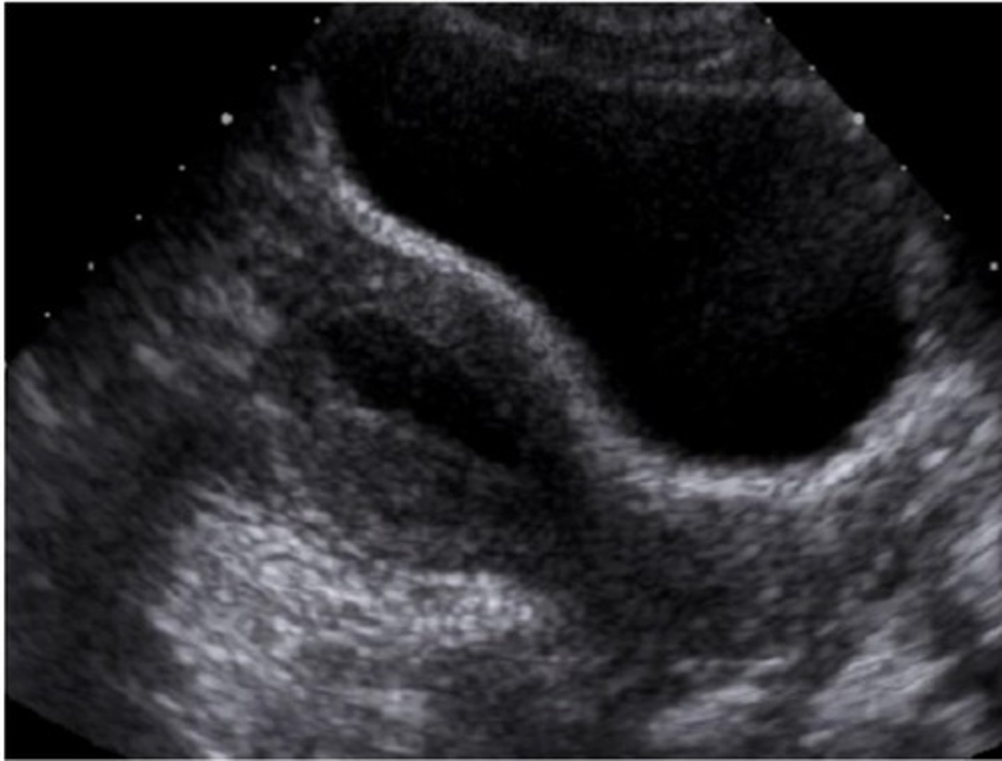
d. Posterior acoustic enhancement
What sonographic characteristic in the image below is most suggestive of fluid in the uterus
a. Marked attenuation of sound
b. Dilation of endometrial cavity
c. Posterior acoustic shadowing
d. Posterior acoustic enhancement
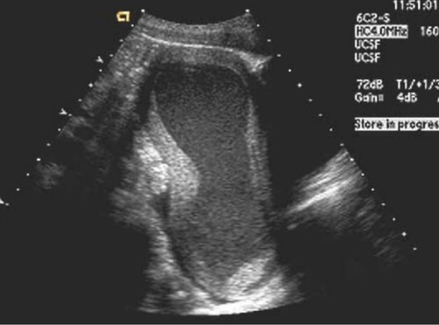
b. Ovarian hyperstimulation syndrome
The size and appearance of the ovary in the image below may be consistent with all of the following scenarios except:
a. Normal post menopausal ovary
b. Ovarian hyperstimulation syndrome
c. Early follicular Ovarian face
d. In adequate response to ovulation induction drugs
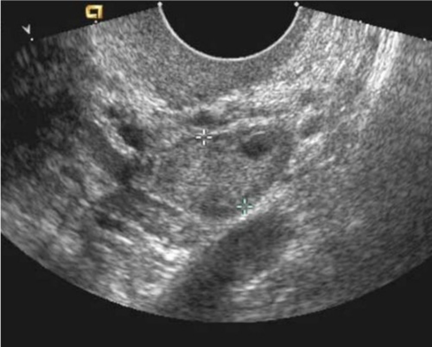
d. Vesicouterine space
The blue arrow and the image below indicates what peritoneal space
a. Rectouterine space
b. Space of retzius
c. Posterior cul-de-sac
d. Vesicouterine space
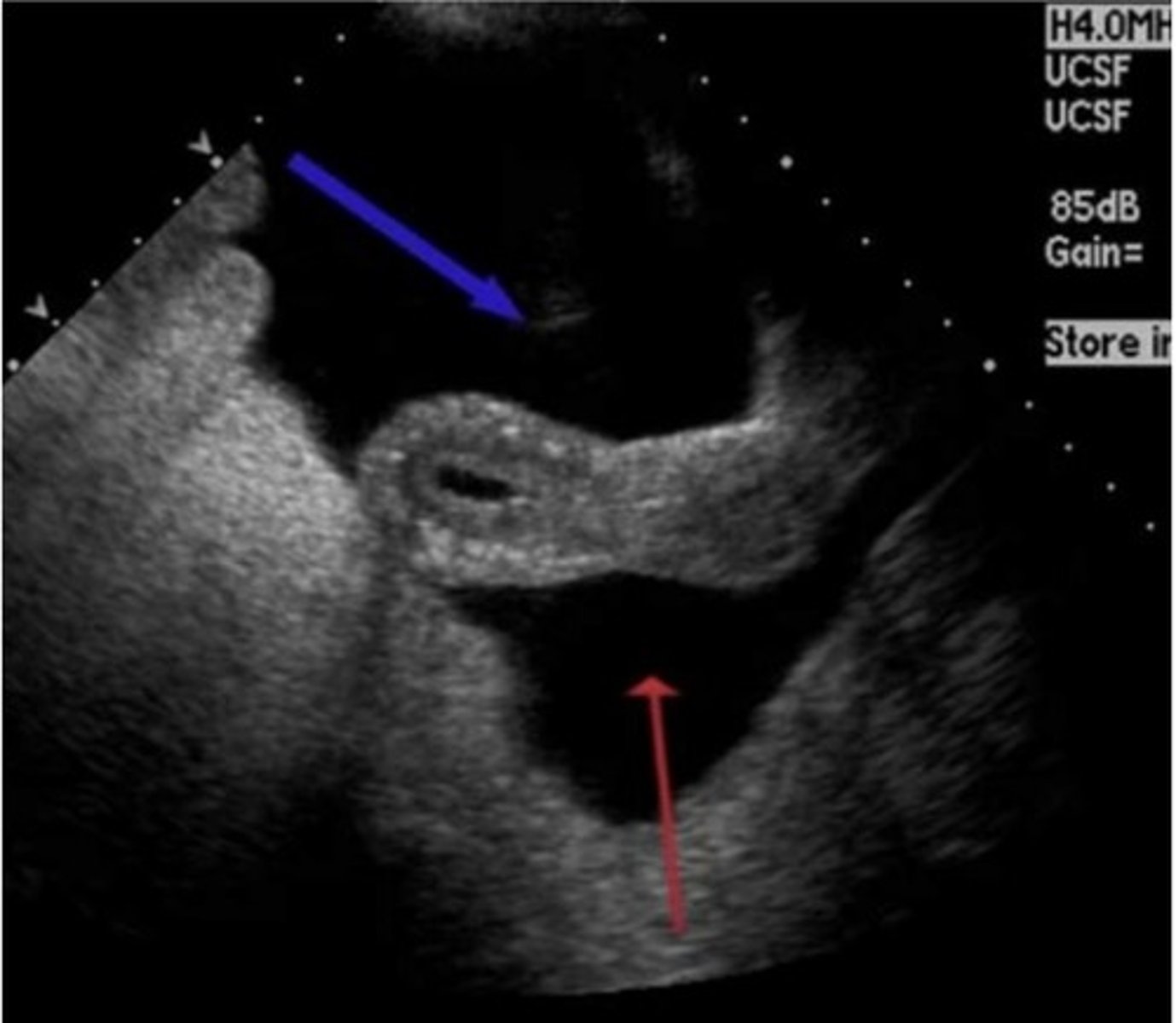
c. Anterior vaginal fornix
The red arrow in the image below is pointing to which anatomic location?
a. Anterior cul-de-sac
b. External os
c. Anterior vaginal fornix
d. Posterior cul-de-sac
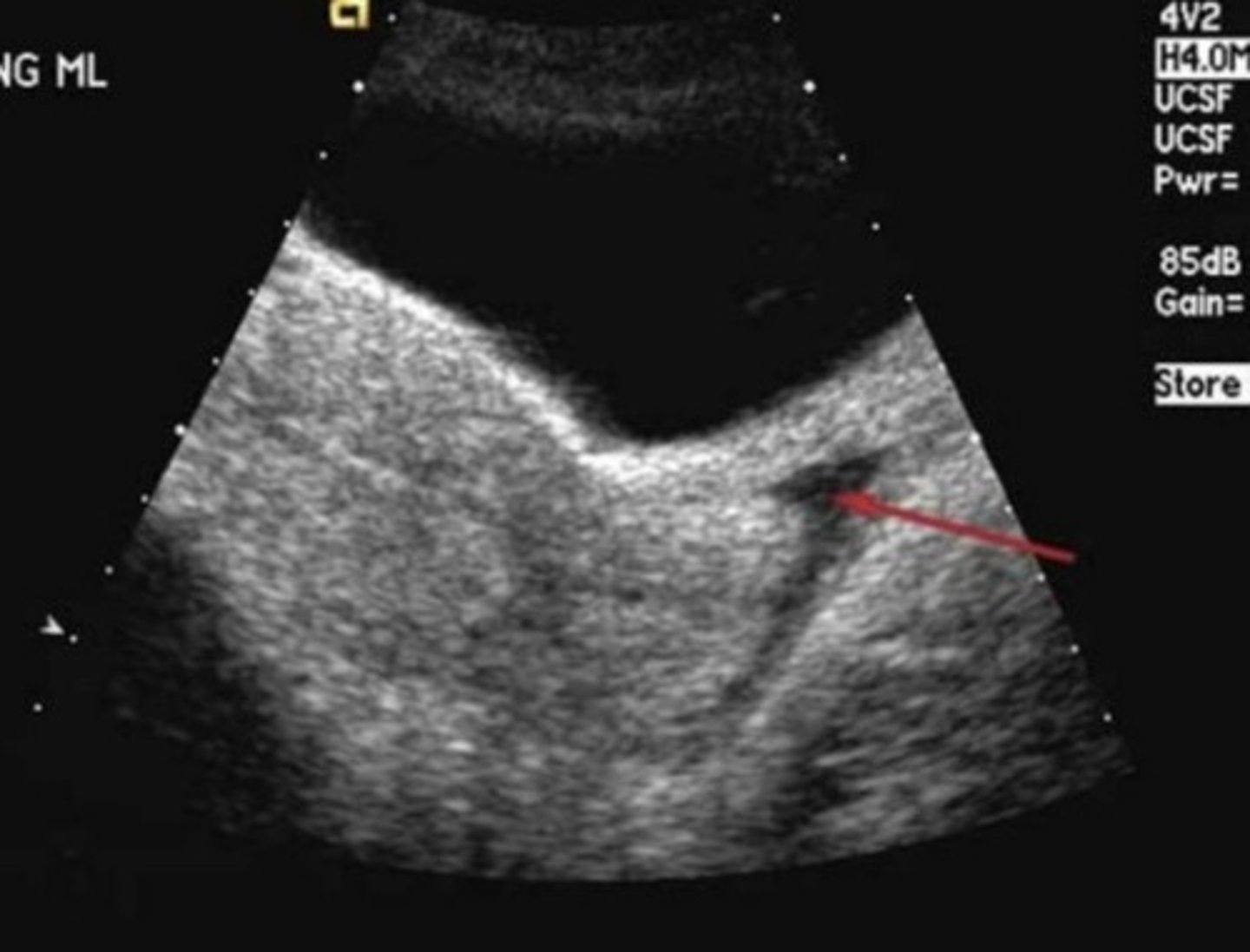
d. Unilateral renal agenesis
Which of the following is most likely to be associated with the finding in the sonographic image below?
a. Gartner's duct cyst
b. Bilateral renal agenesis
C. Tubo-ovarian abscess
d. Unilateral renal agenesis
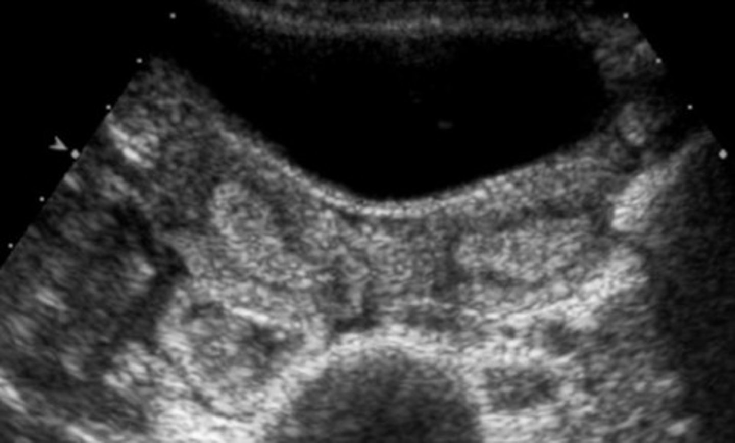
a.Anterior to the iliopsoas muscle
The location of the ovary in the image is:
a.Anterior to the iliopsoas muscle
b. In the true pelvic
c. Adjacent to the iliac vessels
d. In the rectouterine space
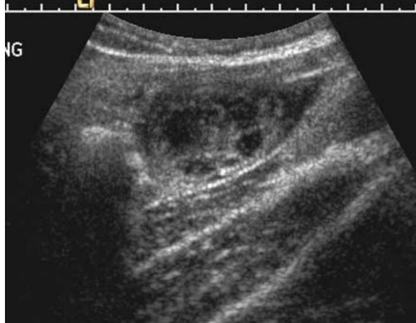
b. A normal uterus
The calipers in the image below are measuring what?
a. The vaginal cuff post hysterectomy
b. A normal uterus
c. An anomalous uterus and cervix
d. A myomatous uterus
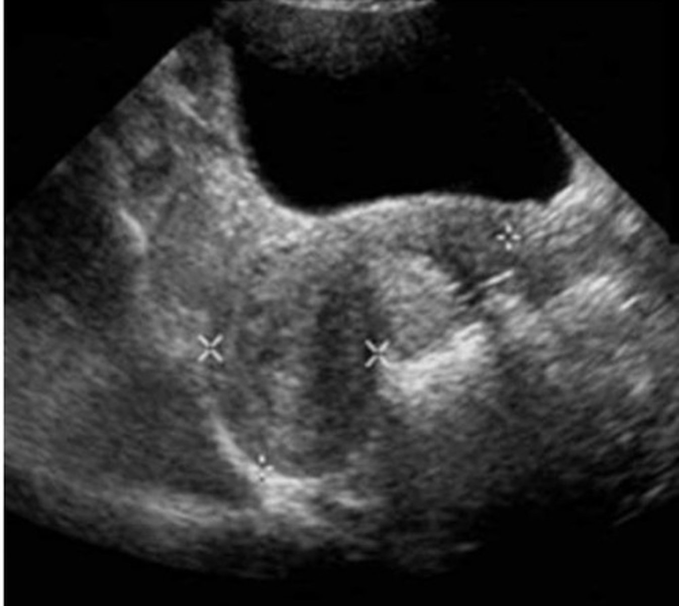
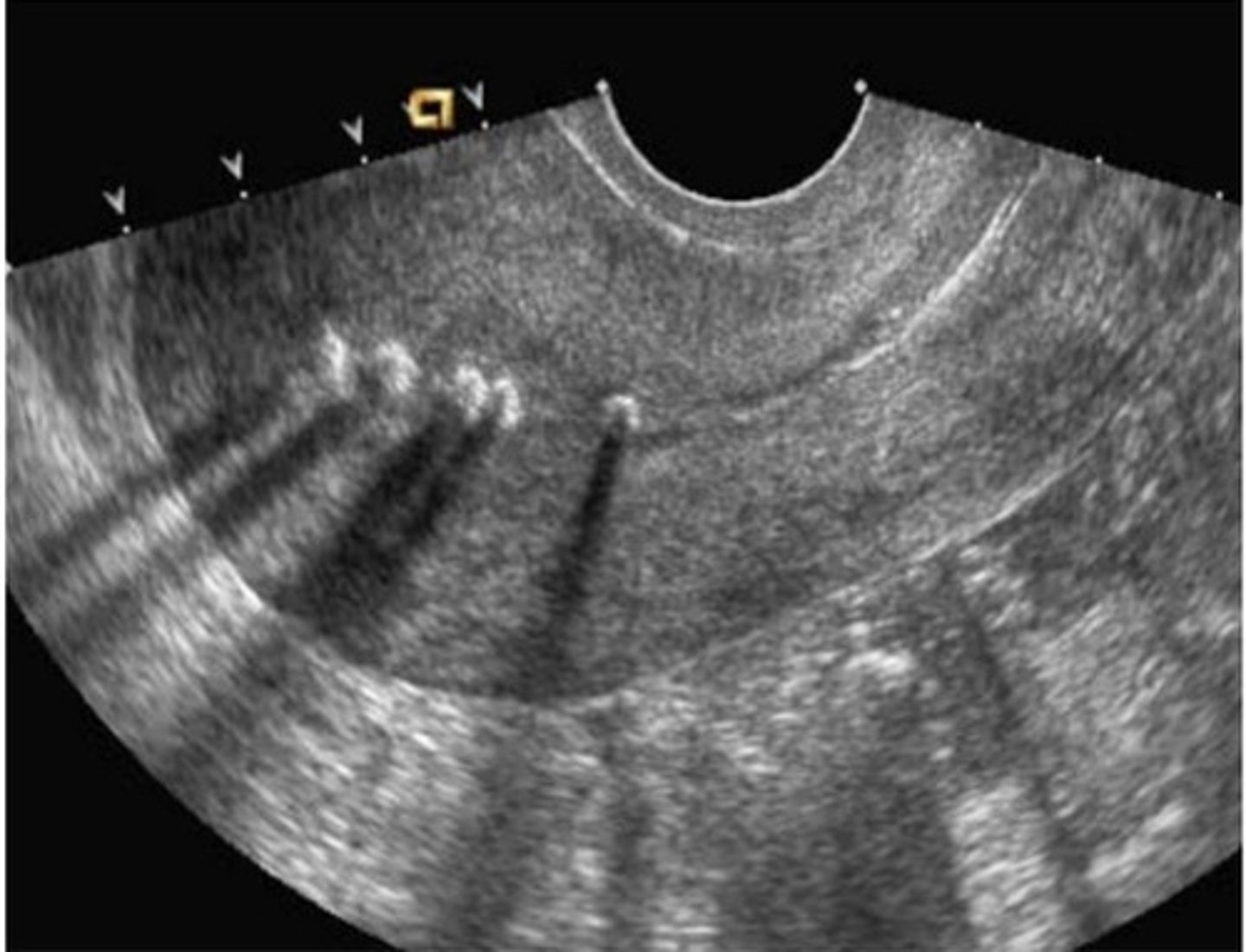
e. A Lippes loop IUD
The strongly shadowing structures in the image below are most likely caused by what?
a. Endometrial polyps
b. Endometrial carcinoma
c. Endometrial calcifications
d. Retained products of conception
e. A Lippes loop IUD