3.4 Coronary circulation and cardiac conduction system
 Call Kai
 Learn
 Practice Test
 Spaced Repetition
 Match
 Flashcards
 Knowt Play
1/54

There's no tags or description
Looks like no tags are added yet.
55 Terms
recap: right atrium receives what blood and pups where?
right atrium receives deoxygenated blood and pumps it to lungs via right ventricle
what does pulmonary arteries do?
carries deoxygenated blood to lungs
what does pulmonary veins do?
returns oxygenated blood from lungs
left atrium receives what blood and pumps where?
receives oxygenated blood and pumps it to the body via the left ventricle

ventricles have ___ at their entry and exits?
valves
what do valves do?
ensure one-way movement of blood by preventing backflow
what are the entry valves?
atrioventricular AV valves
what valve is for right ventricle?
tricuspid valve
what valve is for left ventricle?
bicuspid (mitral) valve
what cause AV valves to close?
increased pressure during ventricular contraction pushes cusps together, papillary muscle contraction ensures cusps don’t prolapse into atria

what are the exit valves?
semilunar valves
semilunar valve for right ventricle?
pulmonary valve
semilunar valve for left ventricle?
aortic valve
what closes the semilunar valves?
decreased pressure during ventricular relaxation causes slight backflow of blood into pulmonary trunk/aorta, filling cup-like cusps and closing valve

coronary circulation:

when aortic valve closes, what happens to right and left coronary arteries?
it’s when they have a pulse of blood flow through them
what is ischemia?
oxygen deprivation caused by blockage of coronary arteries which can lead to necrosis of cardiac muscle tissue
what can cause blockage of coronary arteries?
often due to atheroscerosis (cholesterol plaque buildup in wall of vessel)
what artery is most often blocked in coronary artery disease?
anterior interventricular (left anterior descending) artery most often blocked

what are cardiomyocytes?
cardiac muscle cells
how many nuclei do cardiomyocytes have and what do the cells look like?
1 maybe 2 nuclei and are short, wide branched cells
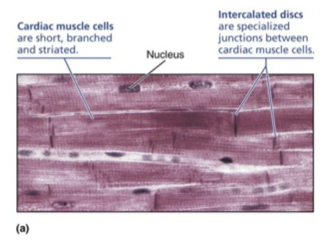
cardiomyocytes are joined by intercalated discs. what are they?
intercalated discs are specialized junctions between cardiac muscle cells
what other cell junctions do cardiomyocytes use?
desmosome and gap junctions
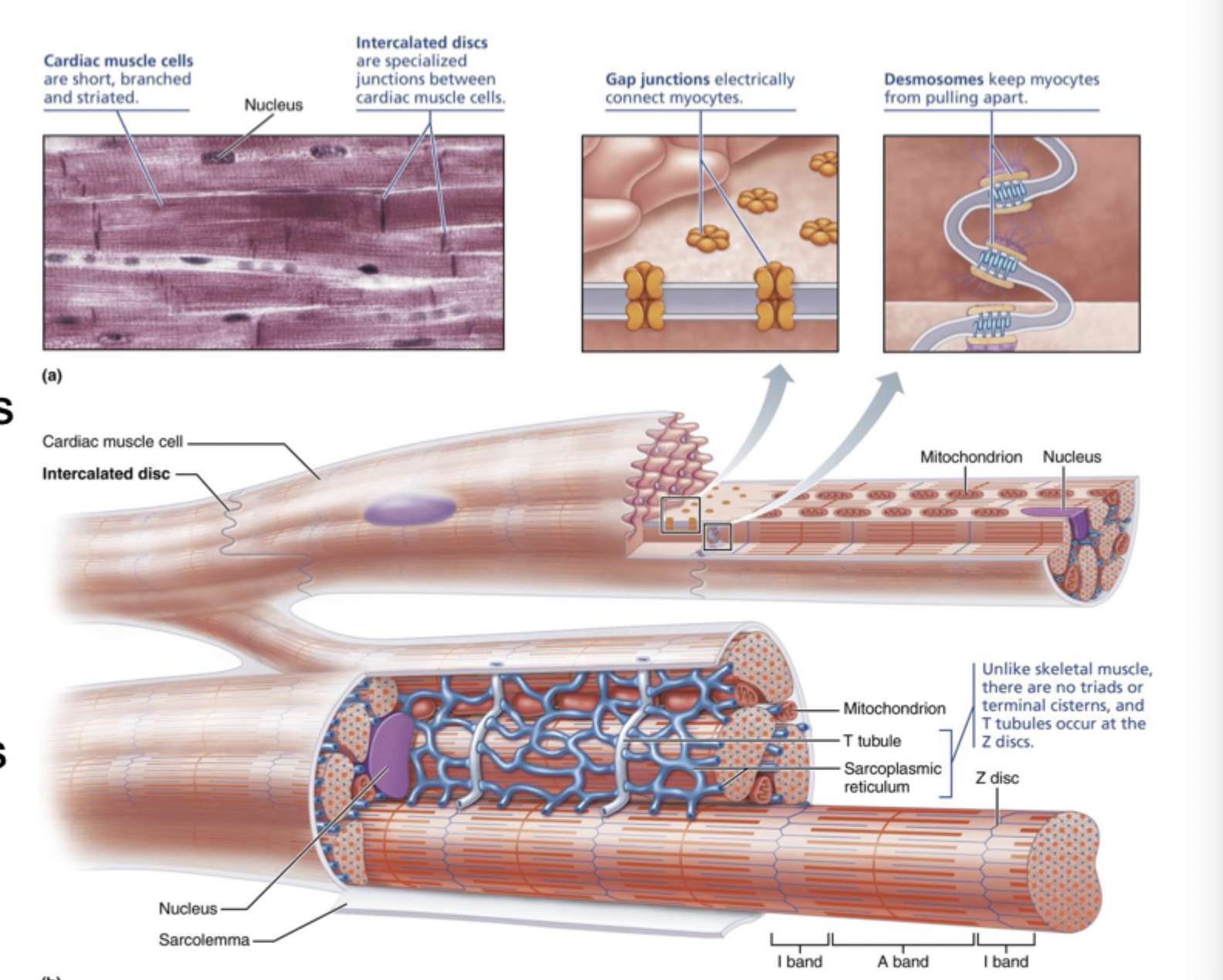
what do desmosomes do in cardiomyocytes?
keep cardiomyocytes joined together

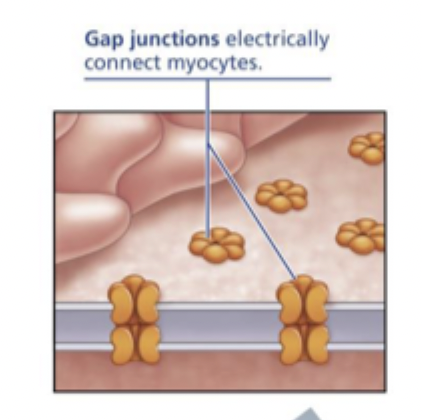
what doe gap junctions allow between cardiomyocytes?
allow transmission of ions and therefor APs between cells so important for cardiac muscle to contract as a unit
how does mitochondria compare in cardiac muscle compared to skeletal muscle?
more mitochondria in cardiac muscle because dependent only on aerobic respiration
how does SR compare in cardiac compared to skeletal muscle?
SR is less elaborate with no terminal cisterns flanking T-tubules in cardiac muscle
what stimulates release of calcium from SR?
influx of calcium from ECF stimulates release of calcium from SR where 10-20% of calcium from ECF, 80-90% from SR
what are pacemaker (nodal) cells?
specialized non-contractile cells that make up the cardiac conduction system (about 1-2% of all cardiomyocytes)
what does autorhythmic mean?
spontaneously generate action potentials
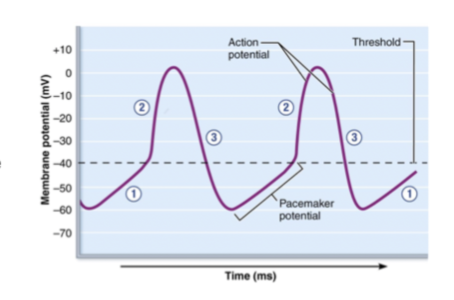
how do pacemaker cells generate spontaneous action potentials? what are the 3 steps?
slow “leaky” Na+ channels allow gradual depolarization called pacemaker potential so never has a stable resting membrane potential
when gradual depolarization reaches threshold, voltage-gated calcium channels causes rapid depolarization of action potential
repolarization occrus when calcium channels close and potassium channels open
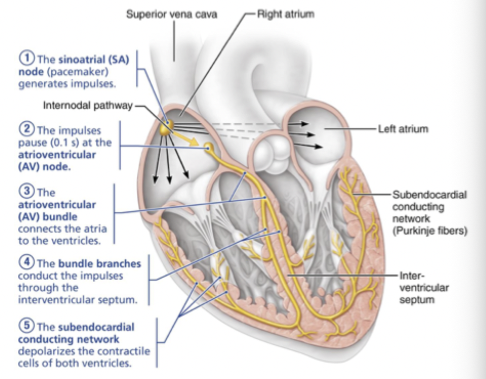
cardiac conduction system: what sets pace of AP signals?
sinoatrial (SA) node so has faster rate of AP generation than other parts of system
what happens at the atrioventricular AV node?
signal from SA node is delayed at AV node which allows atrial contraction to “top off” blood volume in ventricles
cardiac conduction system: what does the AV bundle split into?
splits into right and left bundle branches in interventricular septum
cardiac conduction system: what does the AV bundle do?
connects the atria to the ventricles
cardiac conduction system: what do the bundle branches do?
conduct the impulses through the interventricular septum
cardiac conduction system: what do purkinje fibers do?
purkinje fibers aka subendocardial conducting network, transmit signal through ventricular walls. starts at apex and moves superiorly up walls. Depolarizes the contractile cells of both ventricles
autonomic reg of HR: pacemaker cells generate APs without signals from?
nervous system
how does autonomic nervous system ANS regulate rate of AP generation?
by shifting resting membrane potential relative to threshold
what does sympathetic division of ANS “fight or flight” do?
increases heart rate by making resting potential more positive so closer to threshold
what does parasympathetic division of ANS “rest and digest” do?
decreases heart rate by making resting potential more negative so farther from threshold

how do cardiomyocytes contract?
via sliding filament model
AP events for cardiomyocytes?
Depolarization
Plateau phase
repolarization

AP events: step 1?
depolarization so fast voltage-gated Na+ channels open, causing rapid depolarization
AP events: step 2?
plateau phase: sodium channels close, but slow voltage gated calcium channels open, slowing repolarization
AP events: step 3
repolarization: calcium channels inactivate, potassium channels open, bringing membrane potential back to resting
what is the absolute refractory period for cardiomyocytes?
plateau period makes action potential last about as long as period of contraction so about 200+ ms
tetanus not possible
cardiac muscle must relax before it can be stimulated again
what is electrocardiogram ECG?
composite measure of all action potentials generated by pacemaker and contractile cardiomyocytes
in ECG, what do waves of electrical activity correspond to?
correspond to events in conduction pathway so electrical activity precedes cardiomyocyte contraction/relaxation

ECG: what happens in P wave?
SA node fires, causes atrial depolarization
ECG: what happens in QRS complex?
ventricular depolarization so QRS masks atrial repolarization
ECG: what happens in T wave?
ventricular repolarization

ECG: what is the P-R interaval?
time from beginning of atrial excitation to beginning of ventricular excitation
ECG: what is the S-T segment?
entire ventricular myocardium is contracted because cardiomyocytes are in plateau phase of AP
ECG: what is the Q-T interval?
time from beginning of ventricular depolarization through end of ventricular repolarization
