Neuro - exam 2
 Call Kai
 Learn
Learn Practice Test
 Spaced Repetition
 Match
Match Flashcards
 Knowt Play
1/557

There's no tags or description
Looks like no tags are added yet.
Name | Mastery | Learn | Test | Matching | Spaced | Call with Kai | Chat |
|---|
No analytics yet
Send a link to your students to track their progress
558 Terms
How to test proprioception in LA
placment test, hear elevation, circling, backing, hills, curbs, tail pulls
What do LMN spinal cord lesions look like in LA (5)
SHort strided, trempling, hypotonic, atrophy (unless acute), weakness → recumbency
What do UMN/GP spinal cord lesions looks like in LA
long and lopey, stumbling/tripping, truncal sway, traveling on two tracts, don’t know where their feet are (limb placment, circling, backing, hills/curbs), weakness → recumbency
Causes of infectious spinal cord disease in equine
parasitic - equine protozoal myelitits
viral - equine herpesvirus, EEE/WEE/VEE, west nile virus, rabies
bacterial - discospondylitis, bacterial meningitits
Infectious spinal cord disease in ruminants
parasitic - paralephastrongylus tenuis
viral - rabies, herpesvirus, lentiviral infections
prion - scrapie (sheep), bovien spongiform encephalopathy
Equine protozoal myelitis - pathogen/transmission
exposed via ingestion of opossum feces with CNS migration of protozoan parasites (sarcocystic neurona and neopspora hughesi)
Equine protozoal myelitis (EPM) - how common is it
high seroprevalence but low clinical disease
under 1% develop neurologic disease
EPM - s/s
vary widely
Asymmetrical spinal cord is most common (ataxia, atrophy, asymmetric)
Occ just poor performance
acute or slowly progressive
no age, sex, breed predisposition
EPM - dx
MRI (can only do head in equine) OR
consistent s/s + prove exposure to organism (western blot, IFAT, SAG titers)
need antibodies titers in CSF (intrathecal Ab production)
Best: surface antigen (SAG) 4/3/2 titer on serum and CSF
csf ratio <100 suggests EPM (on exam and Navle)
EPM - management
Anti-protozoal therapy
ponazuril, diclazuril, sulfadiazine and pyrimethamine
labelled for 4 weeks tx, but usually months needed
EPM - prognosis
most improve or stabilize with tx BUT some progress, relapse or reinfected, or never fully heal
Equine Herpesvirus Myeloencephalopathy - s/s
respiraotry and abortions most common but also CNS disease
What virus cuases Equine Herpesvirus Myeloencephalopathy
EHV-1 and rarely EHV-4
Equine Herpesvirus Myeloencephalopathy - epidemiology
Almost all horses infected as foals, latent in trigeminal ganglia and lymph nodes
in foals - subclinical infection and shedding → spread
in adutls - shedding = clinical disease
EHM - pathophysiology
Respiraotry epithelial infection to lyph nodes in 24-48 hrs, shedding up to 3 weeks
viremia (leukocytes) - 2 weeks
infection of CNS endothelial cells
vasculitis → microthrombi + local hemorrhage
disseminated ischemic CNS necrosis
EHM - dx
variable, onset 6-10 days after exposure
fever first, but usually gone by time of neuro sings
± respiraotry disease (on farm)
Ataxia (pelvic>thoracic) → tetraparesis/recumbency
back end signs common - incontinence, loss of tail/anal tone, analgesia
EHM - dx
CSF - increase protein, ± mononuclear pleiocytosis (only indicates inflammation)
Acute and convalescent serum titers - measure at start and 4 weeks later
signs + shedding/viremia - PCR on nasal swabs/buff coat, usually positive at least 10-14 days after infection, can shed intermittently - test more than once
EHM - treatemnts
Supportive care
antiinflammatories (steroids but not for long, NSAIDS)
anti virals - valacyclovir
anticoagulants
EHM - prognosis
variable , better if standing
poor if recumbent
EHM - prevention and outbreak maangement
vx - killed or MLV, neither prevent but limit nasal shedding
outbreak management - early dx, treat clinical cases, prevent spread (isolate, quarantine, vx UNEXPOSED only)
West Nile Virus - type, season, clinical severity
flvivirus
summer/early fall - year round i temperate climates
clinical severity varies - subclinical → fatal encephalitis
West Nile virus - s/s
fever, depression, ataxia (symmetric or asymmetric), often muscle fasiculations, ± other prosencephalon/cranial nerve deficits
any age/sex/breed (rare in foals)
acute onset
WNV - dx/managemnt
S/s, CBC, CSF - not pathogenomonic
IgM capture ELISA - serum or CSF, acute infection even in vx animals
tx - supportive care, hyperimmune plasma
WNV - prognosis and prevention
28% mortaility
vx - effective, boost 1st year than annually before mosquitos (q 6 months in warm climates), improves prongosis in vx animals
EEE/WEE/VEE - type of virus, s/s, dx
alphavirus
s/s - ataxia/recumbency + prosencephalic signs, young or unvaccinated, any breed/sex, acute onsent, rapid progression
dx - csf often has high wbc but not always, serology, IHC on tissue
EEE/WEE/VEE - tx, prognosis, prevention
tx- supportive
prognosis - very poor, basically all die
prevention - vx (q6 months in warm climates)
S/S of rabies timeline to death
dead in 3-5 dyas but max of 10-14
Bacterianl meningitis and discospondylitis prevelence inequine
uncommon
Bacterial meningitis - occirs in what horses and s/s
occurs - neoatal foals w/ bacterial sepsis - any organisms
adult horses with immunodeficiences - any organsims
adult hoses with lyme disease (Borrelia burgdorferi)
s/s - fever, ataxia, NECK PAIN (waxing/waning multifocal signs common)
Equine acterial meningitits - dx and tx
dx - csf tap → increased wbc (pleiocytosis) + protein, can culture
tx - antimicrobials (ensure CNS penetration)
Equine discopondylitits - what horses, s/s
equine - often neonates or adults with immunodeficiency or reent vx
s/s - fever, ataxia, NECK PAIN, waxing/waning multifocal sigsn
Equine discospondylitis - dx, tx
dx -imaging/sampling, brucellosis testing in adults
tx - long term abx
Cervical vertebral stenosis myelopathy - what is it and signalment
developmental orthopedic disease of the spine
aka wobblers
young, big, male horses (thoroughbred, warmbloods, quarter horses)
CVSM - risk factors
unknown
genetic factors - no clear inheritance pattern, genetic predisposition
diet/environment factors - too much carbs → too rapid growth, low copper + high zinc?
trauma → abnormal biomechanics
CVSM - pathogenesis
unknown
developmental abnormalitis of the cervical spin → spinal cord compression → neuro deficits
one or multiple sites
can be overt malformaiton with vertebral canal stenosis, OR subtle dynamic instability
CVSM - clinical presentation
Ataxia (c1-5 or C6-T2 or both) - often pelvic limbs worse
slowly progressive acute onsent
± signs of spinal nerve root damage (cervical muscle atrophy, cervical pain, cervical hypalgesia/analgesia)
CVSM - dx
radiographs - suggestive but not definitive, facet joint arthritis, collapse discs, subluxation, narrow canal
sagittal ratios can be helpful
cervical myelography - 2d or CT
CVSM - tx
Sx - fusion ± dorsal decompression, improvemnt in 44-90% but reutnr to atheltic funciton only 12-62%, 6-12 months off work
dietary restricion - foals udner 1 year + stall rest
euth - often safest and most humane
Equine degenerative myelopathy/ Equine neuroaxonal dystrophy
(EDM/eNAD) - what is it
diffuse spinal cord degeneration
eNAD - lesions in specific brainstem nuclei
EDM - + lesions in cervical cord, grey and white mattter (more severe ?)
eNAD/EDM - cause, risk factors
cuase is unknown
genetic predispostion + oxidative damage (vit E deficiency, cooper deficicency? toxins? insectidice exposure? wood preservatives?)
eNAD/EDM - s/s
young horse (but any age), breed predispostion (any but mostly morgans)
c1-c5 ataxia - abnormal stance at rest common + often mentation/behavior changes
variable progression but usually slower
eNAD/EDM - dx and managment
dx - none, no antemortem test
tx - none
Paralephastrongylus tenuis - reported in
main - sheep, goats, llamas, alpacas
others - cattle, deer, moose
rare - equine (cervical scoliosis)
P. tenuis - pathogensis and s/s
ingestion and larvae can go anywhere in CNS
s/s - any age (except neonates), sex, breed
asymmetric spinal cord signs (asymmetric and ataxia), recumbency
mulitfocal localization = common, less common in prosencephalon and brianstem
typiclaly acute
P. tenuis - dx and managemtn
dx - s/s + ruminats, CSF - wbc normal or pleiocytosis (eosinophilia in 90% cases), + MRI if you want
tx - anthelmintics that cross BBB (fenbendazole, ivermectin for non-cns parasitemia, nsaids or 1-2 doses steroids
prevention - deer and slug control ± monthly ivermectin
Thrombotic meningoencephalitis (TME) is cuased by
Histophilus somin, a gram negative pleomorphic coccobacillus
TME - signlament
recently weaned feeder calves (6-12 months), less often diary cattle
often co-infection with other respiraotry pathogens
THE - pathogenesis
Commensal on bovine mucous membranes (pathogenic and non-pathogenic strains, shed in nasal secreitns → inhlaed → bloodsteam → adhere to endothelium → thrombi → ischemia/necrosis
encephalitis → pleural/lung lesions → myocarditis
TME - dx and maangemnt
dx - s/s + feedlot cattle, lesion culte/pcr (@necropsy)
tx - antimicrobials - florfenicol, early tx is needed but hard (rapidly fatal)
prevention - vx in a number of combo cattle vx (variable efficacy if vx at feedlot arrival)
Prion disease - differnt disease in different species
sheep adn goas - scrapie
cattle - bovien spongiform encephalopathy (BSE)
wildlife (mink, deer/elk) - chronic wasting disease
camels, cats
people - creutzfeldt-jakob disease
Prion disease - pathogeneis
misfoled cellular prion protein (PrPc) highly conserved protein, expressed in CNS, repro, GI
transmission - oral exposure to the prion (eating an infected animal), cotact with body fluids (esp repro) direct or indirect via contaminated environment, housing with infected animals (MM contact)
oral cavity → tonsils → gi lymphoid tissue → enteric nerves → autonomic nerves → CNS
REPORTABLE
Prion disease - s/s
adult animals that progressive over months
vary with stage and species (isolation → ataxia puritis/hyperestheisa, weight loss → emaciaiton, recumbency → death
BLV - associated lymphoma - what virus, prevalnce, transmission
oncogenic retrovirus of cattle - infects lymphocytes, persistent infection
US = high prevalence
transmssion via blood - handling procedures and insects
BLV - associated lymphoma - s/s and tx
only 5%
s/s vary with tumor locations
juvenile - lymphadenopathy, abomassum, retrobulbar, uterus, R/atrium, extradural spinal cord (often lumbosacral (ataxia, weakness, recumbency)
tx - none
Equine encephalitits virus - togavirade family vs flaviviridae family
togaviridae - wester, ester, venezuelan
flavirviridae - west nile, japanse ecephaltits
both are posicitve sense, single stranded, and enveloped RNA virus
Togaviridae distribution vs flaviviridae
Easter - easern candade, us states east of mississippi, caribbean islands
wester - wester canada, us states west of mississippi, mexico and south america
veenezuelan - mexico, central and south america
flaviviridae - west nile has world wide distribution
Equine encephalitis viruses - transmission
via arthropod vector
mosquito - takes blood from aviremic bird and carries to equine (dead end hosts)
peak between 73-79
when the enzootic cycle (brids and mosquitoes inw etlands) is broken and we go to epizootics cycle (vituses escpaes to toehr speices) what other speices can get equine encephalitis viruses
alpacas, llamas, sheep, cattle, swine, cats, and dogs
Equine encephalitis virus - overlapping s/s
fever, alterations in mentation, cranial enrve deficits, spinal ataxia
Equine encephaltiits distinguishing s/s
WNV - muzzle and muscle fasciculations, may progress to recumbency, feel better with NSAIDS
EEE - high fevere, rapid progression to recumbency and death
WEE/VEE - generally, milder s/s
Viral encephalitits - dx
serum/CSF titiers - WNV (IgM caputre elisa) positve = acute
cerebrospinal fluide analysis - EE (neutrophilic pleocytosis (non-degenerate) + increased protein concentration), WNV (normal or mononucealr pleocytosis + increased protein)
necropsy - histopath, PCR of nervous tissue
Herpes virus - characterisitics of the virus and host range
DsDNA, enveloped
restricted host range - pecific virus cna only infect it’s species
Where do herpes virus like to live
Epithelial cells
in URT leads to erosions, spread to LP, and infect other cells
Feline herpes virus -1 cuases what diease, transmission ands/s
feline viral rhinotracheitits (FVR)
transimmsion - slaiva, ocular/nasal secretions
s/s - respiraotry, conjunctiivtis
Herepes virus - pathology how does it lead to lysis
attach to host cell memebrane 2) membrane fusion and penetration 3) vrial DNA transloactes to nucleus 4) viral DNA replicates 5) viral protein synthesis 6) capsid assembly 7) egress from nucleus 8) envelope and egress from cell
9) Lysis
Herpesvirus key featue -
latency - viral DNA transloactes to nucleus but no translation/transcription
viral genome - mutliple copeis in circular episomal form in nucleus
How do horses get infected with herpes viurs
nose or repro fluids/tissue
Cells that house latent herpes virus
CD8+ T lymphocytes - can be infected and cause viremia, some will be latently infected, these cells have shorter life spans
Neuronal cells - can be infected, latency can resul esp in trigeminal ganglion, long life span
Equine herpes virus - reactivation triggers
stressors ( transpot, handling, rehousing, weaning)
corticosteroids (10x doses)
Equine herpes viruses that are primarily respiraotry disease vs reproductive vs neurologic
respiraotyr - EHV-1 through 2 and 4 though 6 and 9
repro - EHV -3, occ EHV-1
neuro - EHV-1 and occ EHV-4
Equine Herpes Myelcoencephalopathy - what virus can cuase it and s/s
OCC EHV-1
Lymphocytic perivascular cuffing of caudal thoracic spina lcord
s/s - fever preceding neurologic signs
± nasal/ocular discahrge, and altered menttion
ataxia (hind limbs most affected) - dog sitting
urine dribbling
loss of tail tone
Equine herpes virus - uterus and CNS
direct damage not common, endothelial cells infected (vasculitis, thrombi and ischemic death of tissue)
Equine herpes virus - dx
PCR to detech viral DNA
samples include - buffy coat, nasal swabs/nasopharyngeal swabs, aborted fetus, placenta
CSF analysis - xanthochromia (yellow)
EHV previont
Vx for respiraotyr disease caused by EHV-1 and 4
pregan mares should be vaccianted using killed vacines at 5,7,9 months to prevent abortions
Not labelled for protection agianst EHM
at this time vx is not recommended in the face of an outbreak
How long does it take for rabies to show s/s
2-6 weeks
Ga rabies requirements for LA
annual vx only requied for itnerstate movement, suggested for horses with public contact
The bony orbit is made of
5-7 bones depending on spcies
frontal, sphenoid, palatine, zygomaitc, lacrimal
variably maxillary, ethmoid, temporal (closed in horses, cattle, sheep, and goats)
The orbit is open floor or closed
open floor
What is nearby the orbit
sinuses, nasal caivty, teeth, ramus of the mandible, masticatory muscles, salivary glands
Extraocular Muscles (6)
medial rectus, lateral rectus, inferior rectus, inferior oblique, superior obliue, superior rectus

what is red, green, blue, yellow, brown, purple
purple - medial rectus
brown - lateral rectus
yellow - inferior rectus
blue - inferior oblique
green - superior oblique
red - superior rectus
Rectus muscles are all inn by _____ except for ____ that is inn by ______
function is _____
inn by CN III except for lateral (CN VI)
rotate globe in direction of their name
Oblique eye muslc superior are inn by and function
INN CN IV
intorsion (pulls 12 o’ clock medially)
Oblique eye muscle inferior are inn by and function
Inn by CN III
extorsion (pulls 12 o’clock laterally)
Extraocular muscles in clude and thier inn and function
retractor bulbi - inn CN VI, retracts globe (bulb)
smooth muscle cone - INN sympathetic, contribute to globe position (tone pushes forward)
eye lid function
protection by
limiting or exlude light
contribute to tear film (outer oil (meibum) layer)
spread tear film and remove foreign material
What muscle closes eye lids and what is it’s INN
orbicularis oculi muscle closes lids
INN CN VII
muscles that open eyelids (type of muscle, function, inn)
levator palpebrae superioris - skeletal muscle, elevates upper (superior) eyelid (palpebrae) innervation by CN II
mueller’s muscle - smooth muscle, assists eyelid opening, inn by sympathetic (fibers of CN V- opthalmic branch)
Eyelid sensation is by
opthalmic and maxillary branches of CN V
palpebral fissure
eyelid opening
Nasolacrimal system - function
makes and removes tears from the ocular surface
orbital lacrimal gland contributes to
tear film (muddle serous (aqueous) layer
Nasolacrimal system - components (7)
eyelid and conjunctival glands, lacrimal and 3rd eyelid glands, puncta (superior and inferio), canaliculi (superior and inferior) lacrimal sac, duct, nasal punctum
thrid eyelid - made of and location
lined by conjunctiva, t-shaped catilage (horizontal and vertical portions)
locaiton - gland located at base of third eyelid, bulbar surface - contributes to tear film (middle serous (aqueous) layer)
third eye lid moves in what direction
ventromedial to dorsolateral
Third eye lid function
makes tears, spreads tears, physical protection to globe
Conjunctiva - palpebral and bulbar and fornix
palpebral - lines eyelids
bulbar - covers the globe (bulb)
fornix - blind sac where they meet
____ cells of conjunctiva contribute to tear film (inner mucin layer)
goblet
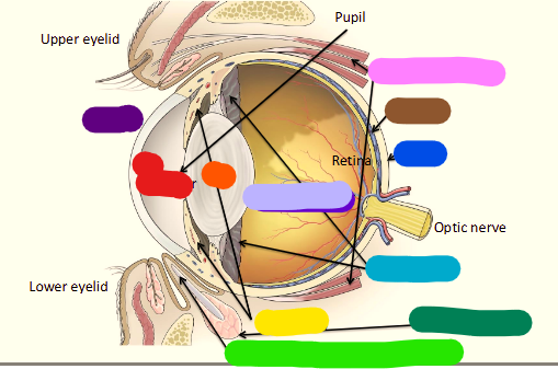
lable
purple - cornea
red - ant chamber
orange - lens
pink - extraocular muscles
brown - choroid
sclera - dark blue
light purple - virteous humor
light blue - ciliary body
yellow - iris (leaflet)
dark green - third eyelid gland
light green - third eyelid (nictitating membrane)
Anterior segments of the eye inculde
The lens and everything beofre it
inlcudes lens and ciliary body, thrid eyelid