NUR 212: Exam 2 (Revised)
 Call Kai
 Learn
Learn Practice Test
 Spaced Repetition
 Match
Match Flashcards
 Knowt Play
1/101

There's no tags or description
Looks like no tags are added yet.
Name | Mastery | Learn | Test | Matching | Spaced | Call with Kai | Chat |
|---|
No analytics yet
Send a link to your students to track their progress
102 Terms
RBC Value
4.2-6.1
Activated Partial Thromboplastin seconds (aPTT)
30-40
low = clotting fast
high = clotting slow
hemoglobin
F: 12-16
M: 14-18
Platelet Count
150,000-400,000
WBC
5,000-10,000
LDL Lab
more than 130 = bad
Troponin Lab
greater than 20 = bad
BNP
heart failure indicator
more than 100 = bad
Myocardial Ischemia
decreased blood flow to the heart → decrease oxygen
cause of angina
Coronary Atherosclerosis (CAD): Manifestation
plaques in vessels
decreased blood flow → decreased oxygen
SOB, angina radiating to the jaw and/or left arm
Coronary Atherosclerosis (CAD): Nursing Interventions
Low cholesterol diet → decrease built up
Lower LDL → with statin
Exercise
watch for ruptures → body will clot as a result → can lead to MI
Statin
Angina Treatment
Nitro and Rest
Oxygenate
Beta-Blocker to decrease workload
EKG & Troponin levels if not stable angina
Acute Coronary Syndrome: Manifestations
Angina not relieved by nitro or rest
emergent
SOB, N/V, Anxiety, increased HR and RR
unstable
no increase in troponin or abnormal EKG
NSTEMI
normal troponin, abnormal EKG
STEMI
ST elevation, elevated troponin
Acute Coronary Syndrome: Nursing Interventions
MONA: morphine, oxygen, nitro, aspirin
oxygenate
give heparin
IV access
EKG leads, troponin levels
prepare for Cath lab
thrombolytics (end in plase) → given if not long wait for cath
STEMI treatments
Thrombolytics → used if you cannot get the patient to CATH within 120 minutes → end in PLASE → stops the blockage in hopes to increase perfusion
Percutaneous Coronary Intervention: balloon inflated in artery (preferred method)
Coronary artery stent or Grafts (CABG) → bypasses blockage
Percutaneous Coronary Intervention: post-op
monitor bleeding (internal as well; don’t forget to check for bruising) and infection
lay patient flat for 4-6 hours
hydrate patient to get AKI dye out of the system used in surgery
Coronary artery stent or Grafts (CABG) : post-op
Watch BP!
low = decreased circulation may be due to donor site
high = suture can burst
Maintain stable fluids
Pain control
What is a sign of a heart valve issue?
murmur
Mitral Valve Prolapse: manifestations/diagnostic
minimal symptoms
mitral click
genetic
echo for diagnostics
Mitral Regurgitation: manifestations
Regurgitating back into the upper left chamber
Systolic murmur
Respiratory symptoms!
SOB, crackles, orthopnea
Left atrial hypertrophy
Mitral Stenosis: manifestations
Thickening of valve → harder for blood to get through → decreased cardiac output
Diastolic Murmur
A-FIB, Fatigue
Linked to strep and rheumatic fever
Aortic Regurgitations: manifestations
Regurgitating back into the lower left chamber
Diastolic Murmur
Visible, bounding pulses
pounding heart → patient will report
Aortic Stenosis: manifestations
Thickening of valve → harder for blood to get through → decreased cardiac output
Systolic Murmur
Syncope→ worsens with activity → think the blood is having trouble getting to the body
Low BP
Valve Disorders: nursing considerations and treatment
fix valve through replacement or suture
diuretics to reduce fluid volume and fluid restrictions if needed
beta-blocker to decreased workload
prevent infection → endocarditis risk
Dilated Cardiomyopathy: Manifestations and Diagnostic
Muscles are stretched thin → loss of pumping function and more blood
Confirmed by severely reduced ejection fraction (less than 50)
Too much fluid
JVD
Lung crackles
Edema
SOB
Hypertrophic Cardiomyopathy
Thick heart muscles → result in arrhythmia
Arrhythmias can be life-threatening
Upon exertion
SOB
Dizziness
Feeling faint/weak
Chest Pressure
Racing heart
genetic → seen in young athletes
Restrictive Cardiomyopathy: Manifestations
Stiff ventricles → the heart doesn’t expand as it should
High diastolic BP → diastolic complications (resting and filling)
Dyspnea, nonproductive cough, and chest pain
Cardiomyopathy Nursing Interventions
Cardiac assessment
oxygenate
Rest
low-sodium diet
CPR education
Beta-Blockers (LOL)
ACE (PRIL)
Diltiazem
LVAD that mechanically pumps the heart for you → improved pumping is the issue here
Infective Endocarditis: manifestations, risk, interventions
Infection of heart valves
Common in IV drug users; a risk for recent dental work, piercings, and tattoos that allow bacteria in
Fever, weight loss, Janeway lesions (red spots on palms/soles)
Long term IV Antibiotics (PICC line) and acetaminophen for fever
Retest after treatment
Myocarditis: Manifestations, Cause, Diagnostic
Inflammation of the heart muscle
Commonly caused by viral infection → vaccines for viral infection and prevention
No symptoms → if they occur, inflammation is severe
Flu-like symptoms
Biopsy to confirm
Pericarditis: Manifestations and Treatment
Access fluid surrounding heart → reducing expansion
PAIN! very painful
improves when leaning forward
Friction Rub, SOB, palpitations, cough
Lean forward to decrease pain, Pericardiocentesis (ACUTE), Pericardial window (Chronic)
Cardiac Tamponade: Manifestation and Treatment
Pericarditis complication
Rapid buildup of fluid compressing the heart
Collapsing cardiac output
Pericardiocentesis → needle to remove fluid
Diminished heart sounds, JVD, decreased arterial pressure
Left-Sided vs. Right-Sided Heart Failure
Left-sided HF → backs up into the lungs
SOB, crackles, orthpnea
Ride-sides HF → backs up into the body
JVD, edema, weight gain
Heart Failure Nursing Considerations
Maintain oxygen status
Low sodium
Daily weight
Fluid restriction
Check HR before meds
Meds: diaretics, vasodilators, beta blockers, RAAS
Nitro
for Angina
3 times every 5 minutes
causes headache
check bp and hr before admin
Statin
decreases LDL
CAD treatment
Thrombolytics
end in PLASE
clot buster
given if STEMI patient cannot make it to CATH in time
Acute Pulmonary Edema: Manifestations
acute fluid buildup in lungs
“I feel like I’m drowning”
Pink, frothy sputum
DVT: manifestation, risk, diagnostic
blood clot in a deep vein
commonly seen in the lower legs
redness, swelling
risk: not moving around (bed rest), long drives, long plane rides
USE DOPPLER FOR DIAGNOSIS
Hemorrhage: manifestations
feeling tired
altered mental status
bad headache
syncope
orthostatic hypotension
High HR really low BP
Hemorrhage: nursing considerations
order CT stat to ensure no intracranial bleed
Give fluids! Oxygen, whole blood, apply pressure to the bleeding if you can see the source
Assess LOC
Anemia symptoms
fatigue, weakness
pale
Tachy
SOB
Dizziness → syncope
cold intolerance
headaches
Iron Deficiency Anemia: Manifestations and Treatment
low RBC production due to low iron (intake or blood loss)
RBCs carry oxygen
SOB, fatigue, pale
Low hemoglobin, ferritin, and small RBCs
IV Iron (Ferrous Sulfate), Oral Iron (black stool), increase iron diet (spinach, meat, eggs), find bleeding source
Vitamin B12 Deficiency (Pernicious Anemia): Manifestations and Treatment
Low B12 and/or no intrinsic factor
smooth, beefy-red tongue and neurological numbness
recent gastric by pass surgery → wont have intrinsic factor
SUBQ B12 (if no intrinsic factor), B12 Diet (eggs, meat, fish)
Sickle Cell Disease: Manifestations
sickle shape RBC → stick together → clotting
PAINFUL
Swelling
low hemoglobin → very low baseline
Sickle Cell: Interventions
reduce infection risk → huge risk
avoid dehydration, stress, extreme temps, and intense physical exertion
IV fluids, supplemental oxygen, and aggressive IV opioid pain control
Long-term maintenance uses hydroxyurea
Thalassemia: Manifestations and Treatments
RBC are shaped oddly → body attacks them
These patients will have frequent transfusion → iron overload risk →iron chelation therapy to get extra iron off
chronic fatigue, weakness, developmental delays, and splenomegaly (enlarged spleen)
Heparin-Induced Thrombocytopenia: manifestations and nursing considerations
Response to Heparin → body forms small clots everywhere → platelets working everywhere → increase clotting risk still since they are being used up
Stop heparin
Immune Thrombocytopenic Purpura: Manifestations and Treatment
body attacks platelets → cannot get transfusion → body will destroy them
very low platelet count
Easy bruising (ecchymosis), petechiae on the trunk and extremities, epistaxis (nosebleeds), and heavy menses
Corticosteroids to suppress the immune system
huge bleeding risk → monitor this
Hemophilia: Manifestations and Considerations/Treatment
long clotting time due to a missing clotting factor 5
Delayed, prolonged, or persistent bleeding following minor cuts, dental work, or trauma → often seen in kids
IV factor for treatment
DO NOT USE HEPARIN (blood thinners on these patients)
head injury? get CT STAT due to bleeding riskl
Disseminated Intravascular Coagulation (DIC): manifestations and treatment
life-threatening
The body creates clots, all of which consume platelets → bleeding risk → causes hemorrhage
Spontaneous bleeding from multiple sites simultaneously (IV insertion sites, mucosal gums, nose, urinary catheter hematuria, petechiae)
Low platelets + elevated D-dimer + prolonged clotting time
Septic patient, pregnant patient
Heparin therapy and blood transfusion
Ferrous Sulfate
IV Iron
anemia
Steroids: Hemo treatment
ITP
They cannot get transfusion
Reduce immune system reaction
Hydrozyura
long term sickle cell treatment
4 C’s (Burns)
cool it → tap water or saline
clean it → mild soap
cover it → antimicrobial ointment and absorbent dressing
comfort it → pain meds
Classification of burns 1-6th degree
first → epidermis only (like a sunburn)
second → entire epidermis and top of dermis → BLISTER and WET
third → complete damage of epidermis and deep into dermis → usually white/yellow and dry → may require skin grafting
All the way through epidermis and dermis (requires skin grafting)'
white/black/brown, dry and leathery, nonblanchable, no pain
fourth → same as third but goes in subcutaneous tissue
fifth → same as third but goes to muscle
sixth → same as third but it goes into bone
Rule of 9s
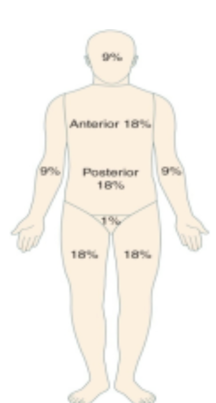
Effects of burns
Stop GI and Kidney function
Infection risk
Hypothermia
Increased potassium, low sodium
cardiovascular and respiratory effects → increased HR, plasma leakage (hypovelmia), decreased cardiac output, low RR status
compartment syndrome
burn causing so much swelling it pushes on nerves and blood vessels
Escharotomy or Fasciotomy
Upper airway VS lower airway (burns)
upper → edema causing reduced oxygenation
lower → carbon monoxide → pink face → give them 100 oxygen stat → pulse ox will be normal
What to do for burns?
NG tube and Foley
IV pain meds → GI not working
Oxygen
Fluids
wash burn if chemical
EKG if electrical
Parkland fluid resuscitation formula burns
2mL LR patients weight (Kg) * TBSA burn (rule of 9s)
give first half in the first 8 HOURS
give second half over the next 16 HOURS
FROM BURN! if you see patient 1 hour after burn you will only have 7 hours to get first half in
Osteoarthritis: manifestations and treatment
Damage to cartilage from use
Asymmetrical joint pain
Worsens with activity, improves with rest
Stiffness, deformities
Pain meds, NSAIDS, corticosteroid injection (limited), replacements, mobility aids, rest
Rheumatoid Arthritis (RA): manifestations and treatment
Chronic autoimmune inflammation of cartilage
Symmetric affect
Does not improve with rest
Swan-neck hands
Morning stiffness
NSAID, Biologicals, Corticosteroids (for exacerbation)

Lupus: Manifestations and Treatment
butterfly rash + photosensitivity (UV light trigger) and multi-system organ involvement
remission and flare-ups
Managed with hydroxychloroquine (Plaquenil), topical steroids, avoiding sun exposure, and decreasing stress
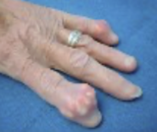
Gout: manifestations and treatment
buildup of urate crystals in joint spaces
PAINFUL!
swelling, redness, warmth
once you have what is in the photo → it will not go away
Colchicine (acute), Alloprinol (chronic), Low-purine diet (no meats, selfish, alcohol)
Fibromyalgia: manifestations
chronic central pain and stiffness
brain frog and GI issues
HIV / AIDS: Manifestations and Treatment
Acute: Flu-like symptoms
Progression: swollen lymph nodes, severe weight loss, chronic diarrhea, and opportunistic diseases
CD4 below 200 = AIDS
Viral load below 75 → low transmission risk
Antiretroviral therapy, prep for prevention
Flu vs common cold: flu interventions
Flu: Fever, aches, chills, tiredness, sudden onset → severe muscle aches
spreads by droplets
antiviral within 48 hours after symptoms (Tamiflu) → or end in VIR
vaccine
Sinusitis: manifestations and treatment
inflammation of the sinus cavity
facial pain/pressure, nasal congestion, postnasal drip, headache, fatigue
Differentiated from seasonal allergies by the presence of fever, bad breath, and thick green or yellow nasal mucus
Steroid nasal sprays, antibiotics, moisturize nasal passages, avoid triggers, warm compress
Meningitis: Manifestations
infection of the meninges
High fever, severe throbbing headache, nuchal rigidity (stiff neck), photophobia, rash, and confusion
Positive Kernig sign (pain when extending the knee) and Positive Brudzinski sign (hip/knee flexes when neck is flexed)
Meningitis: Nursing Considerations/Interventions
high-dose steroid
reduce fever → Acetaminophen
Lumbar puncture to test CSF fluid → cloudy if bacterial cause
Requires droplet isolation precautions for at least 24 hours post-antibiotic initiation
Herpes Zoster (Shingles)
reaction to chickenpox
Severe pain, burning, and vesicular rash
give antiviral agents → end in vir
Fungal Skin Infections (Tinea / Ringworm)
Superficial fungal infection (dermatophytosis) creating characteristic circular lesions
Red, itchy, ring-shaped rash with raised borders and a clear center
Can occur on the scalp, body, groin, foot, and nails/toenails
Biologics
RA treatment
Colchicine vs Allopurinol
Colchicine → acute gout
Allopurinol → chronic gout
Hydroxychloroquine
lupus treatment
Antivirals
end in VIR
herpes, HIV, Flu
Osletmivir (Tamiflu)
antiviral
take within 48 hours of symptoms
for flu
IV opoids
burns
sickle cell
What does the liver primarily do?
Metabolism!
Hepatic Dysfunction : Manifestations
jaundice → high bilirubin level above 2
Ascites → fluid buildup in the abdominal cavity
Varices → swollen veins
Itching
edema
Hepatic Cirrhosis & Portal Hypertension
Long-term liver disease → hard scar tissue blocking blood flow through the liver → causing high pressure in veins (Portal Hypertension)
excess fluid around → fluid overload
Ascites
esophageal varices
liver and spleen enlargement
edema
Ascites: manifestations
fluid in the peritoneal cavity caused by portal hypertension
Abdominal distension, rapid weight gain, abdominal striae, distended abdominal veins, umbilical hernia, and shortness of breath
Ascities: Nursing Intervention
paracentesis → needle to drain fluid
TIPS (Transjugular Intrahepatic Portosystemic Shunt) → helps prevent buildup
IV salt-poor albumin → pulls fluid out
low-sodium diet
diuretics
daily weights
Esophageal Varices: manifestations
fragile, dilated veins in the esophagus due to poor hematic function
prone to rupture → life-threatening hemorrhage
Hematemesis (vomiting blood), melena (dark, tarry stools), shock (hypotension, tachycardia), and general physical deterioration
Esophageal Varices: Nursing Considerations
oxygen
fluids
stop bleeding
IV Octreotide → decrease bleeding
Sengstaken-Blakemore Tube → emergent, not preferred as the balloon can restrict airflow
Vasopressin
Endoscopic Sclerotherapy
Esophageal Banding
Hepatic Encephalopathy: Manifestations
nervous system dysfunction → result in hepatic dysfunction → cannot filter toxic ammonia
Mental status change, altered LOC, motor disturbances, progression to coma
Asterixis (flapping tremor of the hands when wrists are extended) and Apraxia (deterioration of handwriting or inability to draw simple figures)
Hepatic Encephalopathy: Treatment
Lactulose → poop out ammonia → very strong laxitive
LIMIT PROTEIN AND SALT
Viral Hepatitis (Hep A, B, C, D, E)
flu-like symptoms, jaundice, dark tea-colored urine, clay-colored stools
Transmission:
Hep A&E → facal-oral
Hep B&C → blood, mucus membrane, sexual contact
Hep D → only if you have B
Vaccine for A&B → only ones that have vaccines but Hep B stops you from getting D
Liver Biopsy (Key Diagnostic Procedure): What should the patient do?
exhale and hold your breath for at least 10 seconds → so your lungs are small and the needle doesn’t puncture
Goes on the RUQ
No coughing or straining
Lie on your right side afterward!
Acute Kidney Injury and Chronic: Manifestations
Oliguria → low urine production
Fluid overload
Elevated Creatinine and BUN
Fluid & Electrolyte Imbalances
high potassium
Anemia
Polycystic Kidney Disease (PKD): Manifestations
Cyst on the kidney that can compress it
Flank or abdominal pain, hematuria, hypertension, and palpable enlarged kidneys
can travel to brain and heart
Kidney diagnostics: eGFT
eGFT → shows damage
want it above 90
Below 15 is really bad → lost 90% kidney function
Treatments for Renal Dysfunction
Sodium Polystyrene (Kayexalate) → removes potassium
Epoetin Alfa & Ferrous Sulfate → anemia and low RBC
Diuretics (Furosemide / Bumetanide)
Dialysis
transplant
Low sodium, fluids protein diet
Hemodialysis (HD)
Acts as the kidney to get waste out
Vascular Access Types: Arteriovenous (AV) Fistula
should fill a thrill
hearing bruit
do not take BP, blood drawl or anything from that arm
Peritoneal Dialysis (PD)
In abdomen
Warm dialysate fluid is instilled into the abdominal cavity via a Tenckhoff catheter (Infuse → dwell → drain)
What comes in must come out
Hepatic (Liver) Dysfunction Lab Profile
low
Albumin
high
Ammonia: greater than 45
AST & ALT: greater than 34
PT time → bleeding risk
Renal (Kidney) Dysfunction Lab Profile
Low
H&H
Calcium
High
BUN: greater than 20
Creatinine: greater than 1.3
Potassium: greater than 5
Phosphorus: greater than 4.5