Lecture 13 - ER and Proteins
 Call Kai
 Learn
 Practice Test
 Spaced Repetition
 Match
 Flashcards
 Knowt Play
1/16

There's no tags or description
Looks like no tags are added yet.
Name | Mastery | Learn | Test | Matching | Spaced | Call with Kai | Chat |
|---|
No analytics yet
Send a link to your students to track their progress
17 Terms
Describe the endoplasmic reticulum. What are its two parts?What are cisternae?
Networks of membrane-enclosed tubes and flattened sacks called cisternae
Largest organelle in most eukaryotic cells
The ER has two contiguous domains that perform different functions:
Rough ER - studded with ribosomes, more flattened sheet-like
Smooth ER - no ribosomes, more tube like
What is made in the smooth endoplasmic reticulum (SER)
Major site of phospholipid and cholesterol synthesis
Abudant in cells with active lipid metabolism
Steroid hormones are synthesized from cholesterol in the ER
Some are converted to glycolipids or sphingomyelin in the Golgi apparatus
Where are lipids made and why? What are flipases?
Most lipids are synthesized in the membrane of the ER
Cannot be made in an aqueous environment because they are highly hydrophobic
More specifically they are made in the cytosolic portion of the ER bilayer
This would lead to only the outer layer growing
Flipases are enzymes that help phospholipids “flip” from the cytosolic layer to the lumenal layer

Describe the rough endoplasmic reticulum functions
Protein synthesis for membrane-bound, lumenal proteins or proteins that need to leave the cell
Folding and ensuring inocrrectly folded proteins are removed
Packaging of exported proteins in vesicles
Addition of glycosylation and glycolipid anchors
Glycosylation carries information (like a label on a package)
What is in the cytosol?
Contiguous with the inside of the cell
Nucleus
Cytosol
What is the lumen?
Contiguous with the outside of the cell
ER
Golgi
Inside of vesicles
Outside of cell membrane

Describe protein synthesis in the ER
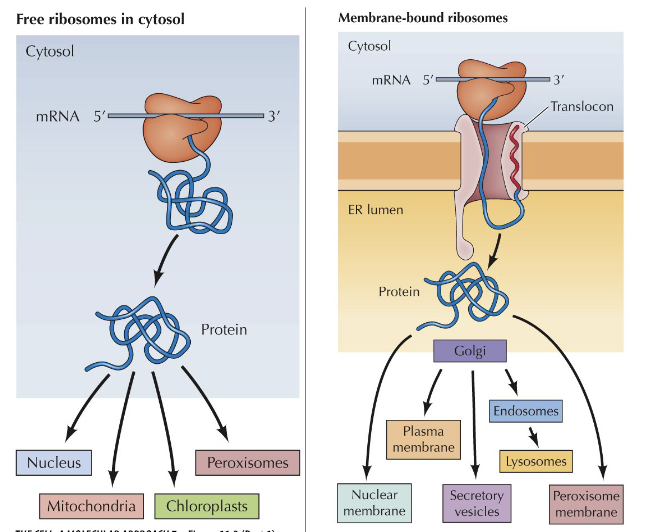
Proteins are made in one location depending on where they will end up
Ribosomes can be free or membrane bound
Domains of membrane proteins that are exposed on the cell surface correspond to regions of polypeptide chains that are exposed to the ER and golgi lumen
What are the two different methods of protein translation?
Translation on Free Ribosomes
Proteins are free to move nucleus and cytosol. Sometimes also mitochondria, chloroplasts, and peroxisomes
Translation on ER-bound ribosomes
Proteins will end up inside the ER, so can only go where it is continuous with the ER
How do proteins get in the ER?
Contranslation Translocation - ribosomes bind to ER while they are translating the protein, so it actively dumps protein into the ER lumen
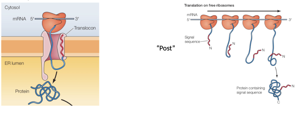
Post translational relocation - Free ribosomes translate proteins, which are then transported to ER lumen afterwards
How do free ribosomes know when to associate with the ER membrane to complete cotranslation relocation?
It’s in the mRNA they are translating
As the amino acid polypeptide chain is being translated, it will contain a segment called the “signaling sequence” that signals this protein needs to be translated with the ER, and so the ribosome must relocate

What are the different steps of cotranslation?
Step 1 - A signal recognition particle (SRP) binds to the signal sequence on the polypeptide chain
Step 2 - The SRP protein binds to an SRP receptor that is attached to a translocon
Step 3 - The ribosome binds to the translocon, which causes the SRP protein to unbind to the SRP receptor. Ribosomes binding to the translocon also causes the translocon to open
Step 4 - Translation resumes. Signal peptidase cleaves the signal sequence off the growing polypeptide chain
Step 5 - Continued translating causes the growing polypeptide chain to grow into the lumen of the ER
Step 6 - The finished product is “spit out” into the lumen of the ER

Now that the protein is in the ER lumen, what happens next?
Folding and quality control
Inside the lumen there are chaperone proteins, like “BiP” that bind to the newly translated polypeptide chain and aid in the folding process. These chaperones do not unbind until the protein is properly folded. BiP stays in the ER lumen

What is protein glycosylation - a type of protein modification. What is it function?
Inside the ER we can add sugars to proteins - protein glycosylation
This will happen on asparagine residues while translation is happening
Glycosylation:
helps prevent protein aggregation
Promotes protein folding
Helps with protein sorting

What are lipid anchors - a type of protein modification
Inside the ER, we can add lipids anchors to newly formed proteins
Protein is held in the membrane by a C-terminal hydrophobic sequence
Anchors are assembled in the ER
C-terminal sequence is cleaved and exchanged for the lipid anchor
What prevents/help proteins from misfolding?
Chaperones - they dont just help proteins fold correctly, but they also detect when a protein is misfolded
A chaperone sends a misfolded protein to the ER membrane, where it:
Binds to a ubiquitin ligase complex
Gets tagged with ubiquitin
And then sent off into cytosol to be broken down by a Proteasome
This process is known as ERAD - ER-Associated Degradation

What is the unfolded protein response
If too many unfolded proteins accumulate in the ER, they will bind to receptors on the ER membrane
These receptors iniate pathways that lead to increased production of:
Increased production of chaperones
Increased production of proteasomes
Limit the number of new unfolded proteins entering the ER
If cells can’t do this apoptosis will occur