K103 Exam 2- IUI Yost
 Call Kai
 Learn
Learn Practice Test
 Spaced Repetition
 Match
Match Flashcards
 Knowt Play
1/321

There's no tags or description
Looks like no tags are added yet.
Name | Mastery | Learn | Test | Matching | Spaced | Call with Kai | Chat |
|---|
No analytics yet
Send a link to your students to track their progress
322 Terms
Tissue
Cells
- Closely associated, similar structure and function
- Characteristic shapes and arrangement in each tissue
Function
- Specialized to perform specific function or group of functions
Organs
- Closely associated tissues that carry out specific function
Systems
- Closely associated organs that carry out specific functions
- Humans have 11 organ systems
Animal Tissues (4 major types)
1. Epithelial
2. Connective
3. Muscle
4. Nervous tissue
Epithelial Tissue Characteristics
Epithelial cells are tightly packed
- Continuous sheet
- Attached to basement membrane
- Helps with the integrity of the tissue
Epithelial Surfaces serve as a barrier (exposed to the environment)
- Everything that goes in or out of the body must cross it
- Ex) In: Food and water, Out: waste, urine.
- Regulates exchange between body and environment
Epithelial Functions
- Protection, absorption, secretion, sensation
Epithelial Cell Contacts (Tight Junctions)
- Anything that goes thru between the cells is being highly regulated
Epithelial Cell Contacts (Desmosomes)
- A specialized structure that helps hold cells tightly together, they act as strong spot welds between neighboring cells.
- Provide mechanical strength, prevent tissues from being pulled apart.
- Usually used for skin
Epithelial Cell Contacts (Gap Junctions)
- Specialized intercellular channels that directly connect the cytoplasm of adjacent animal cells, allowing for rapid passive exchange of ions, small molecules, and second messengers.
Epithelial Polarity
- Means the epithelial cells are structurally and functionally asymmetric (different parts of the same cell have different structures and jobs).
Types of Differences:
- Asymmetry ( because they have three distinct surfaces the apical (inside of an organ), Basolateral (includes lateral and basal sides), and the basal membrane (attaches to the basement membrane).
- Distribution of organelles (planar polarity- cells are aligned in the same direction across the tissue plane)
- Electrical Charge Distribution (neurons)
- Differences in membrane (apical and basolateral membranes)
Epithelial Circulation and Intervention
- Avascular (no blood vessels to get its own blood supply, instead it uses oxygen and nutrients from the blood vessels in the connective tissue underneath it)
- Innervated (it has nerve supply, epithelial tissue contains or is closely associated with nerve endings, that is why our skin can feel pain, touch, temperature, and pressure).
Epithelial tissue doesn't have blood vessels, but does have nerves.
Epithelial tissue can be regenerated if damaged
Epithelial Glands
A group of epithelial cells that are specialized to make and release a substance.
Epithelial tissue lines organs, and covers surfaces like skin. But some of these cells don't just cover surfaces they change their function, and become a gland (a structure that produces and secretes something). For example a sweat gland.
Epithelial Exocrine Glands
- Release their secretions onto a surface or into a body cavity, usually use ducts (tubes). Examples). Sweat Glands (release sweat onto skin), Salivary Glands (release saliva into mouth).
EXO: Exit to Surface.
Epithelial Endocrine Glands
- Release hormones directly into the blood or interstitial fluid. They do not use ducts. Examples). Thyroid glands and Adrenal glands.
Epithelial Membranes
protective tissue layer made of epithelial cells plus connective tissue that covers or lines parts of the body.
Epithelial Mucous Membrane
- Lines body cavities that open to the outside
- Produces mucus to keep surfaces moist (THICK)
- Found in Digestive system, Respiratory system, urinary system, and reproductive system.
- Goblet Cells secrete mucous onto the surface (prevents drying and lubricates)
Epithelial Serous Membrane
- Lines body cavities that do not open to the outside (lines internal body cavities).
- Produce serous fluid to reduce friction between organs (THIN)
- Found around heart, lungs, and abdominal organs
- Ex). Pleural sac- This surrounds each lung, and contains a small amount of fluid, this reduces friction during breathing, and allows lungs to expand smoothly. Pericardial Sac- This cavity surrounds the heart and helps reduce friction as the heart beats, and protects the heart.
Simple Squamous Epithelium Cells
- A type of epithelial tissue made of simple (one single layer of cells), and squamous (thin, flat, scale-like cells).
- Because it is thin, substances can pass through it very quickly.
Simple Squamous Epithelium Cells Function + Example Locations
Function:
- Rapid diffusion because it is thin
- Processes: absorption, filtration, and secretion.
Example Locations:
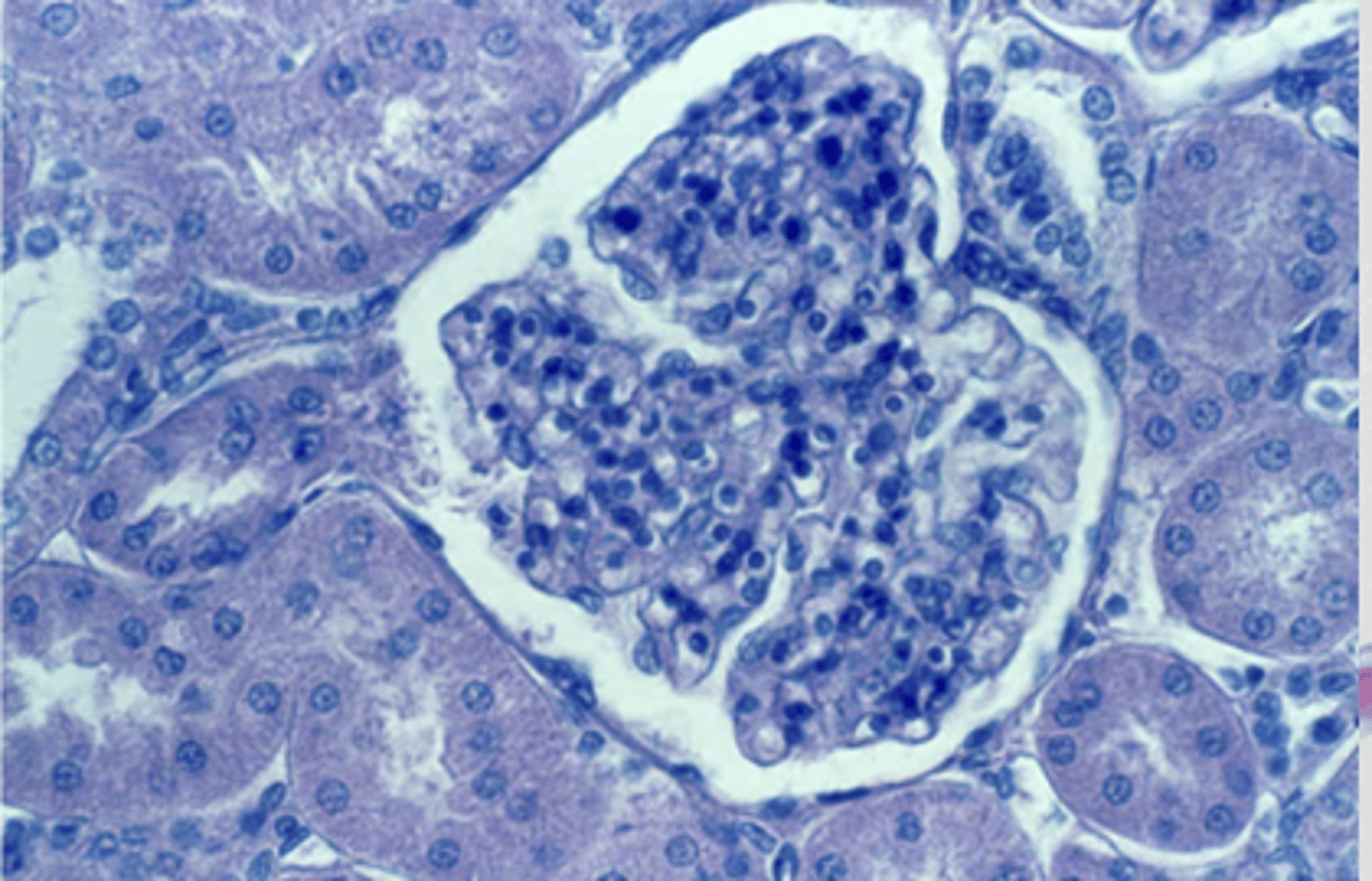
- Kidney Glomerulus (blood is filtered to make urine)
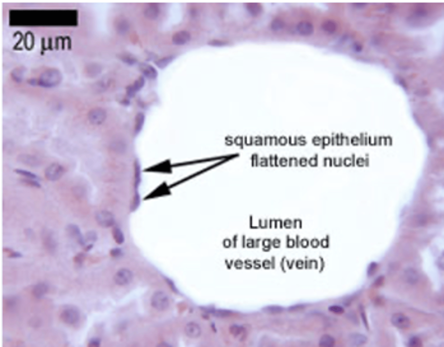
- Blood vessels (allows easy exchange between blood and tissues)
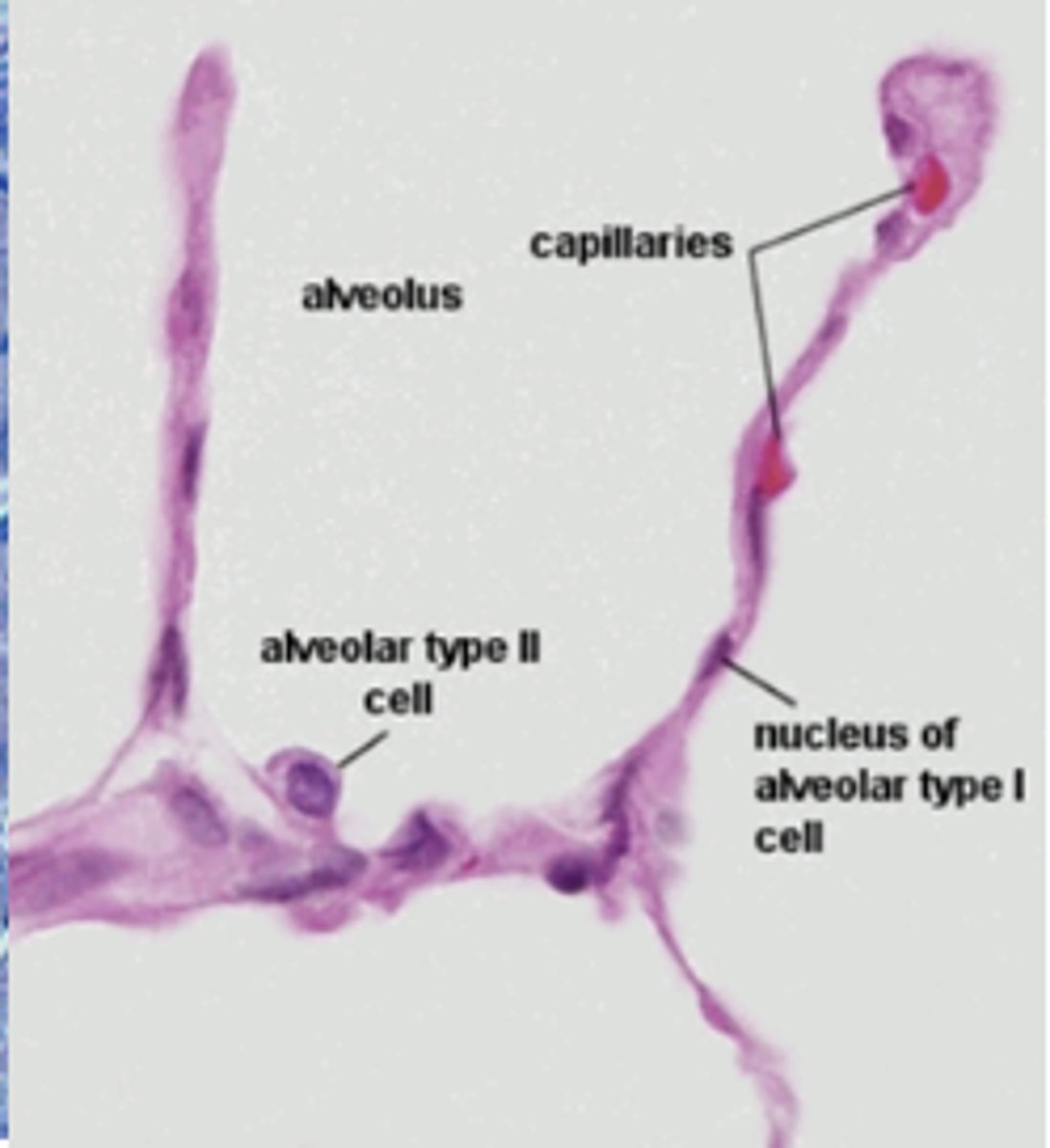
- Lung Alveoli (Oxygen moves from air, into blood, and carbon dioxide moves out).
*needs to be thin so these functions happen quickly*
Glomerulus tissue
simple squamous epithelium
Lung (alveolus) Tissue
simple squamous epithelium
Blood vessel
simple squamous epithelium
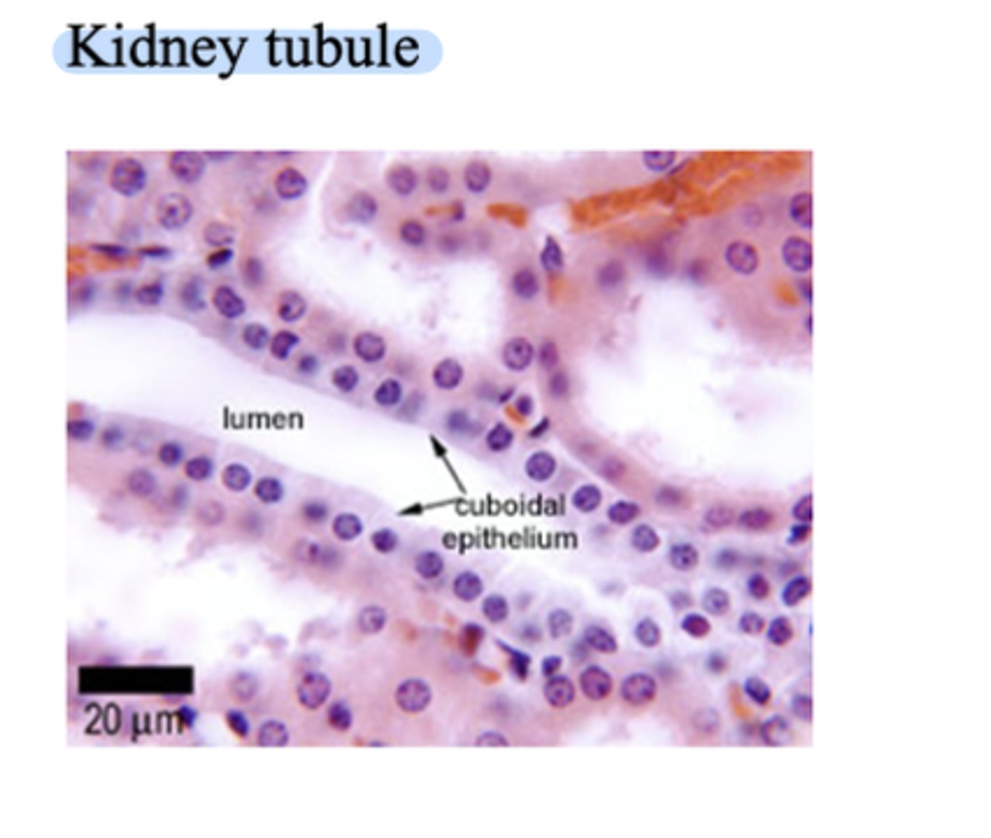
Simple Cuboidal Epithelium Cells
- A type of epithelial tissue made up of a single layer of cube-shaped cells (cells that are about as tall as they are wide).
- Function: Secretion and Absorption
- Location: Often lines small tubes and ducts, like kidney tubules. In the kidney tubules it absorbs water and nutrients in the kidneys, and secretes hormones in glands.
Simple Columnar Epithelium Cells
- A tissue made of one single layer of tall, rectangular (column-shaped) cells that line certain organs.
- Locations: Digestive Tract, Uterus, and small airways of the lungs.
- Function: Absorption, secretion, and movement (if ciliated)
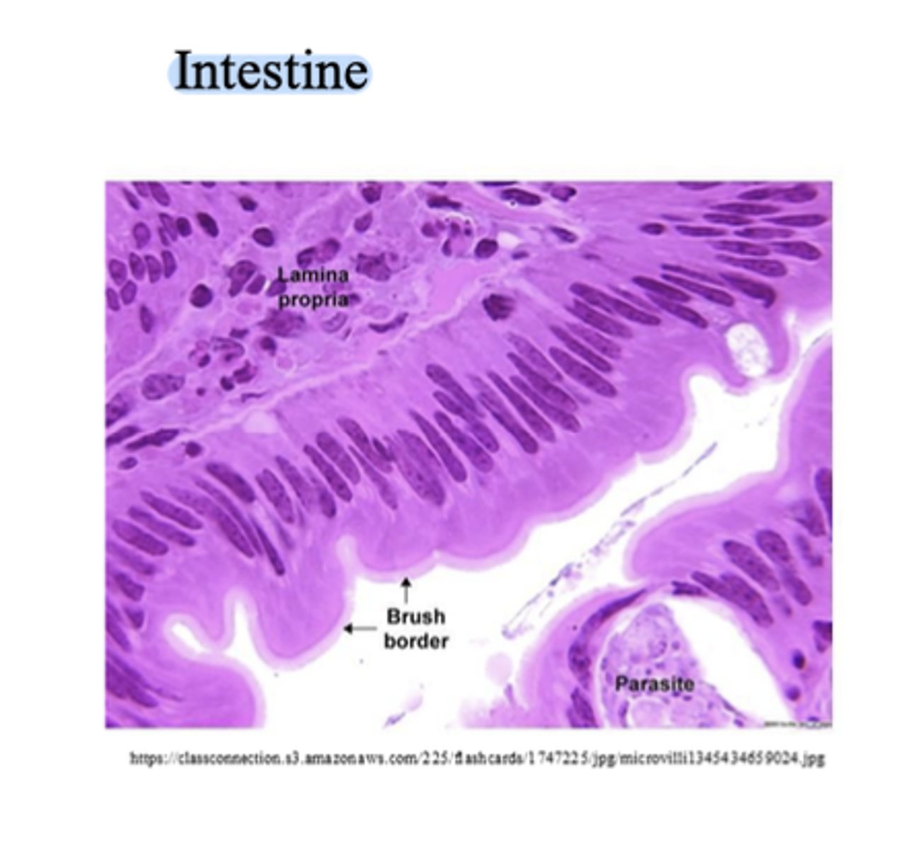
Two Types of Simple Columnar Epithelium Cells
- Non-ciliated: No cilia, Often contains goblet cells (mucus-secreting cells), found in stomach, small and large intestine. Non-ciliated = absorption and secretion.
- Ciliated: Has cilia, cilia move in a coordinated way they help move substances along the surface. Found in uterus, fallopian tube, and small airways. Function example: Move egg toward uterus Ciliated = movement.
stratified squamous epithelium
- Stratified = many layers of cells
- Squamous = flat, scale-like cells
- Mainly found in areas that need strong protection, (example: epidermis)
- The bottom layer, the base, has living cells that divide, as the cells move upward they get flatter, become older, and makes the top layer non-living cells.
Function: protection
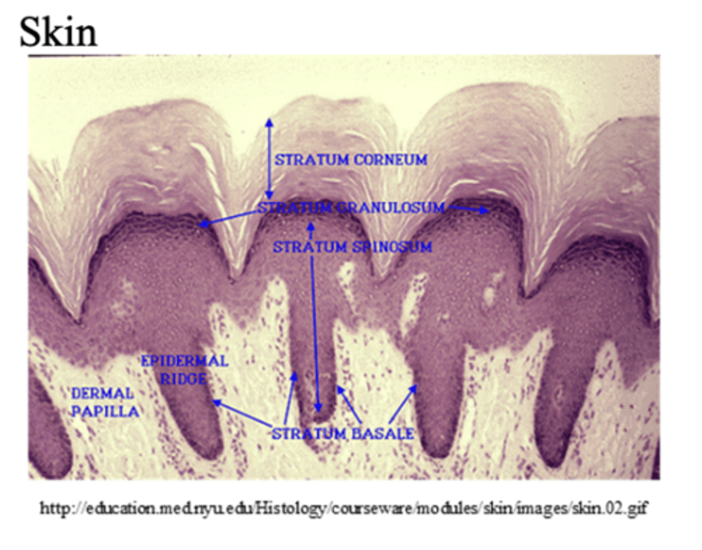
Two Types of Stratified Squamous Epithelium
- Keratinized Stratified Squamous Epithelium: Has keratin, a tough protein, inside cells. The surface cells are non-living and dry, this makes the surface waterproof. EXAMPLE: epidermis of skin.
Keratinized = dry surface (skin)
- Nonkeratinized Stratified Squamous Epithelium: No keratin layer, and surface cells are alive and moist. EXAMPLE: Oral cavity or esophagus. Function to protect against friction and keep surface moist.
Nonkeratinized = wet/ moist surfaces
Stratified Cuboidal Epithelium
- Stratified = multiple layers
- Cuboidal= cube-shaped cells
- Function: secretion and protection. (Help ducts not collapse)
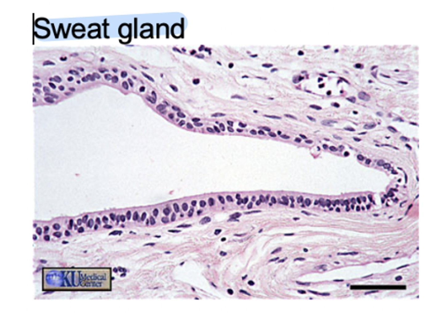
- Location: Mainly in the ducts of larger glands, such as salivary gland, sweat gland, mammary gland.
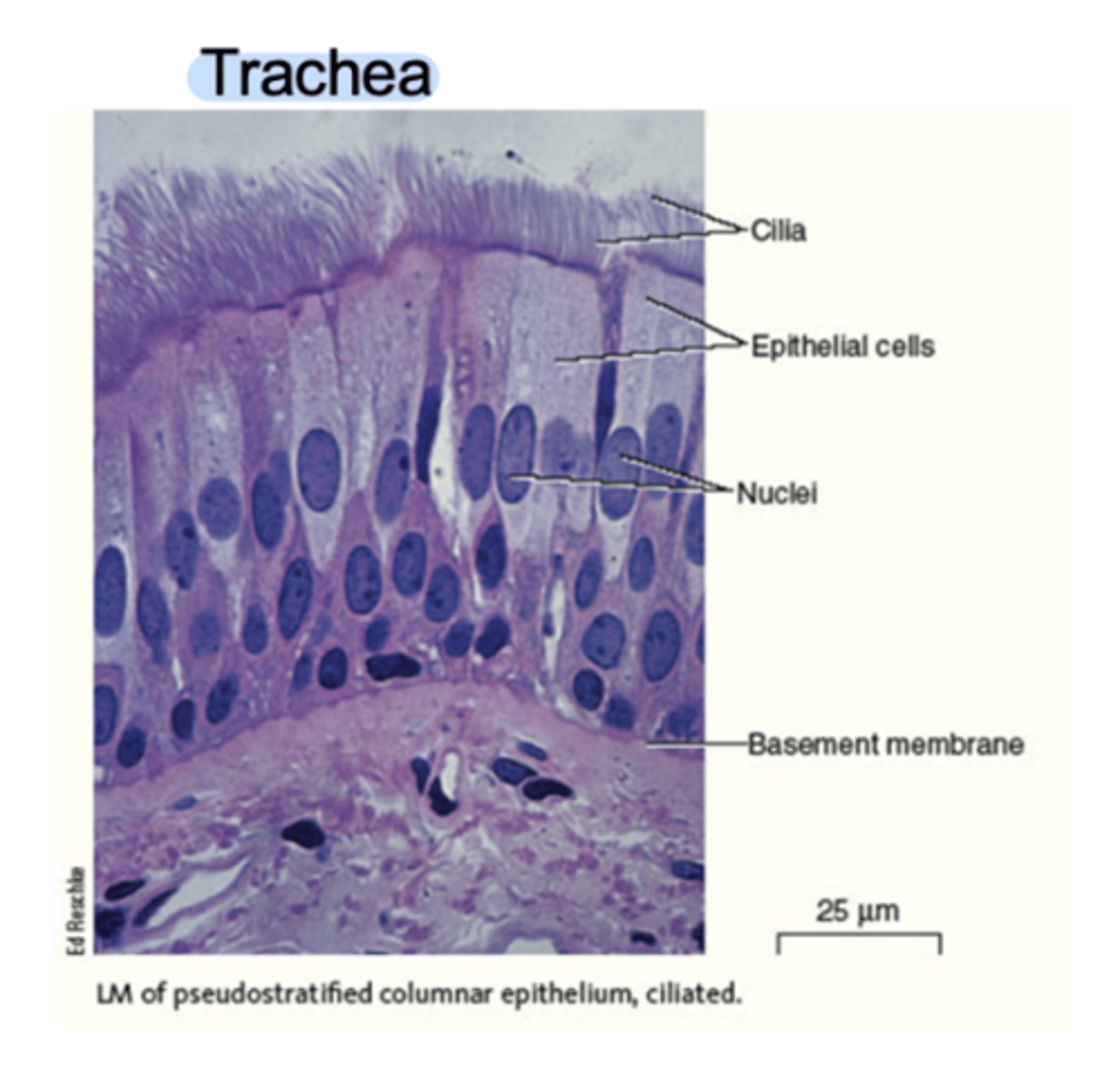
Pseudostratified Epithelium
- Looks like it has multiple layers, but it is actually only one layer of cells (pseudo= false)
- It looks layered because all cells are attached to the basement membrane, but not all the cells reach the surface, so the nuclei are at different heights, which make it look like there are several layers.
Two types of Pseudostratified Epithelium
- Ciliated: Contains cilia on the apical surface, and often contains goblet cells (mucus-secreting cells). Function to secrete mucus, and propulsion (movement of mucus and trapped particles by cilia). Location: Lining of trachea, lining of bronchi.
- Non-ciliated: Lacks cilia, Function: Absorption and Secretion. Location: Epididymis, and parts of the urethra (in males).
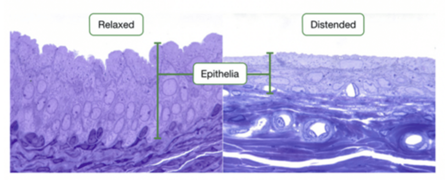
Unique Epithelium
- A special type of stratified epithelium that can change shape when stretched (distension) and return to its original form due to elasticity.
- Cuboidal or columnar shape at the base, and when it stretches it becomes a squamous shape. (It is the same cell, just can become a different type) because shape determine what type of cell it is.
- Location: Bladder, uterus, and upper urethra (allows us to fill bladder).
Connective Tissue
- One of the four main tissue types in the body. Its main role is to connect, support, protect, Insulate, and bind other tissues together.
- Distensible: Can expand in all directions, example urinary bladder.
- Extensible: Can be pulled apart linearly. Examples: Tendons.
Connective Tissue Structure
- Has a few cells
- has a large amount of extracellular (intercellular- in between the cells) substance (Extracellular means outside of the cells), called the matrix. Because connective tissue has few cells, and large space between them, and this large space is filled with matrix (which is both outside and inbetween the cells).
MATRIX (Extracellular Substance): It is secreted by connective tissue and determine the tissue's structure and properties. It has two main components:
1. Ground Substance (fluid, gel-like, or solid material)
2. Fibers (Threadlike proteins)
Examples of Matrix Types
Fluid → blood, vitreous humor of the eye
Gel-like → cartilage
Solid/mineralized → calcified bone matrix
Types of Connective Tissue Fibers
1. Collagen Fibers: Thick, tough, fibrous protein strands, Very strong, Remain intact when tissue is stretched. Function: Provide strength and resistance to stress (prevent tearing).
2. Elastic FIbers: Made of the protein elastin, Thin and branching, Form networks. Function: Allow structures to stretch and return to their original size and shape. These are found in places that expand and recoil, like large arteries.
3. Reticular Fibers: Thin, branched collagen fibers, Contain glycoproteins
Form a delicate supporting network (meshwork) for joining other tissues or holding organs together. Function: Provide structural support for soft organs and help hold tissues together.
Loose Connective Tissues (Areolar Tissue)
Structure: Elastic and collagen fibers (unorganized and run in all directions, making the tissue flexible but still strong). Semifluid matric (the gel-like substance between the cells, it acts as a reservoir for fluid and salts).
Function: Cushions organs, and supports surrounding tissues, allowing for independent movement without tearing.
Location: Subcutaneous layer (under the skin), and around organs, nerves, and blood vessels.
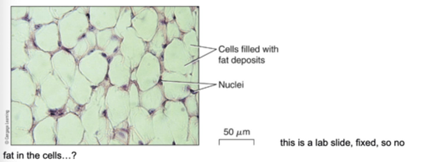
Loose connective proper (Adipose Connective tissue)
- Have a large middle vacuole, which is filled with fat (lipids) so the cells look hollow.
- Structure: Large vacuole which stores fat, very little extracellular matrix because the fat takes up most of the space, contains reticular fibers (thin support fibers).
- Function: Cushion organs, support, insulate, and energy storage.
- Location: Under the skin, around many organs.
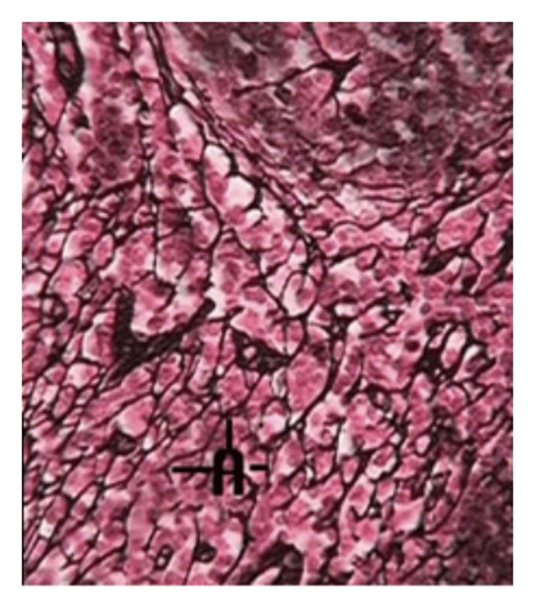
Reticular Connective Tissue
- A network of thin fibers (reticular fibers)
- Forms a mesh-like internal support system
- Function: supports and holds organs together, creates a framework inside organs.
- Location: Liver, spleen, and lymph nodes.
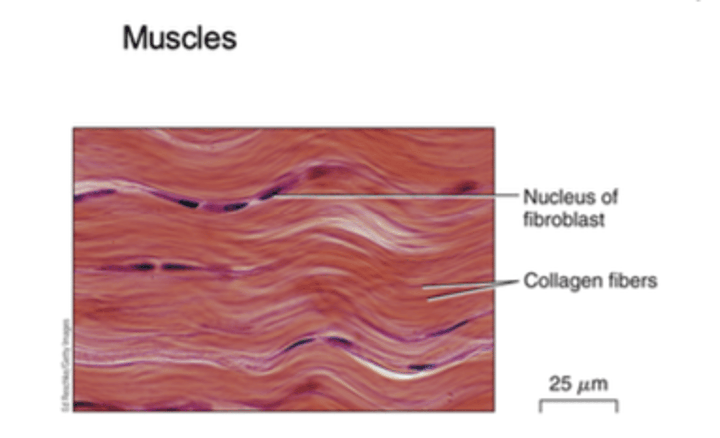
Dense Connective Tissues - Dense Regular Connective Tissue
- Made of mostly collagen fibers, and the fibers run parallel all in the same direction, very strong and built to handle force in one direction.
- Function: Support and transmission of force (it transfers the force when muscles contract).
- Location: Tendons (connect muscle to bone)
- Ligaments: Connect bone to bone
- Fascia: connective tissue that separates muscles
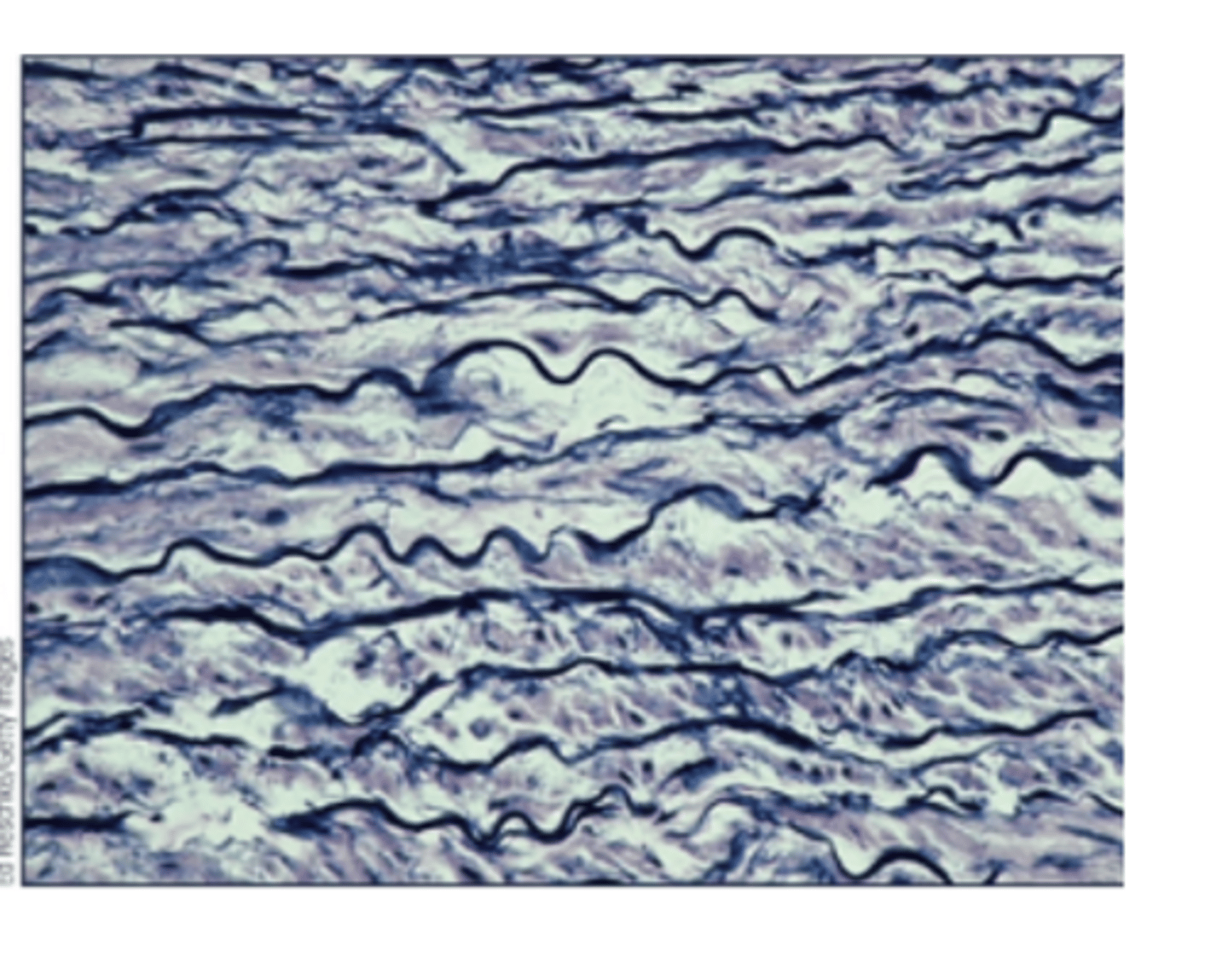
Elastic Connective Tissue
- Made of mostly elastic fibers, fibers run parallel, and designed to stretch and snap back.
- Function: Allows structures to return to original size and shape following expansion, (extension or distension).
- Location: Lungs and walls of large arteries
Supporting Connective Tissue (Cartilage)
- Made of chondrocytes (cartilage cells)- there are three types of cartilage
- cells sit in small spaces called lacunae
- Surrounded by a firm matrix made of collagen fibers or elastic fibers.
- Function: Structural support, helps form the early skeleton in embryos (notochord)
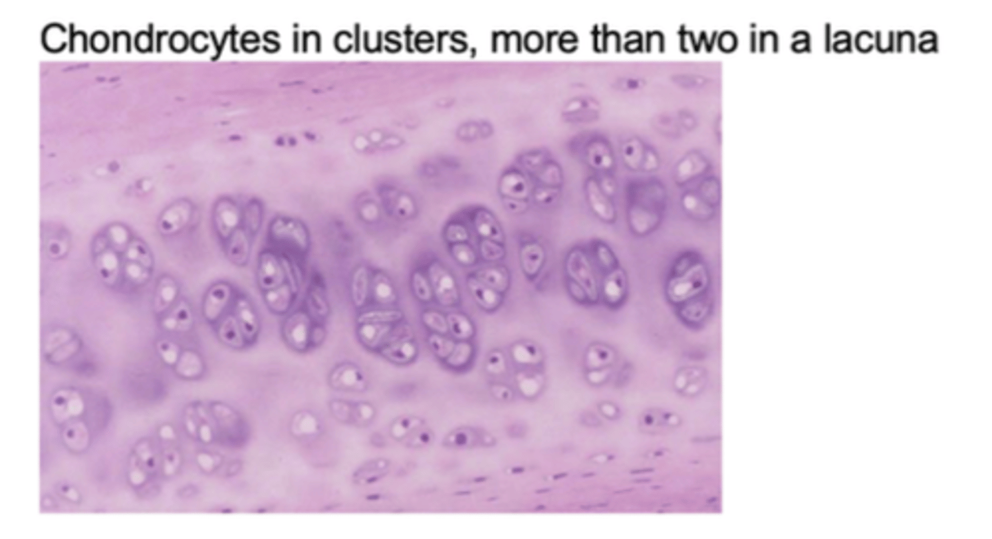
Hyaline Cartilage
- Fiber: mainly collagen.
- Function: provides support and stiffness, reduces friction in joints, and resists compression.
- Example locations: Ends of bones, trachea, rib cage, and nose.
- Chondrocytes in clusters, more than two in a lacuna
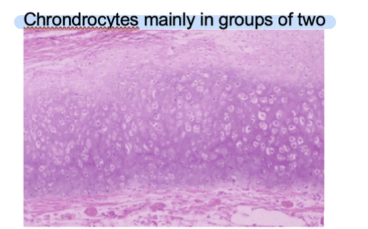
Elastic cartilage
Fibers
- Mainly elastic but also collagen
Function
- Support and flexibility
Location
- Pinna, epiglottic, parts of larynx
- Chondrocytes mainly in groups of two
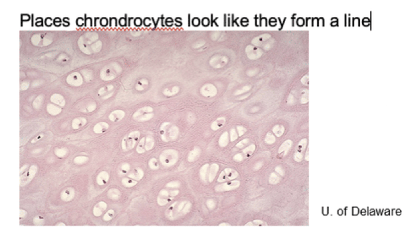
Fibrocartilage
Fibers
- Collagen fibers thicker than in hyaline cartilage
Function
- Strength, resists compression, absorbs shock
Location
- Intervertebral disease, pubic symphysis, knee joint
- Important for childbirth
Deutrostomes
- A large group of animals that share a specific pattern of embryonic development, specifically, the opening in the embryo becomes the anus, and the mouth forms second.
Deuterostomes shared ancestral features
- Multicellular
- Triploblastic- Three germ layers (ectoderm, mesoderm, and endoderm)
- Bilateral symmetry
- Coelomates
- Fully lined by mesoderm, inner and outer
- Main phyla: echinodermata, chordata, and hemichordata
Phylum Echinodermata
- Include sea stars, sea urchins, etc.
- Habitat is marine
- Symmetry- bilateral, then radial as adults
- Body surfaces- oral and aboral
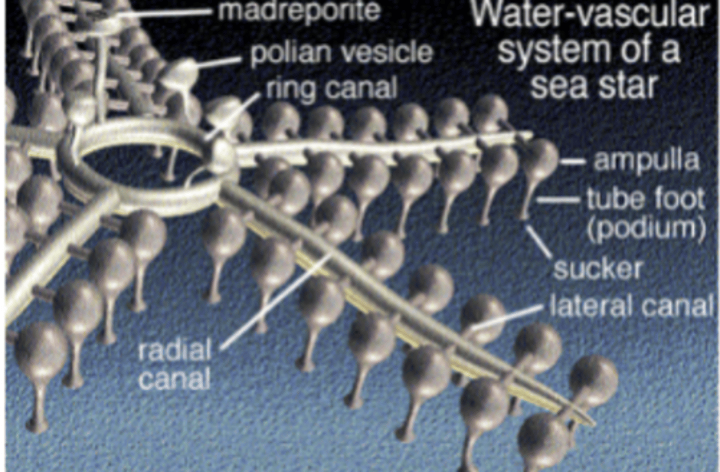
Echinodermata (Water vascular system)
- Derived, defining characteristic feeding, gas exchange,
- close it off, muscle tissue push against it, create activity
- Madreporite
- Stone canal
- Ring canal
- Ampullae- Tube feet
Major Classes of Echinodermata
Asteroidea
- Sea stars
Crinoidea
- Sea lilies and feather stars
Echinoidea
- Sand dollar and sea urchin
- Body: round, flat, no arms
- Skeleton: plates flattened and fused forms solid shell
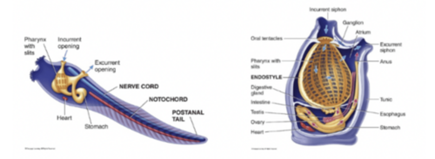
Endostyle
- A ciliated, mucus-secreting groove located on the floor of the pharynx (throat region) in some chordates.
- Involved in transporting food to the esophagus
Phylum Chordata
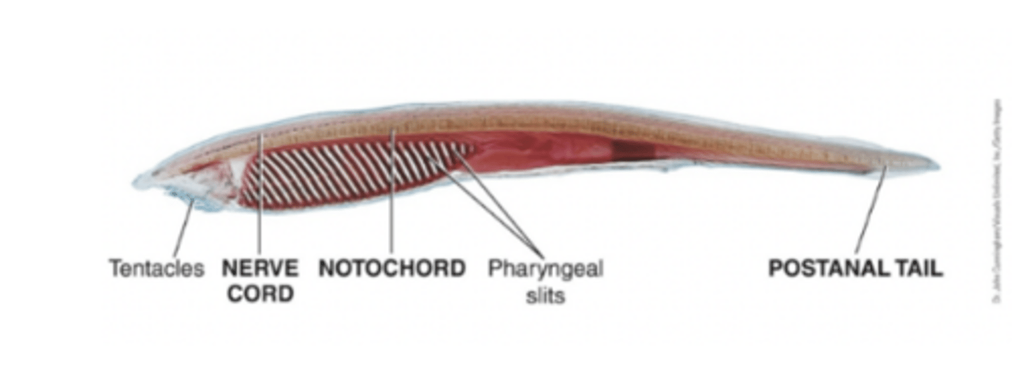
Animals in Chordata share 4 key features (at least at some stage of life):
- Notochord (flexible support rod)
- Dorsal hollow nerve cord
- Pharyngeal gill slits
- Post-anal tail
Some chordates are vertebrates (with backbones), and some are invertebrates (no backbone).
Subphylums of Chordata
Kingdom: Animalia
Phylum: Chordata
Subphylum: Urochordata (tunicates)
Subphylum: Cephalochordata (lancelets)
Subphylum: Vertebrata (animals with backbones)
(chordata) Subphylum Urochordata- invertebrate chordates
- Sea squirts and relatives; also called tunicates.
Body Covering:
- Have a tough outer covering called a tunic. Which is made of a carbohydrate material. It has two opens called siphons: One brings water in, one lets water out.
Chordate Features in tunicates: Larva (all chordate features) and swims, Adults only gill slits and sessile, feeding- suspension feeders.
(chordata) Subphylum cephalochordata - invertebrate
- small, fish-like marine animals commonly called lancelets.
- Body plan: Adults- all four chordate features, no paired fins, jaws, sense organs, heart, head, or brain.
- Filter Feeders
- Genus: Branchiostoma (latin for gills plus mouth)
- Originally amphioxus (latin for sharp on both sides)
Chordates: Hagfish
- They are not vertebrate
- Marine only
- Cranium: brain enclosure, clade cranial
- Teeth: Keratin on tongue (no jaws)
- Notochord: flexible cartilage rod
- Fins: no fins, long dorsal fold
- Feeding: scavenger or predator, nutrients across gills and skins, and tongue rasps off pieces of carcass
- Defense: secretes slime
- Body tonicity
Chordates: Lamprey
Jawless, eel-like fish with a skull (craniates).
Among the oldest living vertebrate lineages.
Live in marine, freshwater, and estuary habitats.
Many are parasitic, attaching to fish and feeding on blood.
Have a primitive vertebral column (notochord remains, cartilage instead of true bone).
Chordates: Jawed fishes- Cartilaginous Fishes
Habitat: Mostly marine (some freshwater species).
Skeleton: Cartilage (hardened with calcium salts), not true bone.
Teeth: Replaceable throughout life.
Scales: Placoid (tooth-like, non-overlapping).
Fins: Paired.
Mouth: Ventral (under the head).
Gills: Exposed slits (side in sharks, ventral in rays).
Senses:
Well-developed brain
Strong smell (nostrils for olfaction)
Electroreceptors (detect prey)
Lateral line (detect vibrations)
Buoyancy: No swim bladder → must keep swimming or use pectoral fins for lift.
Cloaca: One opening for digestive, excretory, and reproductive systems.
Reproduction: Sexual dimorphism; males have claspers to transfer sperm.
Chordates: Jawed fishes- Bony Fishes
Habitat: Freshwater and marine.
Skeleton: Calcified bone.
Teeth: Can regrow if lost.
Scales: Overlapping, flexible (dermal bone origin).
Fins: Paired, supported by bone/cartilage rays.
Mouth: Anterior (front-facing).
Gills: Internal, covered by an operculum (lid).
Swim bladder: Controls buoyancy (gas in = rise, gas out = sink).
Reproduction: Mostly external fertilization (oviparous).
Two Main Types of Bony Fish:
Ray-finned fish
Most modern fish (e.g., catfish).
Fins supported by thin rays.
Lobe-finned fish
Fleshy, paddle-like fins with bones at the base.
Some have lungs.
Ancestors of land vertebrates (tetrapods).
DNA supports this evolutionary link.
Oviparous
- Animals that lay eggs outside the body
- The embroyo gets nourishment from the yolk
Ovoviviparous
- Mothers keep their eggs inside their body
- Embryo still gets food from the yolk
- Eggs hatch inside her, and she gives birth to live young
Viviparous
- The embryo develops inside the uterus
- It gets nutrients directly from the mother's blood supply (often through the placenta)
- It is birthed live
Chordates Amphibians
- Class Amphibia
- Meaning: Having two lives (one in water and one on land)
- Metamorphosis: many complete it, and some adults like salamanders may retain larval characteristics (pedomorphosis)
- Respiratory organ: lungs, gills, or skin
- Heart: Three chambers
Three orders of amphibians
1. Urodela- Visible tail, salamanders, mud puppies, and newts
2. Anura- No tails visible in adult, frogs and toads
3. Apoda- No feet, worm like caecilians (internal fertilization)
Idk what this is
Chordates: Reptiles
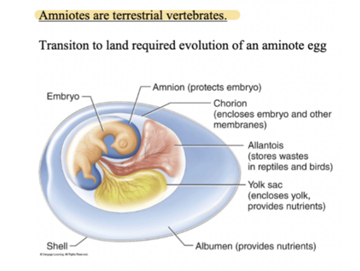
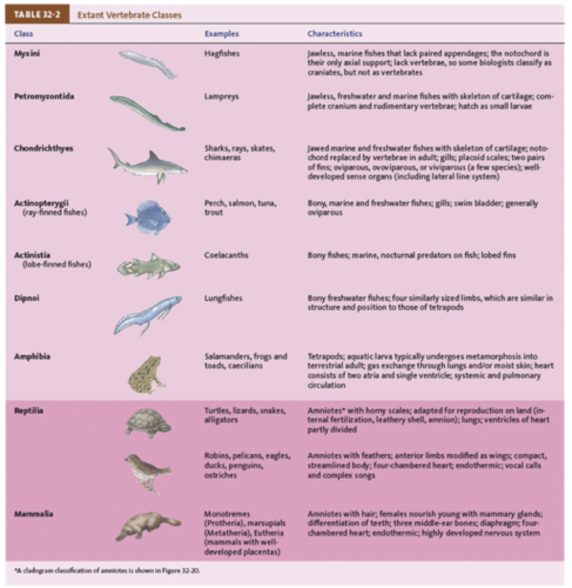
- Class Reptilia
- First to move onto land
- Hard shelled egg
- Body surface: Hard scales (watertight)
- Respiration: Lung divided into many chambers
- Circulation: Three chambered heart, separates oxygen rich and oxygen poor blood
- Ectotherms: Body temperature changes with the surrounding environment
- Well developed to locate prey
Four Orders of Reptiles- 1. Testudines
- Turtles, terrapins, and tortoises
Key Characteristics:
1. Shells- used for protection, has two parts the carapace (top, dorsal part), and the plastron (bottom, ventral part). The shell is made of bony plates and covered by scales (scutes) made of keratin. '
2. Skeleton- Vertebrae and ribs are fused to the shell, the pectoral girdle (shoulder bones) and pelvic girdle (hip bones) these lie inside the rib cage.
3. Mouth- They have no teeth, their jaws are covered by a thorny beak, beak is made of keratin.
Four orders of Reptiles- Squamata
- lizards, snakes, and worm lizards,
- Komodo dragon (close relative of snakes, very infectious bites, they eat rotting food so the bacteria gets to you).
- Snakes- Their jaws as loosely articulated, and hinge construction is key to eating large prey. Their skin is elastic so they can swallow large prey. They have pit organs in vipers, detects heat from endothermic prey. They molt even as adults.
Four orders of reptiles- Crocodillia
- Crocodiles, alligators, caimans, and gavials
- Swamp rivers along sea coasts
- Crocodiles- large slender snout, and large visible bottom tooth, largest living reptile, and have a heart with four chambers, good divers.
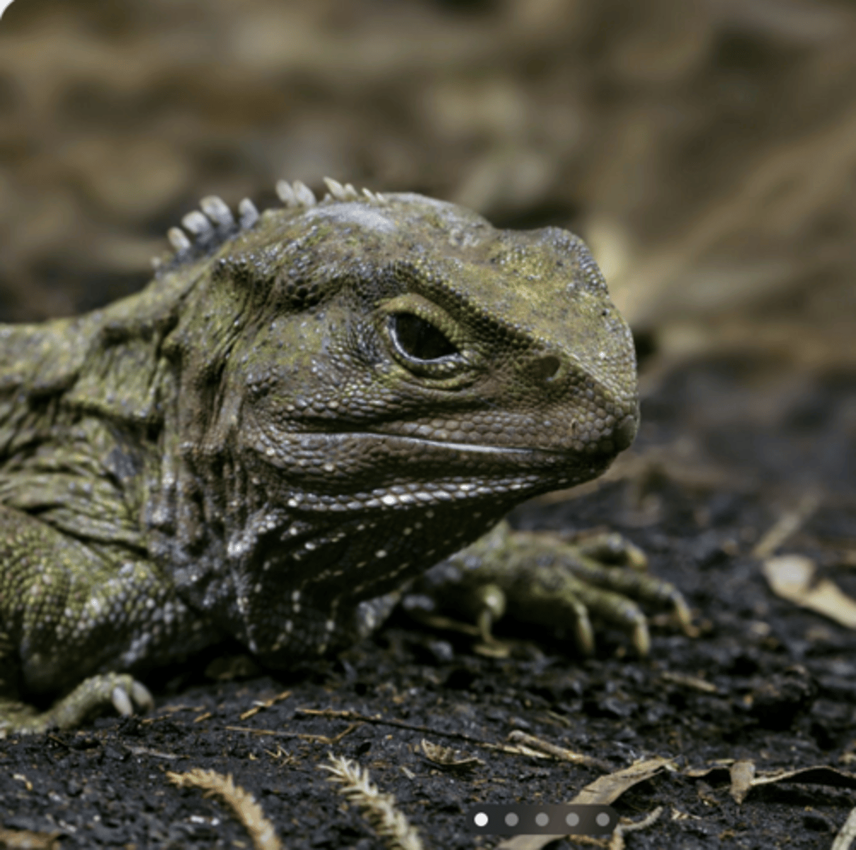
Four orders of reptiles- Sphenodonta
Sphenodontia is a small and ancient order of reptiles. Today, it has only one living representative:
The Tuatara
Clade 1 of modern bird- Paleognaths
- Ostriches, kiwis, cassowaries, and emus
- Flightless
- Flat sternum- no ridge for attachment of wing and muscles
- Vestigial wings
- Legs- well developed for running
- Toes- 3 forward (kiwi), 2 forward (ostrich)
Clade 2 Modern birds- Neognathae
- Most modern birds
- most fly
- Kneeled sternum
- Wings- well adapted for flight
- Legs- passerine (four toes adapted for perching 3 forward and 1 backward)
Characteristics of Modern Birds
- Reproduction- lay eggs, oviparous
- Scales- legs with scales
- Feathers- derived from scales
- Endothermic- shared with mammals
- Nitrogenous waste
- Respiration- lungs with air sacs (one-way air flow system)
- Heart- 4 chambered
- Bills- Adapted for specific food types
- Digestive system- crop: food storage, Gizzard: muscular food grinding
Chordates
- Class mammalia
- Key derived characters: Hair, mammary glands, differentiation of teeth (incisors, canines, premolars, molars), one opening in the temporal bone
- Middle ear bones- reptiles and birds have one, mammals have three malleolus, incus, stapes
- Changes in cochlea: Further evolved in mammals providing more sensitivity in higher frequencies
Clade Prototheria (This is a part of Class mammalia)
- Duckbilled platypus and spiny anteaters (echidnas)
- Monotremes: egg-laying mammals
- Reproduction: carrying eggs in the abdominal punch that forms after fertilization
- Nourishment of young: mother secretes milk from mammary glands onto body surfaces, no nipples.
Clade Theria (This is a part of class mammalia)
- Bear live young
Clade Theria (two groups)
1. Metatheria- kangaroos and opossums, Marsupials (pouched mammals)
2. Eutheria- Placenta, well developed offspring
Extant vertebrate classes
Primates
a group of mammals that includes humans, monkeys, apes, lemurs, and tarsiers.
Primate Cladogram
Three suborders:
a. Prosimii- Lemurs
b. Tarsiiformes- Tarsiers
c. Anthropoidea - New and old world monkeys
- Hominoids- Lesser apes, great apes, and humans
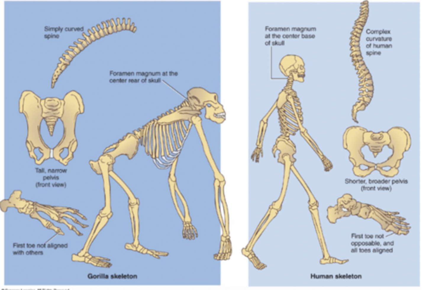
Skeletal differences between humans and gorillas
Toe alignment
1. Humans - first toe aligned and not opposable
2. Gorillas - first toe not aligned and is opposable
Pelvis
1. Humans - short, broad
2. Gorillas - long, oval
Vertebral column
1. Humans - 4 curves
2. Gorillas - one simple curve
Foramen magnum
1. Humans - base of skull
2. Gorillas - rear of skull
Jaw
1. Humans - u-shaped
2. Gorillas - rectangular
Pronounced facial feature
1. Humans - chin
2. Gorillas - supraorbital ridge
comparison of old world and new world monkeys
Tail
1. Old World - no prehensile tail
2. New World - prehensile tail (use tail as another limb to grasp things such as tree branches)
Nose
1. Old World - narrow, with downward nostrils
2. New World - flat, widespread nostrils
Terrestrial vs arboreal
1. Old World - both
2. New World - arboreal
Quadrupedal movement
1. Old World yes
2. New World - no
Platyhelminthes
- Flat and soft-bodied worms
- Bilateral
- Acoelomate
- Have simple Organ Systems
Platyhelminthes classes
1. Turbellaria- Free-living flatworms, have surface cilia which help them glide over surfaces, and rhabdites which release mucus.
2. Trematoda- Parasitic flatworms, they have sukers and hooks they attach to the hosts organs.
3. Cestoda- Tapeworms, parasitic, live in intestines and have suckers and hooks
How different invertebrates are covered and protected: Nematodes
- phylum nematoda (roundworms)
- They have a non-hardening cuticle, thick and tough, works like an exoskeleton, cannot stretch in diameter, only bends side-to-side.
- Since it doesn't grow with the worm- they must shed it (ecdysis).
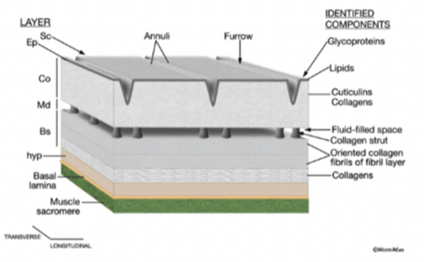
How different invertebrates are covered and protected: Earthworms
- Phylum: Annelida
- Thin, flexible cuticle, made of collagen and carbohydrates, gel-like, can stretch in length and diameter, do NOT shed it.
How different invertebrates are covered and protected: Arthropods
- Phylum: Arthropoda, examples: Insects, crabs, and spiders.
- They have a hard exoskeleton, made of chitin, this can be sometimes strengthened with minerals like calcium, and muscles attach to the inside of the exoskeleton.
- They must molt (ecdysis) to grow.
How different invertebrates are covered and protected: Mollusks
- Phylum: Mollusca, examples: clams, snails, and nautilus.
- They have an external shell, secreted by tissue called the mantle, mostly made of calcium carbonate.
- Pearls form when: A foreign particle gets trapped, the mollusk coats it with shell material repeatedly.
Syncytial Epidermis
- A fused outer tissue where many cells share one continuous cytoplasm, seen in parasitic flatworms to help them survive inside a host.
- Human heart is a syncytium
Human Integumentary System
Skin + Hair + Nails + Glands
Human integumentary system functions
- Protection from mechanical trauma (stretching, abrasion, pressure)
- Protection from Pathogen ( Biological protection)- There are immune cells in the skin, Langerhans cells (in the epidermis, they engulf bacteria, and help activate immune responses), and macrophages (in the dermis).
- Protection from environmental factors- UV Protection, water and salt barrier, skin blocks water (polar molecule, and salt ionic molecule).
- Excretion- sweat removes some nitrogenous waste, water, and salt
- Temperature Regulation- Sweating cools the body, and blood vessel dilation releases heat, blood vessel constriction conserves heat
- Cutaneous Sensation- The skin detects pain, touch, pressure, temperature, and vibration.
- Metabolic Functions: Vitamin D production, and activates some topical steroid medications.
- Blood reservoir- about 5% of the total blood volume is stored in skin, and can be redirected during exercise or shock.
Epidermis
Superficial layer (on top of dermis)
Made of multiple strata (layers)
Avascular (no blood vessels)
Mostly keratinocytes
Vertebrate Skin
Skin has 2 main layers:
Epidermis (outer layer)
Dermis (underneath)
Epidermal Cells- Keratinocytes
- Make up about 90% of the epidermis
- Produce keratin: Tough protein, Resists mechanical stress (stretching, friction, pressure)
- Help form the water barrier
- Produce a precursor to Vitamin D
- UV light converts it → Vitamin D
👉 Basically: Keratinocytes = protection + strength + Vitamin D role
Epidermal Cells- Melanocytes
📍 Located in the stratum basale (bottom layer of epidermis, at junction with dermis)
- Produce melanin
- Melanin protects DNA from UV radiation
- Transfer melanin to keratinocytes
👉 More melanin = more UV protection
Epidermal Cells- Merkel Cells
📍 Found in stratum basale
Function: Light touch sensation
Help you detect:
- Fine details
- Shapes
- Texture
- Slow adapting → continue responding while stimulus is present
👉 Important for precise touch (like reading Braille)
Epidermal Cells- Langerhans Cells
- Immune cells in epidermis
- Derived from bone marrow
- Function: Detect pathogens, Phagocytic, Activate immune response
👉 Basically: Skin immune defense
Dermis
- The dermis is the thick layer beneath the epidermis.
- It contains: Blood vessels, Sensory receptors, Hair follicles, Sweat glands
⚠️ The epidermis has no blood vessels, so the dermis nourishes it.
2 Layers of the Dermis
1. Papillary Layer
2. Reticular Layer
Papillary Layer
- The first layer of the Dermis.
Structure:
- Made of loose connective tissue
- Contains 3 fiber types: Collagen (strength), Elastic fibers (stretch), Reticular fibers (support)
Functions:
- Capillaries- Supply nutrients to: Epidermis, Upper dermis
- Epidermal ridges- Upward projections of papillary layer, Form fingerprints, Increase surface area for attachment
- Meissner (Tactile) Corpuscles- Detect light touch, Detect shape & texture, Rapidly adapting (respond quickly, stop quickly), Found especially in fingertips.
- Blister Formation: If the epidermis separates from the papillary layer, fluid fills the gap → blister
Reticular Layer
- The second layer of the Dermis.
Structure:
- Dense irregular connective tissue
- Mostly collagen → Gives skin strength and protection
- Contains elastic fibers → Allow stretching
⚠️ If elastic fibers tear → stretch marks
Pacinian (Lamellated) Corpuscles:
- Detect deep pressure
- Detect vibration
- Located deep in dermis
- Rapidly adapting
1st Degree Burns
Involves:
- Only epidermis
Signs:
- Redness
- Mild pain
- No blisters
- No permanent damage
Example: Mild sunburn
Healing: 3–7 days, no scarring