Practical 4
 Call Kai
 Learn
 Practice Test
 Spaced Repetition
 Match
 Flashcards
 Knowt Play
1/73

There's no tags or description
Looks like no tags are added yet.
74 Terms
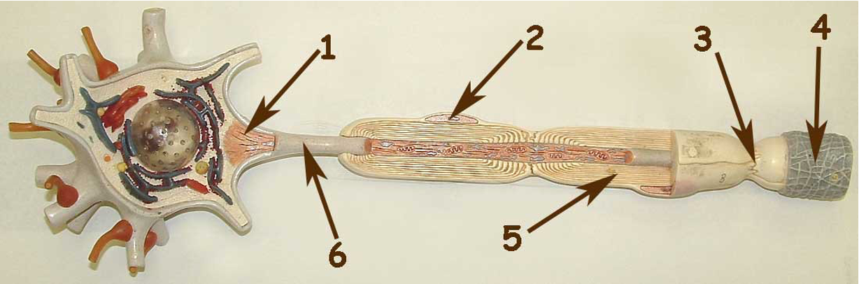
A Typical Multipolar Neuron
1.Axon Hillock
2.Nucleus of Schwann Cell
3.Node of Ranvier
4.Endoneurium
5.Schwann Cell
6.Initial Segment of Axon

Cell Body of a Typical Motor Neuron
1.Axon Terminal
2.Dendrite
3.Axon Hillock
4.Axon (Initial Segment)
5.Nissl Bodies

Motor Neuron End View
1.Nucleus of Schwann Cell
2.Endoneurium
3.Neurolemma (Sheath of Schwann)
4.Axon (Composed of Neurofibrils) Myelin Sheath
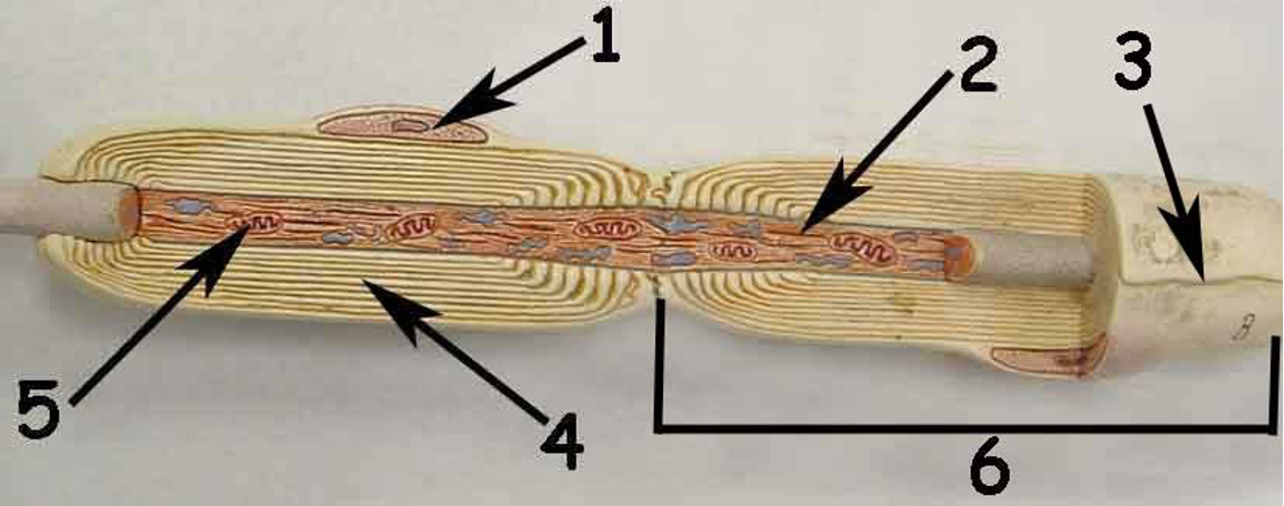
Motor Neuron Showing Schwann Cells
1.Nucleus of Schwann Cell
2.Axon
3.Neurolemma (Sheath of Schwann)
4.Myelin Sheath
5.Mitochondria
6.Schwann Cell

Brain Model One Midsagittal Section
1.Corpus callosum
2.Corpora quadrigemina
3.Cerebellum (Arbor vitae)
4.Fourth ventricle
5.Spinal cord
6.Medulla oblongata
7.Pons
8.Pituitary gland
9.Optic chiasma
10.Hypothalamus
11.Thalamus
12.Septum pellucidum
13.Fornix
14.Pineal Body (Gland)

Brain Model Two Midsagittal Section
1.Temporal lobe
2.Frontal lobe
3.Anterior Commissure
4.Interthalamic adhesion
5.Parietal lobe
6.Olfactory Nerve (I)
7.Pineal Body
8.Occipital Lobe
9.Cerebral aqueduct
10.Pons
11.Medulla oblongata
12.Cerebellum
13.Mammillary Body
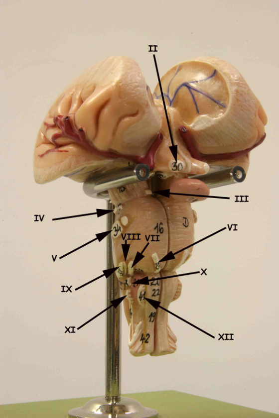
Cranial Nerves II - XII
II. Optic
III. Oculomotor
IV. Trochlear
V. Trigeminal
VI. Abducens
VII. Facial
VIII. Vestibulocochlear
IX. Glossopharyngeal
X. Vagus
XI. Spinal Accessory
XII. Hypoglossal

Cranial Nerves II - XII, Side View
II.Optic
III.Oculomotor
IV.Trochlear
V.Trigeminal
VI.Abducens
VII.Facial
VIII.Vestibulocochlear
IX.Glossopharyngeal
X.Vagus
XI.Spinal Accessory
XII.Hypoglossal
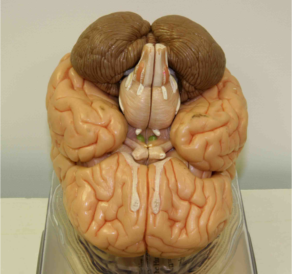
Cranial Nerves II – XII, View
I.Olfactory (bulb)
II.Optic
III.Oculomotor
IV.Trochlear
V.Trigeminal
VI.Abducens
VII.Facial
VIII.Vestibulocochlear
IX.Glossopharyngeal
X.Vagus
XI.Spinal Accessory
XII.Hypoglossal

Silver Ventricle Model (Lateral View)
1.Anterior Horn
2.Choroid Plexus
3.Lateral Ventricle
4.Posterior Horn
5.Fourth Ventricle
6.Inferior Horn
7.Cerebral Aqueduct
8.Third Ventricle
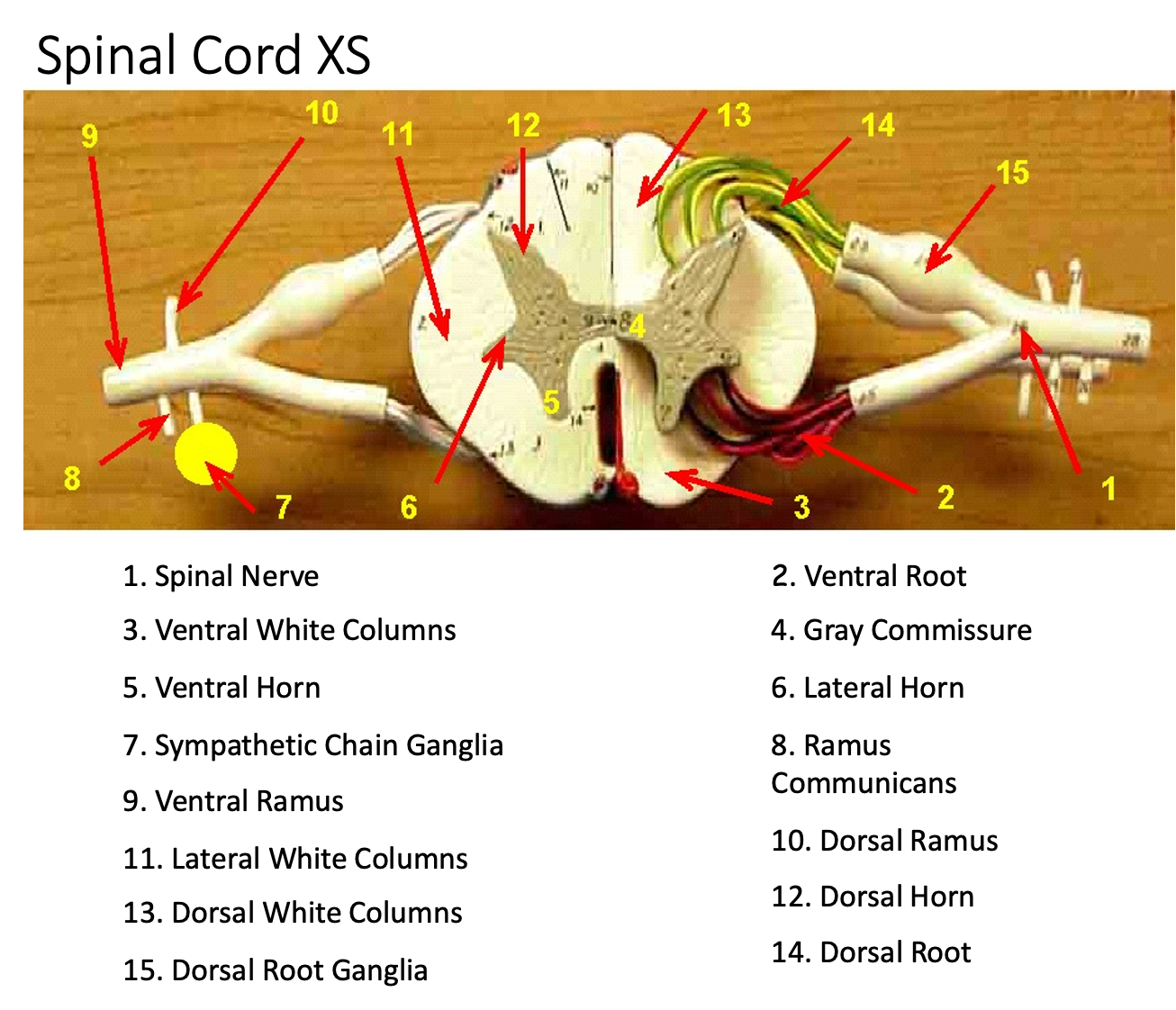
Spinal Cord XS
1. Spinal Nerve
3. Ventral White Columns
5. Ventral Horn
7. Sympathetic Chain Ganglia
9. Ventral Ramus
11. Lateral White Columns
13. Dorsal White Columns
15. Dorsal Root Ganglia
2. Ventral Root
4. Gray Commissure
6. Lateral Horn
8. Ramus Communicans
10. Dorsal Ramus
12. Dorsal Horn
14. Dorsal Root

Spinal Cord XS With Vertebrae
1.Pia Mater
2.Dura Mater
3.Spinal Nerve
4.Ramus Communicans
5.Ventral Ramus
6.Dorsal Ramus
7.Dorsal Root Ganglion
8.Dorsal Root
9.Ventral Root

Human Torso Showing Structure of Spinal Cord
1.Dorsal Root
2.Sympathetic Chain Ganglia
3.Intercostal Nerve
4.Dorsal Root Ganglion
5.Dura Mater
6.Posterior Median Sulcus

Spinal Cord Inferior
1. Ilioinguinal nerve
4. Cauda equina
2. Genitofemoral nerve
5. Conus medullaris
3. Femoral nerve
6. Sciatic nerve

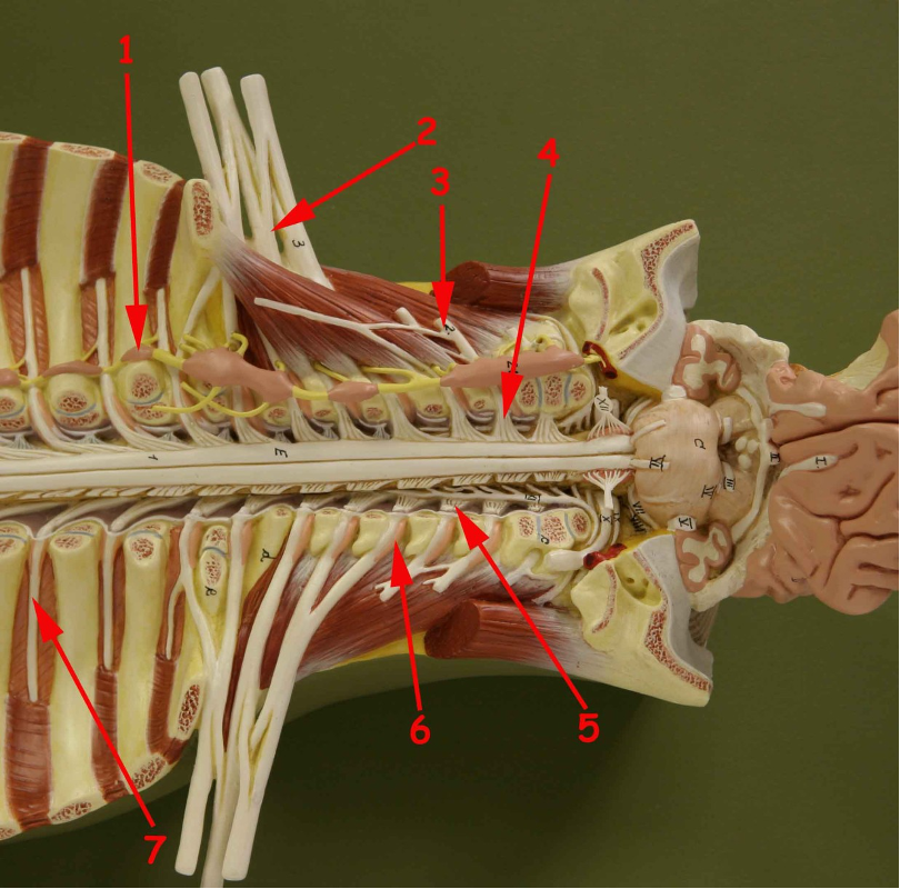
Spinal Cord Superior
1.Sympathetic chain ganglia
2.Brachial Plexus
3.Cervical Plexus
4.Ventral Root
5.Dorsal Root
6.Dorsal Root ganglion
7.Intercostal nerve

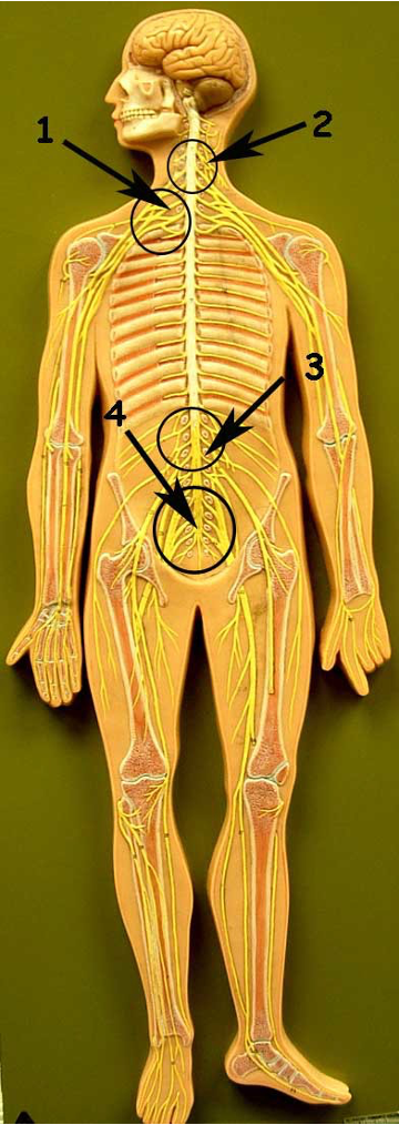
Nerveman Showing Plexuses
1.Brachial Plexus
2.Cervical Plexus
3.Lumbar Plexus
4.Sacral Plexus
5.The ventral Rami of spinal nerves T2 - T12 pass anteriorly as the Intercostal nerves. The ventral rami of all the other spinal nerves form complex networks of nerves called plexuses.

Nerveman Superior View
1.Phrenic Nerve
2.Cord (Lateral)
3.Median Nerve
4.Radial Nerve
5.Ilioinguinal Nerve
6.Ulnar Nerve
7.Femoral Nerve
8.Musculocutaneous Nerve

Nerveman Inferior View
1.Femoral Nerve
2.Pudendal Nerve
3.Sciatic Nerve
4.Saphenous Nerve
5.Tibial Nerve 6.Common Fibular Nerve
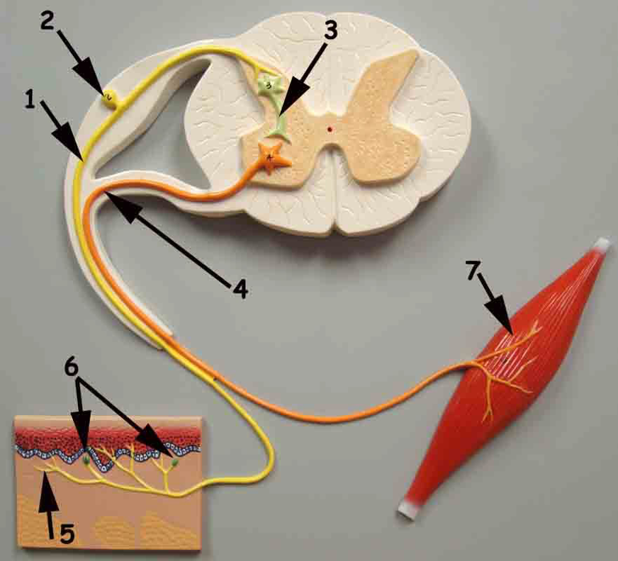
Neurons on Basis of Function
1.Sensory Neuron (Afferent)
2.Ganglion
3.Association Neuron (Interneuron)
4.Motor Neuron (Efferent)
5.Dendrites
6.Receptors
7.Effector (Muscles and Glands)
cell body (soma)
in neurons, that portion of the cell that contains the nucleus; the cell body, as opposed to the cell processes (axons and dendrites)
dendrites
one of many branchlike processes that extends from the neuron cell body and functions as a contact for incoming signals (synapses) from other neurons or sensory cells
axon
single process of the neuron that carries an electrical signal (action potential) away from the cell body toward a target cell
axon hillock
tapering of the neuron cell body that gives rise to the axon
initial segment of axon
myelin sheath (Schwann cell vs. Oligodendrocyte)
lipid-rich layer of insulation that surrounds an axon, formed by oligodendrocytes in the CNS and Schwann cells in the PNS; facilitates the transmission of electrical signals
glial cell type in the PNS that provides the myelin insulation for axons in nerves
glial cell type in the CNS that provides the myelin insulation for axons in tracts
node of Ranvier
gap between two myelinated regions of an axon, allowing for strengthening of the electrical signal as it propagates down the axon
endoneurium
loose connective tissue surrounding inidividual axons
synapse
specialized areas of contact
axon terminal
At the end of the axon where there are usually several branches extending toward the target cell, each of which ends in an enlargement
Nissl bodies
unipolar cells
have only one process emerging from the cell.
Human unipolar cells have an axon that emerges from the cell body, but it splits so that the axon can extend along a very long distance. At one end of the axon are dendrites, and at the other end, the axon forms synaptic connections with a target. Unipolar cells are exclusively sensory neurons
bipolar cells
two processes, which extend from each end of the cell body, opposite to each other. One is the axon and one the dendrite
not very common.
They are found mainly in the olfactory epithelium (where smell stimuli are sensed), and as part of the retina.
multipolar cells
all of the neurons that are not unipolar or bipolar. They have one axon and two or more dendrites (usually many more)
Medulla oblongata: function
Reflexive, involuntary control of heart, breathing & blood vessels
Pons: function
Timing of subconscious breathing
Cerebellum: subparts and function
subparts: arbor vitae; L/R cerebellar hemispheres
function: Subconscious control & coordination of voluntary muscle
midbrain: subparts and function
subparts: corpora quadrigemina
superior colliculi
inferior colliculi
function: Filter and relay visual (superior colliculi) & auditory (inferior colliculi) stimuli; reflexive control of eye movement, focusing lens and pupil diameter
Hypothalamus: subparts and function
subparts: mammillary bodies
function: Control of autonomic nervous system (ANS) & pituitary gland; regulates involuntary body functions, homeostasis
Thalamus: subparts and function
subparts: interthalamic adhesion = intermediate mass
function: Crude (raw) perception of sensation & emotions; relay afferent/efferent impulses to/from the cerebrum
Pineal gland: function
Endocrine gland – secretes one hormone (melatonin)
Pituitary gland: subparts and function
subparts: infundibulum
functions: Endocrine gland – secretes many hormones
subpart of cerebrum- longitudinal fissure: function
Divides cerebrum into cerebral hemispheres
subpart of cerebrum- left cerebral hemisphere: function
Dominates speech sounds and in understanding sequential, rational & analytical concepts
subpart of cerebrum- right cerebral hemisphere: function
Dominates in non-speech sounds (melodies, laughing, etc.), in spatial perception & in holistic, artistic & emotional concepts
subpart of cerebrum- frontal lobe: function
Voluntary control over muscles, learning, planning, higher psychological functions
subpart of cerebrum- parietal lobe: function
Perception of sensations of touch, temperature, taste & and body position
subpart of cerebrum- temporal lobe: function
Perception of auditory sensations & related speech center
subpart of cerebrum- occipital lobe: function
Perception of visual sensations
subpart of cerebrum- cerebral cortex, cerebral tracts, white matter vs. grey matter, and fornix: function
Will (choice), intelligence, memory, awareness, personality
subpart of cerebrum- corpus callosum and anterior commissure: function
Communication between the two cerebral hemispheres
subpart of cerebrum- gyri & sulci: function
Increase surface area of cerebrum for exchange of nutrients/wastes to/from blood vessels in the pia mater
Functions of olfactory nerves (I)
Sensory – carries impulses associated with smell
Functions of optic nerves (II)
Sensory – carries impulses associated with vision
Functions of oculomotor nerves (III)
Motor – inferior oblique, superior/inferior and medial rectus muscles of eye, moves eye in eye socket, movement of eyelid, shape of lens
Functions of trochlear nerves (IV)
Motor – superior oblique eye muscle moving eye in eye socket
Functions of trigeminal nerves (V)
Both – Sensory: nerves of face, Motor: muscles of mastication
Functions of Abducens nerves (VI)
Motor – lateral rectus eye muscle moving eye in eye socket
Functions of facial nerves (VII)
Both – Sensory: taste at anterior portion of tongue, Motor: facial expression muscles, tear glands, salivary glands
Functions of Vestibulocochlear nerves (VIII)
Sensory – balance/equilibrium & motion in the vestibule and semicircular canals, hearing in the cochlea
Functions of Glossopharyngeal nerves (IX)
Both – Sensory: general throat sensations, taste at posterior portion of tongue, pressure receptors in carotid artery, Motor: muscles of larynx, pharynx, control of blood pressure
Functions of vagus nerves (X)
Both – Sensory: pharynx, larynx, impulses from many viscera (internal organs), Motor: pharynx, larynx, heart rate many visceral organs
Functions of spinal accessory nerves (XI)
Motor – voice production, neck movement, motor control of many thoracic and abdominopelvic viscera
Functions of Hypoglossal nerves (XII)
Motor – movement of tongue
dura mater
thick fibrous layer and a strong protective sheath over the entire brain and spinal cord. It is anchored to the inner surface of the cranium and vertebral cavity; part of meninges
arachnoid mater
a membrane of thin fibrous tissue that forms a loose sac around the CNS; part of meninges
pia mater
a thin fibrous membrane that follows the convolutions of gyri and sulci in the cerebral cortex and fits into other grooves and indentations; part of meninges
epineurium
The outer surface of a nerve is a surrounding layer of fibrous connective tissue
perineurium
layer of fibrous connective tissue surrounding fasicles
endoneurium
loose connective tissue surrounding individual axons
neuron fascicle
small bundles of nerve or muscle fibers enclosed by connective tissue
cauda equina
long bundle of nerves that resembles a horse’s tail in the vertebral column
posterior median sulcus
posterior midline of the spinal cord
4 of the spinal nerve plexuses