Urinary System Test
 Call Kai
 Learn
 Practice Test
 Spaced Repetition
 Match
 Flashcards
 Knowt Play
1/138

There's no tags or description
Looks like no tags are added yet.
139 Terms
The urinary system includes:
•Two kidneys
•A ureter for each kidney
•A urinary bladder
•A urethra
What makes up the upper urinary tract ?
kidneys and ureturs
What makes up the lower urinary tract?
Urinary bladder and urethra
Renal Anatomy
•retroperitoneal organs, lie one on each side of the spine between the peritoneum and back muscles
•has a lateral convex and a medial concave border
Right vs Left kidey
•The liver displaces the right kidney inferiorly so:
right kidney is lower than the left kidney and has a slightly shorter ureter
LOCATION & CHARACTERISTICS
•Near the posterior body wall
•Below diaphragm
•Protected by ribs
•Between 1st & 3rd Lumbar Vertebrae
•With deep inspiration the kidney moves downward 1inch
Anterior to right kidney
adrenal gland, liver, MORISON’S POUCH, 2nd part of the duodenum and rt colic flexures
Posterior to right kidney
diaphragm, psoas muscle, quadratus lumborum, transverse abdominis muscles, 12th rib & nerves
Anterior to left kidney
adrenal gland, spleen, stomach, pancreas, lt colic flexure & jejunum coils
Posterior to left kidney:
same as right but also add the 11th rib
INDICATIONS FOR EXAM
•Uncontrollable Hypertension – Renal Artery Stenosis
•Urinary tract infection (UTI)
•Palpable mass
•Elevated serum creatinine, BUN levels
•Severe flank pain
•Hematuria
•Proteinuria
•Oliguria
Normal Measurements
length of adult kidney: 9-12 cm
thickness of bladder: 3-6 mm
Size and shape of kidneys
•Measurements 9-12 long, 5 wide & 2.5 cm thick
•Bean Shaped
•Smooth outer contours surrounded by reflected echoes of perirenal fat
Sonography of kidney
Cortex is hypoechoic compared to the hyperechoic sinus.
•Echogenic renal sinus due to collecting system. (pelvis, calyces, renal vessels, fat & lymphatics)
More sonolucent is the cortex and pyramids, cortex being more echogenic than the pyramids
arcuate vessels
Arcuate vessels
bright echoes at corticomedullary junction
Kidney in SAG plane
smooth contours of renal capsule
Always want to see both upper and lower poles in the sagittal view.
Kidney in TRV plane
upper and lower pole are round
hilum is indented by sinus
Always want to document the medial and lateral aspects in the transverse view.
Four layers surround the kidney
Pararenal fat
Perirenal fascia
Perinephric fat
Renal capsule
Pararenal fat
•Pararenal body
Perirenal fascia
•Perinephric fascia
•Gerota’s Fascia
What is another name for the perinephric fascia ?
Gerotas fascia
Perinephric fat
Adipose capsule
Renal capsule
•True capsule
•Fibrous capsule
Anterior Pararenal Space contains (WO?)
•Pancreas
•Duodenal sweep
•Ascending and transverse colon
Perirenal Space contains (WO?)
•Kidneys
•Adrenal glands
•Perirenal fat
•Ureters
•Renal vessels
•Prevertebral vessels
•Retroperitoneal nodes
Perirenal Space is contained by
GEROTA’S FASCIA SEPARATING IT FROM THE PARAENAL (ant & post)
Posterior Pararenal Space
between the renal fascia and transversalis fascia, it contains fat and is of variable size.
Does not contain solid organs.
Sonographic Appearance of the Renal Capsue
Hyperechoic thin, continuous, highly reflective line visualized along periphery of kidney
Sonographic Appearance of renal crotex
•Midgray or medium- to low-level homogeneous echo pattern
•Hypo- to isoechoic to normal liver or spleen
Sonographic Appearance of Renal Medulla (what we see as the pyramids)
•Triangular, round, or blunted anechoic areas when urine filled and are otherwise not visible
•Anechoic pyramids have a distinctive and readily identifiable appearance
Sonographic Appearance of Renal sinus
•Hyperechoic, ovoid central portion of the kidney
Scanning the kidneys (Sagittal)
See: superior to inferior (upper pole to lower pole)
Scan: medial to lateral
Document: SAG Medial, Mid and Lateral
Scanning the kidneys (Transverse)
See: medial to lateral
Scan: superior and inferior (upper pole to lower pole)
Document: TRV UP, Mid, LP
Sonographic Appearance of the Pediatric renal cortex
•Hyperechoic relative to cortex in adult kidney
Sonographic Appearance of the Pediatric renal medulla
•Easily visible
•Anechoic
Sonographic Applications
•Renal size
•Detection and composition of renal masses and cysts
•Urinary system obstruction
•Renal abscess
•Renal hematoma
•Enlarged ureters
•Urinary bladder masses
•Renal transplantation
•Doppler evaluation of renal blood flow abnormalities
•Ultrasound-guided biopsies of renal parenchyma or masses
•Ultrasound-guided fluid aspirations
What indicates renal disease ?
1) Increase in Blood nitrogen Urea (BUN)
2) Increase in creatinine
3) Decrease in glomerular filtration rate
Associated Tests
•Intravenous pyelogram (IVP): an x-ray exam that uses dye to outline the kidneys, ureters and bladder.
used to diagnose why a patient has blood in their urine, or pain in their side/lower back.

Intravenous pyelograms (IVP) can identify
• Kidney stone
• Enlarged prostate
• Tumors in the kidney, ureters or urinary bladder
• Scarring from UTI
• Birth defects of the urinary tract.
• Can identify ectopic kidneys
Physiology of the Urinary System
excretory organs that maintain the body’s chemical equilibrium through excretion of urine, a waste product
Ureters carry urine to the urinary bladder for temporary storage
Urine is excreted from the bladder to outside of the body through the urethra
Functions of the Kidneys
•Blood pressure regulation
•Renin-angiotensin system influences blood pressure & blood
volume.
•Urine production
•Homeostasis (maintenance of normal body physiology)
•Detoxification
•Removes waste from blood
•Maintenance of the correct balance of pH, minerals, iron, and salt levels in blood
•Adjusts the amount of water & electrolytes leaving the body so it equals the amount entering the body: Maintains salt & H20
What is the functional unit of the kidney ?
nephron
Functions of the nephrons
•There are over a million microscopic nephrons in each kidney.
•Nephron moves metabolic products from areas of high concentration to areas of low concentration though osmosis
•Renal parenchyma consists of two areas:
•Cortex
•Medulla (what we see as pyramids)

Renal cortex
•Outer portion
•Contains the renal corpuscle and the proximal and distal convoluted tubules of the nephron

Medulla
•Inner portion
•Consists of 8 to 18 medullary pyramids, which contain the loops of Henle

Medullary pyramids
Apex
•Narrow tip
•Sits within a minor calyx
Base
•Abuts the renal cortex
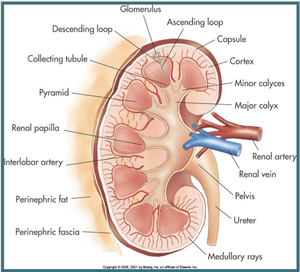
Columns of Bertin
•Bands of cortical tissue
•Separate the medullary pyramids

Renal lobes
•Portion of the kidney that consists of a single pyramid bordered by arteries and veins

Renal sinus
•Central portion of the kidney
•Houses the renal artery and vein, fatty fibrous tissue, nerves, and lymphatics

•Infundibulum
•Minor and major calyces
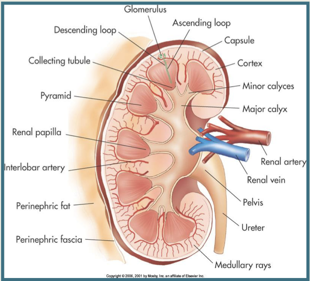
•Renal pelvis
•Upper expanded end of ureter
•Receives urine from major calyces

Renal hilum
•Medial portion of the renal sinus, where the renal artery enters the kidney and the renal vein and ureter exit
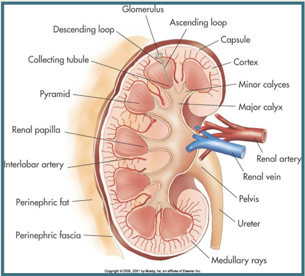
•Kidneys have 3 distinct regions:
Cortex: area from capsule to base of pyramids
Medulla: area where pyramids are located
Pelvis: flat and funnel-shaped area continuous with the ureter. Pelvis is located in the sinus.
What are the three structures that come out of the kidney ?
lymphatics, veins, ureters
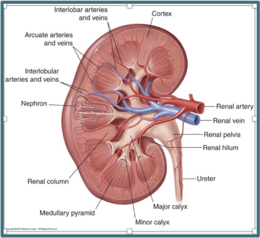
•Main renal arteries
•Arise from the abdominal aorta
•Segmental
•Interlobar
•Arcuate
•Interlobular

•Renal veins
•Drain into inferior vena cava
•Left renal vein receives blood from left suprarenal and gonadal veins
quadratus lumborum
wider cranial/cephalad than caudad
Arises from the 13th rib and attaches to the vertebral transverse process before fusing into the iliac crest.
The quadratus lumborum is ..
posterior to the kidney
psoas
frequently made of both the psoas major and minor.
Originate from the lumbar vertebrae and transverse process and insert into the lesser trochanter of the femur.
The psoas is..
posterior to the ureter
Ureter Anatomy
•Ureters begin in the kidney as the renal pelvis and end posteriorly at the trigone portion of the urinary bladder
What are the three layers of tissue the ureter is composed of ?
•inner mucosal layer
•medial layer of longitudinal and circular smooth muscle
•outer fibrous layer
What does ureter peristalsis do ?
transports urine to the urinary bladder
Urinary Bladder Anatomy
•Elastic, muscular sac that collects urine from ureters before it is excreted through the urethra
•Consists of the posterior base (trigone area) and neck, which communicates with the urethra
•Bladder wall is 3-6 mm in thickness
What are the four layers of tissue the bladder consists of ?
•Inner mucosa
•Submucosa
•Muscularis
•Outer serosa
PVR: Post Void Residual Volume
•Amount of urine left in the bladder after the patient voids
•Volume = length x width x height x 0.52
Normal bladder wall should be
3-6mm
post void: less than 20 ccs
Urethra Anatomy
consists of a membranous, hollow canal that conveys urine from the bladder to outside
Male urethra length
20cm
female urethra length
3.5 cm
•Male urethra is composed of three parts:
•Prostatic urethra
•Membranous urethra
•Penile urethra
•Female urethra is composed of
the membranous urethra, which also pierces the urogenital diaphragm
Nephron includes
a
renal corpuscle,
renal tubule
vascular component
What do nephrons do ?
filter the blood and produce urine
Where is blood filtered through ?
the renal corpuscle
Filtered fluid then passes through the renal tubule…the tubule takes substances that are needed by the body and returns the substance to the blood & anything that is not needed is passed through the collecting ducts as urine.
Renal Corpuscle
network of capillaries called the Glomerulus,surrounded by Bowmans Capsule.
Glomerulus & Bowmans Capsule
part of the filtration process. Blood flows into the glomerulus through a small afferent arteriole and out through an efferent arteriole. Opening at bottom of Bowmans Capsule where filtrate passes into the renal tubule.
•Renal Tubule (first part)
filtrate flows through the coiled proximal convoluted tubule
•Renal Tubule (second part)
filtrate flows into the loop of Henle (descending and ascending branches), reabsorption of fluid, sodium, and chloride occurs in the proximal convoluted tubule & the loop of Henle
•Renal Tubule (third part)
filtrate flows into the distal convoluted - then drains into the collecting ducts
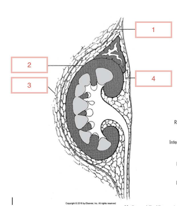
1
Gerotas Fascia

2
fibrous capsule

3
pararenal fat

4
perirenal

1
glomerulus

2
descending loop

3
collecting tubule

4
pyramid

5
renal papilla

6
interlobar artery

7
perinephric fat

8
perinephric fascia

9
medullary rays

10
ureter

11
pelvis

12
renal vein

13
renal artery

14
major calyx

15
minor calyces