Anatomy and Physiology: Levels of Organisation, Tissues, and Bone Physiology
 Call Kai
 Learn
Learn Practice Test
 Spaced Repetition
 Match
Match Flashcards
 Knowt Play
1/53

Earn XP
Description and Tags
Comprehensive vocabulary flashcards covering anatomical levels of organisation, directional terminology, epithelial and connective tissues, glandular mechanisms, muscle and neural tissues, and bone histology, growth, and repair.
Name | Mastery | Learn | Test | Matching | Spaced | Call with Kai | Chat |
|---|
No analytics yet
Send a link to your students to track their progress
54 Terms

Anatomical Position
The standard reference posture for the human body, characterised by an individual standing upright, facing forward, feet together, arms resting at the sides, and hands supinated with the palms facing forward.

What is Negative Feedback
A homeostatic control mechanism that opposes variations from the normal set point to restore balance.
What is Positive Feedback
A control mechanism that exaggerates or enhances variations from normal, creating a self-reinforcing cascade of chemical reactions.
Retroperitoneal
The anatomical position of organs located outside and posterior to the peritoneal cavity lining within the abdominal region, specifically including the kidneys and the pancreas.

Epithelial Tissue Classification
The categorization of covering and lining tissues based on layer number (simple having one layer, stratified having multiple layers) and cell shape (squamous, cuboidal, columnar, or transitional).

State the sectional plane
Transverse plane

State the sectional plane
Sagittal plane

State the sectional plane
Frontal plane
Simple Squamous Epithelium
A single layer of flattened, delicate cells that reduces friction and facilitates absorption and secretion; found lining blood vessels, alveoli of the lungs, and the peritoneal cavity.

Stratified Squamous Epithelium
A multi-layered tissue with flattened superficial cells that provides robust physical protection against abrasion, pathogens, and chemical attack; found in the epidermis of the skin and the surface of the tongue.

Transitional Epithelium
A stratified epithelium capable of significant stretching, unfolding, and recoiling without damage; lines organs subject to volume changes such as the urinary bladder.

Pseudostratified Ciliated Columnar Epithelium
An epithelial lining where cell nuclei sit at varying distances from the basement membrane, giving a layered appearance, that secretes mucus and utilizes cilia to propel it; found in the nasal cavity, trachea, and bronchi.

Merocrine Secretion
(Merely vesicles) Glandular secretion where secretory products are discharged exclusively through exocytosis from vesicles.

Apocrine Secretion
A portion off the cytoplasm is shed) glandular secretion involving the shedding of a portion of the apical cytoplasm packed with secretory material, after which the cell regrows, exemplified by mammary glands.

Holocrine Secretion
(Whole cell bursts) glandular secretion where the entire cell bursts and dies to release its cytoplasmic contents, requiring continuous cell division of stem cells to replace lost cells, exemplified by sebaceous glands.

Mesenchyme
The embryonic stem-cell connective tissue that is the first connective tissue to appear in an embryo and gives rise to all other connective tissue types.
Dense Regular Connective Tissue
A connective tissue packed with parallel bundles of collagen fibres aligned along lines of stress, providing firm attachment and conducting the pull of muscles; forms tendons, aponeuroses, and ligaments.

Dense irregular connective Tissue
Highly branched bundles of collagen fibres to provide strength in multiple directions to prevent over expansion in organs like the urinary bladder.

Elastic connective tissue
High concentration of elastic fibres which stabilise by cushioning shocks as well as permitting expansion and compression like in elastic ligaments and blood vessel linings

Hyaline Cartilage
A supportive cartilage containing closely packed collagen fibres within a glassy matrix and chondrocytes in lacunae; provides stiff but flexible support and reduces friction at synovial joints, the ribs-sternum connections, and nasal septum.


State the type of cartilage
Hyaline cartilage
Elastic cartilage
Supportive cartilage made up of chondrocytes in lacunae and elastic fibres in the matrix, which tolerate distortion with the ability to return to its original shape without damage
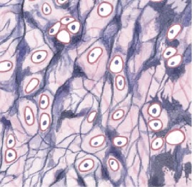

State the type of cartilage
Elastic cartilage
Fibrocartilage
Your chondrocytes in lacunae in a fibrous matrix that resist compression and prevent bone to bone contact


State the type of cartilage
Fibrocartilage
Serous Membrane
A two-layer tissue consisting of a mesothelium and loose areolar tissue that lines ventral body cavities and produces a thin, watery transudate to minimize organ friction.

Mucous membrane
Two layers of tissues (columnar epithelial tissue & areolar tissue) coated with secretions of mucous glands and line the respiratory tract

Cutaneous membrane
Three layers of tissues (areolar, deep connective tissues and stratified cuboidal epithelial tissue) that cover the surface of the body (skin)

Synovial membrane
Two layers of tissues (squamous epithelial and areolar tissue) which line synovial joints as well as secrete synovial fluid to reduce friction

Superficial Fascia
A subcutaneous tissue layer situated between the skin and underlying organs, composed of areolar connective tissue and adipose tissue.
Deep Fascia
A dense connective tissue layer that forms a tough, fibrous internal framework bound to capsules, tendons, and ligaments around muscles and visceral organs.
Subserous fascia
Skeletal Muscle Tissue
Striated, voluntary muscle tissue consisting of very large, multinucleated cells measuring approximately 100μm in diameter and up to 60cm in length, innervated and stimulated by motor neurons.
Cardiac Muscle Tissue
Striated, involuntary muscle tissue located in the heart wall, composed of branched cells measuring 10–20μm×50–100μm connected by intercalated discs and regulated by pacemaker cells.
Smooth Muscle Tissue
Non-striated, involuntary muscle tissue found in walls of visceral organs, consisting of small spindle-shaped cells (5–10μm×30–200μm) regulated by pacesetter cells and hormonal input.
Neuroglia
Supporting cells of neural tissue that maintain physical tissue architecture, repair frameworks following injury, perform phagocytosis, provide nutrients to neurons, and regulate interstitial fluid composition.

Bone Classification by Shape
The structural classification of osseous elements into six primary categories: flat bones, long bones, short bones, irregular bones, sutural bones, and sesamoid bones.
Diploë
The layer of spongy (trabecular) bone sandwiched between the internal and external tables of compact bone within cranial flat bones.
Diaphysis
The extended tubular shaft of a typical long bone, consisting of a thick outer collar of compact bone surrounding a central medullary cavity.
Epiphysis
The expanded terminal end of a long bone, predominantly composed of an interior network of trabecular bone capped by articular cartilage.
Hydroxyapatite
The primary mineral salt of the bone extracellular matrix, with chemical formula Ca10(PO4)6(OH)2, forming roughly two-thirds of the total matrix mass alongside other calcium salts.
Osteoblast
An immature, matrix-forming bone cell that synthesizes and secretes organic matrix proteins to produce uncalcified osteoid.
Osteocyte
A mature bone cell residing inside a lacuna that maintains protein and mineral matrix turnover and participates in mechanical strain detection.
Osteoclast
A large, multinucleated bone-resorbing cell that secretes acids and proteolytic enzymes to dissolve the mineralized matrix and release calcium.
Osteon
The basic microscopic functional unit of mature compact bone, consisting of concentric lamellae arranged around a vascular central canal oriented parallel to the long axis.
Trabeculae
The branching framework of struts, rods, plates, and arches of bone matrix that make up spongy (trabecular) bone, providing strength against multi-directional stresses without containing osteons.
Periosteum
A dual-layered membrane covering outer bone surfaces (except at joints), consisting of an outer fibrous layer continuous with tendons and joint capsules, and an inner cellular osteogenic layer.
Endosteum
An incomplete cellular membrane lining the internal marrow cavity, central canals of osteons, and the surfaces of trabeculae.
Intramembranous Ossification
The formation of bone directly from embryonic mesenchymal stem cells without a prior cartilage precursor; produces flat bones such as skull bones.

Endochondral Ossification
The developmental process by which a hyaline cartilage model is gradually replaced by bone tissue via primary and secondary ossification centres.
Appositional Bone Growth
The process by which bones expand in diameter and wall thickness, characterized by osteoblasts depositing new bone at the periosteal surface while osteoclasts resorb bone along the endosteal surface.

Fracture Repair
The multistep healing sequence following bone breakage, progressing from fracture haematoma formation to fibrocartilaginous external and spongy internal callus creation, followed by osseous replacement and remodeling.
Trochanter
A massive, rough anatomical projection on a bone designated for tendon or ligament attachment, specific to the proximal femur.
Foramen
An anatomical opening or hole passing through a bone that serves as a passageway for blood vessels and nerves.