Integumentary System (copy)
 Call Kai
 Learn
Learn Practice Test
 Spaced Repetition
 Match
Match Flashcards
 Knowt Play
1/169

There's no tags or description
Looks like no tags are added yet.
Name | Mastery | Learn | Test | Matching | Spaced | Call with Kai | Chat |
|---|
No analytics yet
Send a link to your students to track their progress
170 Terms
Session 1A
Which component of the basement membrane zone anchors the epidermis to the underlying dermis?
A. Type II collagen
B. Type VII collagen anchoring fibrils
C. Elastic fibers
D. Keratohyalin granules
Correct Answer: B.
Why: Type VII collagen forms anchoring fibrils that secure the basement membrane to the papillary dermis and help prevent separation of the epidermis from the underlying tissue.
Session 1A
Which epidermal cell population produces melanin pigment that helps protect against ultraviolet radiation?
A. Keratinocytes
B. Merkel cells
C. Melanocytes
D. Langerhans cells
Correct Answer: C.
Why: Melanocytes synthesize melanin within melanosomes and transfer the pigment to neighboring keratinocytes. The melanin forms a protective cap over the nucleus that helps absorb ultraviolet radiation.
Session 1A
Which layer of the epidermis is composed entirely of dead, keratinized cells and forms the primary barrier against water loss and pathogen entry?
A. Stratum basale
B. Stratum granulosum
C. Stratum lucidum
D. Stratum corneum
Correct Answer: D.
Why: The stratum corneum is the outermost epidermal layer. Its dead, keratinized cells form the skin's principal physical and water-resistant barrier.
Session 1A
Which layer of the skin is primarily responsible for anchoring the epidermis to deeper tissues while also providing vascular support and housing cutaneous nerves?
A. Epidermis
B. Dermis
C. Stratum corneum
D. Dermal–epidermal junction
E. Hypodermis
B. Correct.
The dermis is a vascularized connective tissue layer that provides structural support, contains blood vessels and nerves, and contributes to skin function.
Session 1A
Which epidermal cell type is primarily responsible for detecting light touch and forming a receptor complex with sensory nerve endings?
A. Tactile Epithelial Cell
B. Melanocyte
C. Keratinocyte
D. Intraepidermal macrophage
E. Fibroblast
A. Correct.
Merkel cells are slow-adapting mechanoreceptors located in the stratum basale that form Merkel cell–neurite complexes, allowing the skin to detect light touch and fine tactile stimuli.
Session 1A
Which feature of the stratum spinosum allows it to resist mechanical stress within the epidermis?
A. Keratohyalin granules
B. Lamellar bodies
C. Desmosomes
D. Melanosome
E. Hemidesmosomes
C. Correct.
Desmosomes connect adjacent keratinocytes in the stratum spinosum and, together with cytokeratin filaments, provide resistance to mechanical stress.
Session 1A
Which epidermal layer is composed primarily of flattened, anucleate, keratin filled corneocytes that form the outer protective barrier of the skin?
A. Stratum basale
B. Stratum spinosum
C. Stratum granulosum
D. Stratum corneum
D. Correct.
Session 1A
Significant Problem: A 2-year-old Labrador Retriever presents with superficial abrasions of the paw pads after running on rough concrete. The injuries are confined to the epidermis. Two weeks later, the paw pad epidermis appears intact.
Following superficial epidermal injury, which epidermal structure is most critical for initiating successful tissue regeneration of the footpad of this dog?
A. Keratinized corneocytes
B. Granular keratinocytes
C. Basal keratinocytes
D. Sub-basal fibroblasts
C. Correct.
Successful regeneration of the epidermis depends on the survival of basal keratinocytes that remain attached to the basement membrane. These mitotically active cells function as the regenerative population of the epidermis, proliferating to replace lost cells and producing daughter cells that migrate toward the surface while undergoing progressive differentiation. An intact basement membrane provides structural support, maintains epithelial polarity, and serves as a scaffold for re-epithelialization. In superficial injuries where the basement membrane and basal layer remain intact, the epidermis can regenerate completely. If these structures are destroyed, regeneration is markedly impaired and healing occurs primarily through scar formation rather than restoration of normal epidermal architecture.
Session 1A
Significant Problem: A 2-year-old Labrador Retriever presents with superficial abrasions of the paw pads after running on rough concrete. The injuries are confined to the epidermis. Two weeks later, the paw pad epidermis appears intact.
The paw pad develops a continuous, multilayered epidermis. Despite restoration of the epidermal layers, transepidermal water loss remains abnormally high.
Which stage of epidermal regeneration is most likely defective?
A. Basal keratinocyte proliferation
B. Upward migration of keratinocytes
C. Terminal differentiation of keratinocytes
D. Activation of epidermal immune cells
C. Correct.
The epidermal barrier is restored only after keratinocytes undergo terminal differentiation as they migrate from the stratum spinosum through the stratum granulosum to the stratum corneum. During this process, keratinocytes synthesize keratin, form a cornified cell envelope, and release lipid-rich lamellar bodies that establish the stratum corneum's waterproof barrier. Although basal keratinocyte proliferation and upward migration restore epidermal cellularity, failure of terminal differentiation prevents formation of a functional barrier, resulting in continued transepidermal water loss despite an apparently regenerated epidermis.
Session 1A
Significant Problem: A 2-year-old Labrador Retriever presents with superficial abrasions of the paw pads after running on rough concrete. The injuries are confined to the epidermis. Two weeks later, the paw pad epidermis appears intact.
Further evaluation indicates that the regenerated keratinocytes reach the superficial epidermal layers but fail to establish a normal epidermal permeability barrier. Dysfunction of which process most directly accounts for this defect?
A. Mitosis of basal keratinocytes
B. Keratin filament assembly within the stratum spinosum
C. Desquamation of the stratum corneum
D. Formation of hemidesmosomes at the basement membrane
E. Secretion of lamellar granules within the stratum granulosum
E. Correct.
Lamellar granules (membrane-coating granules) are specialized, Golgi-derived lipid reservoirs synthesized as cells mature. Within the stratum granulosum, these granules migrate to the cell membrane and release their lipid contents (ceramides, cholesterol, and fatty acids) via exocytosis into the intercellular spaces. These lipids act as the waterproof "mortar" between the dead corneocytes; failure to secrete them directly causes abnormal water permeability.
Session 1A
Significant Problem: A 2-year-old Labrador Retriever presents with superficial abrasions of the paw pads after running on rough concrete. The injuries are confined to the epidermis. Two weeks later, the paw pad epidermis appears intact.
Two more weeks pass and the epidermis has now successfully restored normal barrier function. Which epidermal cell population would be expected to have the least impact on the maintenance of this barrier if absent?
A. Basal keratinocytes
B. Differentiating keratinocytes
C. Melanocytes
D. Tactile epithelial cells
E. Intraepidermal macrophages
D. Correct.
Tactile epithelial cells (Merkel cells) function as slow-adapting sensory mechanoreceptors for touch. While they are connected to adjacent keratinocytes, their absence affects sensory perception rather than any protective, diffusion, metabolic, or immunological barrier function.
Session 1A
Significant Problem: A pig suffers a large full thickness skin wound that cannot be closed primarily. To promote healing, the surgeon places an engineered fibroblastic sheet containing living dermal fibroblasts over the wound. Several days later, an epithelial graft is applied over the developing dermal scaffold. The veterinary team later evaluates whether normal structural and functional relationships between the dermis and epidermis have been restored.
Before application of the epithelial graft, what is the most important contribution of the fibroblastic sheet to reconstruction of the wound bed?
A. Differentiate into basal keratinocytes.
B. Produce and organize the extracellular matrix.
C. Form intercellular junctions between adjacent keratinocytes
D. Promote keratinization of the superficial epidermal layers.
B. Correct.
Fibroblasts are the principal cells responsible for producing and organizing the extracellular matrix of connective tissue. Within the developing wound bed, they synthesize collagen and other extracellular matrix components that provide structural support for the reconstructed dermis and create an appropriate substrate for subsequent epithelial repair.
The other options describe functions associated primarily with keratinocytes. Fibroblasts do not differentiate into basal keratinocytes, form junctions between adjacent keratinocytes, or directly undergo the differentiation processes that produce the keratinized epidermal surface.
Session 1A
Significant Problem: A pig suffers a large full thickness skin wound that cannot be closed primarily. To promote healing, the surgeon places an engineered fibroblastic sheet containing living dermal fibroblasts over the wound. Several days later, an epithelial graft is applied over the developing dermal scaffold. The veterinary team later evaluates whether normal structural and functional relationships between the dermis and epidermis have been restored.
Following reconstruction of the dermal scaffold, the extracellular matrix appears normally organized. However, evaluation reveals impaired antigen capture and presentation within the reconstructed dermis.
Deficiency of which cell population most directly explains this functional defect?
A. Histiocytes
B. Mast cells
C. Intraepidermal macrophages
D. Dermal dendritic cells
D. Correct.
Dermal dendritic cells are resident immune cells of the dermis that participate in local immune surveillance and antigen presentation. Deficiency of this population would impair recognition and processing of antigens within an otherwise structurally restored dermis.
Histiocytes are resident tissue macrophages that participate primarily in phagocytosis and innate immune defense. Mast cells are also resident within the dermis and contribute to inflammatory and hypersensitivity responses. Langerhans cells are professional antigen presenting cells, but they are located primarily within the epidermis rather than the dermis.
Session 1A
Significant Problem: A pig suffers a large full thickness skin wound that cannot be closed primarily. To promote healing, the surgeon places an engineered fibroblastic sheet containing living dermal fibroblasts over the wound. Several days later, an epithelial graft is applied over the developing dermal scaffold. The veterinary team later evaluates whether normal structural and functional relationships between the dermis and epidermis have been restored.
Following restoration of the dermal scaffold, the epithelial graft is successfully placed onto the newly reconstructed dermis. Several weeks later, the epithelial graft separates from the underlying dermis when subjected to mild mechanical stress. Keratinocyte proliferation and dermal collagen production are normal.
At which anatomical interface is the defect most likely located?
A. Stratum basale
B. Basement membrane zone
C. Papillary dermis
D. Superficial vascular plexus
E. Cutaneous musculature
B. Correct.
The basement membrane zone forms the specialized interface that mechanically couples the epidermis to the underlying dermal connective tissue. It provides both structural support and an attachment substrate for basal keratinocytes, allowing forces applied to the epidermis to be transmitted to the connective tissue.
In this case, normal keratinocyte proliferation indicates that the epidermis can regenerate, while normal dermal collagen production indicates that the connective tissue scaffold is being restored. Separation of the epidermis from the dermis during mild mechanical stress therefore localizes the defect to the interface between these two tissue compartments, the basement membrane zone.
The stratum basale contains the basal keratinocytes involved in epidermal regeneration and attachment, but it does not constitute the entire epidermal dermal attachment interface. The papillary dermis provides connective tissue support, whereas the superficial vascular plexus primarily supports tissue perfusion. Cutaneous musculature is not responsible for attachment of the epidermis to the dermis
Session 1A
Significant Problem: A pig suffers a large full thickness skin wound that cannot be closed primarily. To promote healing, the surgeon places an engineered fibroblastic sheet containing living dermal fibroblasts over the wound. Several days later, an epithelial graft is applied over the developing dermal scaffold. The veterinary team later evaluates whether normal structural and functional relationships between the dermis and epidermis have been restored.
Further examination shows that the basement membrane zone is structurally intact, but basal keratinocytes detach from it when the graft is subjected to mechanical stress. Which defect most directly explains this finding?
A. Reduced synthesis of collagen III within the papillary dermis
B. Reduced synthesis of collagen IV within the basal lamina
C. Failure of hemidesmosome attachment to the basal lamina
D. Defective anchoring fibrils linking the basal lamina to the dermal connective tissue
C. Correct.
Hemidesmosomes provide strong cell to extracellular matrix adhesion between basal keratinocytes and the underlying basal lamina. They connect the intracellular keratin cytoskeleton of basal keratinocytes to extracellular components of the basement membrane zone, thereby helping the epidermis resist mechanical separation from the underlying tissue.
In this case, the basement membrane zone remains structurally intact, but basal keratinocytes detach from it during mechanical stress. This localizes the defect to the attachment of the basal keratinocytes to the basal lamina, making defective hemidesmosomal attachment the most direct explanation.
Reduced collagen III synthesis would primarily compromise the dermal connective tissue scaffold. Reduced collagen IV synthesis would compromise the basal lamina itself, which is described as structurally intact. Defective anchoring fibrils would impair attachment of the basement membrane zone to the underlying dermal connective tissue rather than specifically causing basal keratinocytes to detach from the basal lamina.
Session 1A
Significant Problem: A pig suffers a large full thickness skin wound that cannot be closed primarily. To promote healing, the surgeon places an engineered fibroblastic sheet containing living dermal fibroblasts over the wound. Several days later, an epithelial graft is applied over the developing dermal scaffold. The veterinary team later evaluates whether normal structural and functional relationships between the dermis and epidermis have been restored.
After the attachment defect is corrected, the epithelial graft remains firmly anchored to the reconstructed dermis. However, metabolically active keratinocytes in the deeper epidermal layers show signs of hypoxia.
Failure to restore which vascular component would most directly explain this finding?
A. Superficial papillary plexus
B. Middle cutaneous plexus
C. Superficial lymphatic capillaries
D. Middle cutaneous venous plexus
A. Correct.
The epidermis is avascular, so epidermal keratinocytes depend on diffusion of oxygen and nutrients from capillaries in the superficial dermis. Restoration of the superficial papillary vascular plexus is therefore critical for supporting metabolically active cells in the deeper epidermal layers. Failure to adequately revascularize this superficial dermal compartment increases diffusion distance and can result in hypoxia despite successful mechanical attachment of the epidermis to the dermis.
Session 1B
Which phase of the hair cycle is characterized by active mitosis of hair matrix cells and elongation of the hair follicle?
A. Telogen
B. Catagen
C. Anagen
D. Desquamation
Correct Answer: C. Anagen
Why: During anagen, hair matrix cells proliferate rapidly to produce the growing hair shaft.
Session 1B
Sebaceous glands utilize which mode of secretion?
A. Merocrine
B. Apocrine
C. Holocrine
D. Endocrine
Correct Answer: C. Holocrine
Why: Sebaceous glands release sebum through the disintegration of entire secretory cells, which is characteristic of holocrine secretion.
Session 1B
Which structure of the equine hoof is primarily responsible for hydraulic shock absorption during locomotion?
A. White line
B. Coronary band
C. Digital cushion
D. Periople
Correct Answer: C. Digital cushion
Why: The digital cushion is a modified subcutaneous structure composed of collagen, elastic fibers, and adipose tissue that helps dissipate concussive forces during weight bearing.
Session 1B
Which layer of the hair consists of overlapping keratinized cells and helps anchor the developing hair within the follicle?
A. Medulla
B. Cortex
C. Cuticle
D. Hair bulb
C. Correct.
The cuticle is the outermost layer of the hair. Within the follicle, its overlapping cells are oriented opposite those of the internal epithelial root sheath, helping firmly implant the developing hair.
Session 1B
Sebaceous glands release their secretory product by which mode of secretion?
A. Merocrine secretion
B. Apocrine secretion
C. Holocrine secretion
D. Exocytosis
E. Diffusion
C. Correct.
Sebaceous glands use holocrine secretion, in which entire secretory cells rupture and release their lipid-rich contents (sebum), contributing to waterproofing, antimicrobial defense, and lubrication of the skin and hair.
Session 1B
Which structure of the equine hoof is primarily responsible for shock absorption during locomotion?
A. Frog
B. White line
C. Coronary band
D. Hoof wall
A. Correct.
The frog is a wedge-shaped, elastic structure of the equine hoof that absorbs concussion during weight-bearing, aids hoof expansion, and helps pump blood through the hoof during locomotion.
Session 1B
Which specialized cutaneous gland of the dog is composed of large polygonal cells that resemble hepatocytes and is androgen responsive?
A. Supracaudal gland
B. Interdigital gland
C. Circumanal gland
D. Circumoral gland
C. Correct.
NO EXPERT RESPONSE PROVIDED :(
Session 1B
A conservation zoo is constructing new exhibits for several species. Before construction begins, the exhibit designers meet with the veterinary team to understand how each species' integumentary anatomy supports its normal behavior and survival. The designers ask a series of questions to ensure each enclosure accommodates these anatomical adaptations. As the consulting veterinarian, determine which anatomical structure best explains each adaptation.
The architects are designing an Arctic fox exhibit where environmental temperatures fluctuate substantially throughout the day. They ask which integumentary adaptation allows the animal to modify the insulating properties of its coat.
A. Cuticle
B. Cortex
C. Medulla
D. Arrector pili muscle
D. Correct.
The arrector pili muscles attach to hair follicles and contract in response to sympathetic stimulation, causing the hairs to stand more erect (piloerection). By increasing the amount of trapped insulating air within the coat, piloerection improves thermoregulation in cold environments. This allows species such as the Arctic fox to adjust the insulating properties of their coat as environmental temperatures fluctuate.
Session 1B
Construction costs require the zoo to retain only one environmental feature within a red river hog enclosure.
Which feature best accommodates the anatomical limitations of the porcine’s integumentary system?
A. Mud wallow
B. Shade structures
C. Misting system
D. Rubbing posts
E. Artificial grooming brushes
A. Correct.
Pigs have a limited ability to dissipate heat through sweating because their eccrine glands are largely restricted to the snout and their apocrine glands contribute minimally to thermoregulation. Mud wallowing provides an important behavioral and anatomical adaptation for heat exchange while also protecting the relatively sparsely haired skin from environmental insults and ectoparasites. Without access to a mud wallow, pigs are more susceptible to heat stress.
Session 1B
The architects propose a feeder design that repeatedly contacts the horn near its base.
Which structural feature of horn anatomy most strongly supports the veterinarian's recommendation to redesign the feeder?
A. Horn growth occurs by deposition of new keratin at the horn tip.
B. Horn growth occurs at the germinative epidermis surrounding the horn base.
C. Horn growth occurs by appositional growth of the cornual process alone.
D. Horn growth resumes only after seasonal shedding.
E. Horn growth occurs uniformly along the entire horn sheath.
B. Correct.
True horns grow continuously throughout life by proliferation of the germinative epidermis located at the base of the horn sheath. Damage or chronic trauma to this region can alter normal horn growth and permanently affect horn morphology. Designing feeders that minimize repeated contact with the horn base helps preserve normal horn development.
Session 1B
The architect proposes replacing the natural substrate in a Przewalski’s horse exhibit with a hard concrete surface. The veterinarian is concerned that repeated loading on this type of surface could increase mechanical trauma and bruising of the ground facing hoof tissues. Which anatomical structure is most directly at risk?
A. Sole
B. Coronary band
C. Periople
A. Correct.
The sole forms much of the ground facing surface of the hoof and protects the underlying sensitive tissues. Repeated loading on a hard, minimally deformable surface can increase mechanical stress on the sole and contribute to solar bruising, pain, and lameness. The coronary band is located at the proximal margin of the hoof and produces much of the hoof wall horn; it does not directly contact the substrate. The periople is a thin layer of softer horn at the proximal hoof wall that protects newly formed hoof horn and is not a primary weight bearing surface. Therefore, of the structures listed, the sole is most directly exposed to mechanical trauma associated with hard footing.
Session 1B
While designing a snow leopard exhibit, the architects want to provide climbing structures that allow the animals to use their claws for traction during climbing.
Which anatomical structure provides the bony support for the claw and helps transmit forces from the claw to the distal digit?
A. Distal phalanx (P3)
B. Claw wall
C. Unguicular process
D. Claw apex
C. Correct.
The ungual process is the distal bony projection of the distal phalanx (P3) that supports the claw and provides an attachment site for the keratinized claw capsule. During climbing, substantial forces are transmitted through the claw to the ungual process and underlying skeleton. Rigid climbing structures are therefore necessary to accommodate normal claw function and climbing behavior in felids.
Session 1B
The zoo is designing an exhibit for a group of goats and wants to accommodate integumentary adaptations involved in chemical communication during the breeding season.
Which specialized structure is most relevant to this behavior?
A. Interdigital glands
B. Horn glands
C. Inguinal glands
D. Circumanal glands
B. Correct.
Horn glands are modified sebaceous and apocrine glands located caudomedial to the horn base in goats. They become particularly active under the influence of testosterone during the breeding season and contribute to the characteristic odor of intact bucks.
Session 1B
While testing flooring materials for a new wild felid exhibit, the architects notice wet paw prints where a felid has walked across a smooth surface. They ask the veterinarian how these wet footprints are produced.
Which mechanism is used by these glands to release their secretion?
A. Merocrine
B. Apocrine
C. Holocrine
A. Correct.
NO EXPERT RESPONSE PROVIDED :(
Session 1B
A 1-year-old Domestic Shorthair cat is placed on a metal examination table during her first veterinary visit. She is nervous, and as she walks across the table, she leaves behind a trail of wet paw prints. You point out to the owner that cats and dogs do not sweat over their entire bodies to cool down the way humans do; instead, they have specialized sweat glands located primarily in their footpads.
The veterinarian explains that the moisture on the examination table originates from specialized sweat glands within the footpads.
Which feature best distinguishes these glands from the sweat glands associated with haired skin?
A. Their ducts open directly onto the epidermal surface.
B. Their ducts open into associated hair follicles.
C. Their ducts empty into sebaceous glands.
D. Their ducts open into the deep portion of hair follicles.
A. Correct.
In cats, specialized sweat glands of the footpads are eccrine (atrichial) glands that secrete by a merocrine mechanism. Their ducts traverse the epidermis and open directly onto the surface of the footpad. In contrast, apocrine (epitrichial) glands of haired skin generally empty into the associated hair follicles.
Session 1C
When examining an unknown skin biopsy, what should be assessed first using low magnification?
A. Hair follicle growth phase
B. Secretory mechanism of glands
C. Overall tissue architecture and orientation
D. Cellular morphology of the stratum basale
Correct Answer: C. Overall tissue architecture and orientation
Why: Histologic interpretation begins with a low-power architectural assessment to identify tissue layers and establish orientation before examining finer details.
Session 1C
Which epidermal layer is absent in hard keratin-producing tissues such as the equine hoof wall?
A. Stratum basale
B. Stratum spinosum
C. Stratum granulosum
D. Stratum corneum
Correct Answer: C. Stratum granulosum
Why: Hard keratin-producing tissues do not undergo the typical keratohyalin phase and therefore lack a stratum granulosum.
Session 1C
Which type of hair follicle arrangement is characteristic of dogs and cats?
A. Simple follicles with one hair per pore
B. Follicular triads
C. Compound follicles sharing a common pore
D. Hairless epidermal follicles
Correct Answer: C. Compound follicles sharing a common pore
Why: Dogs and cats possess compound hair follicles in which multiple hairs emerge through a common epidermal opening.
Session 1C
Which epidermal layer contains prominent keratohyalin granules that contribute to the formation of the skin's waterproof barrier?
A. Stratum basale
B. Stratum spinosum
C. Stratum granulosum
D. Stratum lucidum
E. Stratum corneum
C. Correct.
Stratum granulosum- keratohyalin granules are characteristic of the stratum granulosum and contribute to keratin filament aggregation and the formation of the cornified epidermal barrier.
Session 1C
A skin biopsy from a patient with bullous pemphigoid shows complete separation of the epidermis from the underlying dermis. Damage to which structure is most directly responsible for this finding?
A. Hemidesmosomes within the basement membrane zone
B. Desmosomes between keratinocytes of the stratum spinosum
C. Lamellar granules of the stratum granulosum that contribute to the epidermal barrier
D. Collagen bundles of the reticular dermis
A. Correct
Hemidesmosomes are specialized anchoring structures that attach the basal keratinocytes of the epidermis to the basement membrane. In bullous pemphigoid, autoantibodies target proteins associated with hemidesmosomes, causing the entire epidermis to separate from the underlying dermis and resulting in subepidermal blister formation. Although desmosomes connect adjacent keratinocytes and lamellar granules contribute to the skin's waterproof barrier, neither is responsible for anchoring the epidermis to the dermis. Collagen bundles of the reticular dermis provide tensile strength to the skin but do not directly mediate epidermal attachment.
Session 1C
Which cell type associated with cutaneous sweat glands contracts to help expel glandular secretions into the duct system?
A. Fibroblast
B. Keratinocyte
C. Myoepithelial cell
D. Mast cell
E. Pericyte
C. Correct.
Myoepithelial cell: Myoepithelial cells are contractile cells located between the secretory epithelium and the basal lamina of sweat glands. Their contraction helps propel glandular secretions into the duct and toward the skin surface or hair follicle.
Session 1C
When examining an unknown skin section histologically, which feature is most useful for distinguishing a digital pad from typical haired skin?
A. Presence of a keratinized stratified squamous epithelium
B. Presence of dense irregular connective tissue in the dermis
C. Thick stratum corneum with prominent epidermal pegs and dermal papillae
D. Presence of adipose tissue within the hypodermis
C. Correct.

Session 1C
Significant Problem
Three histologic specimens submitted to the diagnostic laboratory have become separated from their labels during processing. The submitting veterinarians are waiting for preliminary interpretations before making clinical decisions. Your team has been asked to identify the tissue represented by each slide and determine which histologic feature provides the strongest evidence supporting your conclusion.
The signalment described for this mystery slide is from an 8-month-old cat with a non-painful abrasion on its footpad. The abrasion is affecting this layer, indicated by the number 9. What is the most likely reason this injury is not painful?
A. The affected layer contains dysfunctional free nerve endings
B. The affected layer consists of anucleated keratinized epithelial cells
C. The affected layer contains inflammatory mediators
D. The affected layer has nociceptors but are inactive in young cats
Correct Answer: B.
The stratum lucidum is a translucent layer of dead, keratinized epithelial cells present in thick skin such as the paw pad. Because these cells are nonviable and lack sensory innervation, superficial abrasions confined to this layer are typically painless. This specialized adaptation enhances resistance to friction and mechanical stress in weight-bearing regions.

Session 1C
Significant Problem
Three histologic specimens submitted to the diagnostic laboratory have become separated from their labels during processing. The submitting veterinarians are waiting for preliminary interpretations before making clinical decisions. Your team has been asked to identify the tissue represented by each slide and determine which histologic feature provides the strongest evidence supporting your conclusion.
This mystery histological slide is from a 6-year-old gelding. Which statement about the structure indicated by the arrow is most accurate?
A. It is produced by the coronary epidermis and contributes to the strength of the hoof wall.
B. It contains vascular connective tissue that nourishes the surrounding hoof wall.
C. It is formed by the laminar epidermis and anchors the hoof wall to the distal phalanx.
D. It is produced by the periople and forms the shiny outermost layer of the hoof wall.
E. It contains living keratinocytes that migrate downward toward the dermis.
Correct Answer: A.
Tubular horn is produced by the coronary epidermis and forms the primary structural component of the hoof wall. The tightly packed keratinized tubules provide substantial mechanical strength and resistance to compression and wear, allowing the hoof to withstand the considerable forces generated during locomotion.
Session 1C
Significant Problem
Three histologic specimens submitted to the diagnostic laboratory have become separated from their labels during processing. The submitting veterinarians are waiting for preliminary interpretations before making clinical decisions. Your team has been asked to identify the tissue represented by each slide and determine which histologic feature provides the strongest evidence supporting your conclusion.
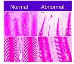
The following histologic section is from the hoof of a horse diagnosed with laminitis (indicated by abnormal). The slide demonstrates disruption of the normal attachment between the hoof wall and the distal phalanx.
Which microscopic structure has been damaged?
A. Horn tubules of the stratum mediums
B. Secondary epidermal and secondary dermal laminae
C. Dermal papillae of the coronary corium
D. Tubular horn of the sole
E. Digital cushion
Correct Answer: B.
The secondary epidermal and secondary dermal laminae form the microscopic suspensory apparatus of the equine hoof. Their extensive interdigitation creates a large surface area that securely anchors the hoof wall to the distal phalanx while distributing weight-bearing forces during locomotion. In laminitis, inflammation and degeneration weaken this attachment, allowing the distal phalanx to rotate or sink within the hoof capsule. Although the horn tubules, sole, and digital cushion all contribute to normal hoof function, they do not directly suspend the distal phalanx within the hoof capsule.

Session 1C
Significant Problem
Three histologic specimens submitted to the diagnostic laboratory have become separated from their labels during processing. The submitting veterinarians are waiting for preliminary interpretations before making clinical decisions. Your team has been asked to identify the tissue represented by each slide and determine which histologic feature provides the strongest evidence supporting your conclusion.
This histological slide comes from a wool-bearing sheep. Which description most accurately distinguishes the histologic organization of gland A from gland B?
A. A secretes by holocrine rupture of entire epithelial cells, while B secretes by apical decapitation releasing cytoplasm into the lumen.
B. A is composed of simple cuboidal to columnar epithelial cells, while B is composed of specialized stratified squamous epithelial cells.
C. A consists of lobules of lipid-filled epithelial cells, while B consists of coiled tubular glands lined by simple cuboidal to columnar epithelium.
D. A secretes by merocrine exocytosis of watery fluid, while B secretes by holocrine breakdown of entire epithelial cells.
Correct Answer: C.
On routine H&E sections, sebaceous and apocrine sweat glands are most reliably distinguished by their microscopic architecture rather than their secretory mechanism. Sebaceous glands are composed of lobules of lipid-filled epithelial cells that progressively mature toward the duct. In contrast, apocrine sweat glands are simple coiled tubular glands lined by simple cuboidal to low columnar secretory epithelium. Although Answer A correctly describes the secretory mechanisms of these glands (holocrine versus apocrine secretion), these processes cannot usually be identified directly on a routine histologic section. Instead, the gland is first identified by its characteristic microscopic appearance, from which its secretory mechanism can then be inferred. Therefore, the histologic architecture provides the strongest evidence for distinguishing these gland types on microscopic examination.
Session 2A
Which structure provides the primary physical barrier against water loss, abrasion, and microbial entry?
A. Hypodermis
B. Stratum corneum
C. Dermal papillae
D. Sebaceous gland
Correct Answer: B. Stratum corneum
Why: The stratum corneum is the major superficial keratinized barrier that limits water loss, resists abrasion, and reduces microbial penetration.
Session 2A
An animal develops moist dermatitis after repeated licking of one area. Which sequence best explains how this can disrupt integumentary homeostasis?
A. Increased moisture strengthens the stratum corneum and reduces microbial growth.
B. Moisture softens the stratum corneum, alters surface microbes, increases inflammation, and predisposes to secondary infection.
C. Moisture reduces immune activity and prevents inflammation.
D. Moisture primarily affects the hypodermis without changing the epidermal barrier.
Correct Answer: B. Moisture softens the stratum corneum, alters surface microbes, increases inflammation, and predisposes to secondary infection.
Why: Chronic moisture can weaken the physical barrier, alter the surface microenvironment, promote microbial imbalance, and increase the risk of inflammation and secondary infection.
Session 2A
Which statement best describes the role of sebum in cutaneous barrier function?
A. Sebum provides the primary blood supply to the epidermis.
B. Sebum lubricates skin and hair, supports waterproofing, reduces water loss, and contributes to antimicrobial defense.
C. Sebum detects pain and temperature changes.
D. Sebum replaces the need for immune surveillance in the dermis.
Correct Answer: B. Sebum lubricates skin and hair, supports waterproofing, reduces water loss, and contributes to antimicrobial defense.
Why: Sebum is a chemical component of the cutaneous barrier that helps lubricate the skin and coat, limit water loss, and support antimicrobial defense.
Session 2A
Why is the resident skin microbiota considered part of the cutaneous barrier?
A. It replaces keratinocytes in damaged skin.
B. It occupies ecological niches and can limit overgrowth of opportunistic organisms.
C. It prevents all inflammatory responses.
D. It produces dermal collagen and elastin.
Correct Answer: B. It occupies ecological niches and can limit overgrowth of opportunistic organisms.
Why: Resident microbial populations help stabilize the skin surface by competing for space and resources, reducing opportunities for potentially harmful organisms to overgrow.
Session 2A
Which situation most directly illustrates interaction between the physical, microbial, and immunologic components of the skin barrier?
A. A superficial abrasion allows microbial entry, triggering inflammation and immune-cell recruitment.
B. A thick hair coat completely eliminates the need for thermoregulation.
C. Vasoconstriction increases heat loss during cold exposure.
D. A sensory receptor directly produces sebum.
Correct Answer: A. A superficial abrasion allows microbial entry, triggering inflammation and immune-cell recruitment.
Why: This sequence combines disruption of the physical barrier, microbial access to underlying tissue, and activation of the immunologic response.
Session 2A
The epidermis is avascular. What does this imply about its maintenance and repair?
A. It does not require oxygen or nutrients.
B. It receives oxygen and nutrients by diffusion from the vascularized dermis.
C. It depends primarily on the hypodermis for sensory input.
D. It can repair independently of blood flow.
Correct Answer: B. It receives oxygen and nutrients by diffusion from the vascularized dermis.
Why: Because the epidermis lacks blood vessels, it depends on diffusion from dermal capillaries for oxygen and nutrients. Adequate dermal perfusion is therefore important for epidermal maintenance and repair.
Session 2A
During heat stress, cutaneous vasodilation supports thermoregulation primarily by:
A. Reducing blood flow to the skin surface to conserve heat.
B. Increasing blood flow to the skin surface to promote heat transfer to the environment.
C. Blocking sweat gland secretion in all species.
D. Increasing keratinocyte mitosis to thicken the epidermis.
Correct Answer: B. Increasing blood flow to the skin surface to promote heat transfer to the environment.
Why: Cutaneous vasodilation brings warm blood closer to the body surface, supporting heat loss through radiation, convection, conduction, and evaporation.
Session 2A
A tight bandage compresses the skin over a distal limb. Which outcome is most likely if the pressure persists?
A. Increased oxygen delivery and faster epithelial repair
B. Reduced microvascular perfusion, ischemia, ulceration, and possible necrosis
C. Increased sebaceous gland secretion and improved barrier function
D. Enhanced sensory perception without tissue damage
Correct Answer: B. Reduced microvascular perfusion, ischemia, ulceration, and possible necrosis
Why: Prolonged pressure can compress dermal microvessels, reducing oxygen and nutrient delivery. Persistent ischemia may lead to tissue breakdown, ulceration, and necrosis.
Session 2A
Which receptor type is most closely associated with detection of deep pressure and vibration?
A. Lamellated corpuscles
B. Sebaceous glands
C. Melanocytes
D. Keratinocytes
Correct Answer: A. Lamellated corpuscles
Why: Lamellated corpuscles are mechanoreceptors specialized for detecting deep pressure and vibration.
Session 2A
An animal has peripheral nerve damage affecting cutaneous sensory fibers to part of a limb. Which prediction is most appropriate?
A. The animal may have reduced touch, pain, or temperature perception and may be at increased risk of unnoticed injury.
B. The animal will have increased melanin production in that area.
C. The stratum corneum will immediately become thicker.
D. Sebaceous glands will stop producing sebum throughout the body.
Correct Answer: A. The animal may have reduced touch, pain, or temperature perception and may be at increased risk of unnoticed injury.
Why: Peripheral sensory nerve damage can reduce perception of touch, pressure, pain, or temperature, weakening protective responses and increasing the risk that injuries will go unnoticed.
Session 2A
Which component of the integument provides the primary physical barrier limiting water loss and entry of microorganisms?
A. Sensory nerve endings surrounding hair follicles
B. Subcutaneous adipose tissue of hypodermis
C. Keratinized cells and extracellular lipids of stratum corneum
D. Vascularized connective tissue of dermis
C. Correct.
The stratum corneum consists of flattened, keratinized cells embedded in a lipid-rich extracellular matrix.
This arrangement limits transepidermal water loss, resists abrasion, restricts entry of microorganisms and environmental substances, and protects viable epidermal and dermal tissues.
Session 2A
Disruption of the stratum corneum is most likely to initiate which sequence of events?
A. Increased microbial access → local immune activation → inflammation
B. Increased sebum production → immediate restoration of epidermal integrity
C. Reduced sensory signaling → increased tissue perfusion → accelerated healing
D. Reduced water loss → decreased inflammation → increased resistance to infection
A. Correct.
Damage to the stratum corneum increases skin permeability.
Microorganisms, irritants, and antigens can gain greater access to underlying tissues.
Local immune cells in the loose connective tissue detect tissue damage or microbial products and release inflammatory mediators.
The resulting inflammation may cause erythema, edema, pain, pruritus, and exudation.
Session 2A
Why is adequate dermal blood flow essential for maintenance of the epidermis?
A. Dermal vessels directly produce keratin for the stratum granulosum
B. Blood circulating through epidermis prevents microbial attachment
C. Epidermis contains large arteries that require continuous filling
Correct response
D. Epidermal keratinocytes receive oxygen and nutrients by diffusion from dermal vessels
D. Correct.
The epidermis is considered generally avascular.
Oxygen, nutrients, hormones, and other substances must diffuse from capillaries in the underlying dermis.
Reduced dermal perfusion can impair keratinocyte turnover, epidermal maintenance, barrier repair, wound healing, and tissue viability because nutrients and oxygen are not delivered to the area.
Session 2A
During exposure to a cold environment, which cutaneous vascular response most directly conserves core body heat?
A. Increased capillary permeability that promotes interstitial edema
B. Vasoconstriction that reduces transfer of core heat to the skin
C. Compression of cutaneous vessels that produces local ischemia
D. Vasodilation that increases blood flow near the skin surface
B. Correct.
Less warm blood reaches the periphery, decreasing heat loss through radiation, convection, and conduction.
This response helps preserve core temperature during cold exposure.
Session 2A
A peripheral sensory nerve supplying an area of skin is completely disrupted. Which outcome is most likely within that area?
A. Loss of sensation automatically increases local immune surveillance
B. Pain sensation is preserved because nociceptors do not require sensory neurons
C. Sensory signals fail to reach the central nervous system despite the possible presence of intact receptors
D. Receptors continue to produce normal conscious sensation because they remain in the skin
C. Correct.
Cutaneous receptors detect mechanical, thermal, or damaging stimuli.
Sensory fibers carry this information to a dorsal root ganglion (sensory ganglion) and then into the spinal cord and on to higher central nervous system centers.
If the peripheral nerve is disrupted, receptor activation cannot be transmitted normally.
Consequences may include reduced touch, pressure, pain, or temperature perception; loss of protective withdrawal responses; unnoticed injury; abnormal posture or weight bearing; and increased risk of self-trauma or tissue damage.
Session 2A
Significant Problem
A 4-year-old Labrador Retriever presents 48 hours after a distal limb bandage was placed for a superficial abrasion. The owner reports that the dog has been licking at the bandage and is now reluctant to bear weight.
On examination:
Skin beneath the bandage is moist, erythematous, and mildly eroded.
There is a small amount of superficial exudate.
The distal paw is swollen and cooler than the opposite paw.
Capillary refill time in the affected digits is delayed.
The dog strongly withdraws when the eroded skin is touched.
Light-touch response is reduced over part of the distal paw.
The dog responds normally to firm pressure applied proximal to the bandage.
No fracture is detected radiographically.
Students must determine how disruption of cutaneous barrier function, altered cutaneous perfusion, and changes in sensory signaling explain the clinical findings.
The moist, reddened, eroded skin beneath the bandage most directly reflects failure of which integrated cutaneous function?
A. Mechanical support by deep dermal collagen only
B. Coordinated physical, chemical, microbial, and immunologic barrier protection
C. Endocrine hormone release into the bloodstream
D. Voluntary motor control of the distal limb
B. Correct.
Normal skin barrier function depends on multiple interacting components:
Physical barrier: keratinized stratum corneum and tight organization of epidermal cells
Chemical barrier: surface lipids, antimicrobial molecules, and local pH
Microbial barrier: resident microbiota that help limit pathogen overgrowth
Immunologic barrier: local immune cells that detect and respond to injury or microbial invasion
The bandage-associated moisture and friction have disrupted this integrated barrier. Once the epidermis becomes macerated and eroded, water balance is altered, microbes can proliferate more easily, and inflammation develops.
Session 2A
Significant Problem
A 4-year-old Labrador Retriever presents 48 hours after a distal limb bandage was placed for a superficial abrasion. The owner reports that the dog has been licking at the bandage and is now reluctant to bear weight.
On examination:
Skin beneath the bandage is moist, erythematous, and mildly eroded.
There is a small amount of superficial exudate.
The distal paw is swollen and cooler than the opposite paw.
Capillary refill time in the affected digits is delayed.
The dog strongly withdraws when the eroded skin is touched.
Light-touch response is reduced over part of the distal paw.
The dog responds normally to firm pressure applied proximal to the bandage.
No fracture is detected radiographically.
Students must determine how disruption of cutaneous barrier function, altered cutaneous perfusion, and changes in sensory signaling explain the clinical findings.
Which consequence is most likely when prolonged moisture under the bandage disrupts the stratum corneum and surface microenvironment?
A. Increased resistance to microbial colonization
B. Decreased local inflammation
C. Maceration, microbial overgrowth, and increased activation of local immune responses
D. Immediate restoration of normal epidermal barrier function
C. Correct.
Moisture trapped beneath a bandage can soften and damage the stratum corneum. This process, called maceration, weakens the physical barrier and alters the local skin surface environment.
Barrier disruption can lead to:
Increased permeability
Microbial overgrowth
Entry of irritants or antigens
Activation of local immune responses
Erythema, exudate, and pain
This explains why the skin beneath the bandage is moist, red, eroded, and painful.
Session 2A
Significant Problem
A 4-year-old Labrador Retriever presents 48 hours after a distal limb bandage was placed for a superficial abrasion. The owner reports that the dog has been licking at the bandage and is now reluctant to bear weight.
On examination:
Skin beneath the bandage is moist, erythematous, and mildly eroded.
There is a small amount of superficial exudate.
The distal paw is swollen and cooler than the opposite paw.
Capillary refill time in the affected digits is delayed.
The dog strongly withdraws when the eroded skin is touched.
Light-touch response is reduced over part of the distal paw.
The dog responds normally to firm pressure applied proximal to the bandage.
No fracture is detected radiographically.
Students must determine how disruption of cutaneous barrier function, altered cutaneous perfusion, and changes in sensory signaling explain the clinical findings.
The affected paw is swollen, cooler than the opposite paw, and has delayed capillary refill. Which interpretation best explains these findings?
A. Reduced cutaneous perfusion can impair oxygen delivery, nutrient delivery, waste removal, and tissue viability
B. Increased keratin production directly increases blood flow to the digits
C. Loss of pain receptors increases oxygen delivery to the skin
D. Resident microbiota eliminate the need for cutaneous circulation
A. Correct.
Cutaneous blood flow supports tissue viability by delivering oxygen and nutrients while removing carbon dioxide, metabolic waste, and inflammatory byproducts.
A tight or poorly positioned bandage can impair:
Venous return
Lymphatic drainage
Arterial inflow
Microvascular exchange
The cool paw and delayed capillary refill suggest reduced peripheral perfusion. If prolonged, inadequate perfusion can worsen tissue injury and delay healing.
Session 2A
Significant Problem
A 4-year-old Labrador Retriever presents 48 hours after a distal limb bandage was placed for a superficial abrasion. The owner reports that the dog has been licking at the bandage and is now reluctant to bear weight.
On examination:
Skin beneath the bandage is moist, erythematous, and mildly eroded.
There is a small amount of superficial exudate.
The distal paw is swollen and cooler than the opposite paw.
Capillary refill time in the affected digits is delayed.
The dog strongly withdraws when the eroded skin is touched.
Light-touch response is reduced over part of the distal paw.
The dog responds normally to firm pressure applied proximal to the bandage.
No fracture is detected radiographically.
Students must determine how disruption of cutaneous barrier function, altered cutaneous perfusion, and changes in sensory signaling explain the clinical findings.
If this dog were exposed to a warm environment, which normal cutaneous vascular response would most directly support heat loss?
A. Vasoconstriction of superficial cutaneous vessels
B. Complete cessation of blood flow to the skin
C. Vasodilation of cutaneous vessels to increase blood flow near the body surface
D. Decreased perfusion of the skin to conserve core heat
C. Correct.
Cutaneous vessels participate in thermoregulation by adjusting blood flow to the skin surface.
During heat exposure, cutaneous vasodilation increases blood flow near the body's surface. This allows heat from the core to move toward the periphery, where it can be lost to the environment.
This response supports heat dissipation while also requiring adequate cardiovascular function to maintain tissue perfusion.
Session 2A
Significant Problem
A 4-year-old Labrador Retriever presents 48 hours after a distal limb bandage was placed for a superficial abrasion. The owner reports that the dog has been licking at the bandage and is now reluctant to bear weight.
On examination:
Skin beneath the bandage is moist, erythematous, and mildly eroded.
There is a small amount of superficial exudate.
The distal paw is swollen and cooler than the opposite paw.
Capillary refill time in the affected digits is delayed.
The dog strongly withdraws when the eroded skin is touched.
Light-touch response is reduced over part of the distal paw.
The dog responds normally to firm pressure applied proximal to the bandage.
No fracture is detected radiographically.
Students must determine how disruption of cutaneous barrier function, altered cutaneous perfusion, and changes in sensory signaling explain the clinical findings.
The dog has reduced response to light touch over part of the distal paw but strongly withdraws when the eroded skin is pressed. Which explanation best fits this pattern?
A. All sensory pathways in the paw are nonfunctional
B. Different sensory modalities use different receptors and neural pathways, so light touch may be reduced while nociception remains active
C. Pain perception requires intact sebaceous glands
D. Thermoreceptors are responsible for all forms of cutaneous sensation
B. Correct.
Cutaneous sensation is not a single pathway. Different receptors and sensory fibers detect different modalities:
Merkel cells and tactile receptors: light touch and sustained pressure
Pacinian corpuscles: deep pressure and vibration
Ruffini endings: stretch
Thermoreceptors: temperature change
Nociceptors/free nerve endings: painful or damaging stimuli
Compression, swelling, or inflammation may alter some sensory fibers more than others. Therefore, light-touch sensation may be reduced in one region while nociceptive withdrawal remains strong at the eroded, inflamed site.
Session 2B
Which integumentary response primarily increases heat loss by bringing warm blood closer to the body surface?
A. Cutaneous vasoconstriction
B. Cutaneous vasodilation
C. Piloerection
D. Sebaceous secretion
Correct Answer: B. Cutaneous vasodilation
Why: Vasodilation increases blood flow near the skin surface, promoting heat transfer from the body to the environment.
Session 2B
During cold exposure, cutaneous vasoconstriction helps maintain body temperature by:
A. Increasing evaporative cooling from the skin surface.
B. Reducing blood flow to the skin surface and conserving core heat.
C. Increasing sweat production.
D. Increasing microbial activity on the skin.
Correct Answer: B. Reducing blood flow to the skin surface and conserving core heat.
Why: Cutaneous vasoconstriction decreases warm blood flow to the body surface, limiting heat loss and helping preserve core temperature.
Session 2B
Which combination best describes the major integumentary mechanisms involved in thermoregulation?
A. Keratinocyte mitosis, melanin synthesis, collagen remodeling, and scar formation
B. Vasomotor responses, evaporative cooling, insulation, and behavioral adaptations
C. Sebum secretion, wound contraction, pigmentation, and lymph drainage
D. Pain perception, hair growth, immune surveillance, and vitamin D metabolism
Correct Answer: B. Vasomotor responses, evaporative cooling, insulation, and behavioral adaptations
Why: Thermoregulation depends on adjustments in cutaneous blood flow, evaporation, insulation from hair and subcutaneous tissue, and behaviors such as seeking shade or cool surfaces.
Session 2B
Why is sweating especially important in horses during exercise or heat exposure?
A. Horses lack cutaneous blood vessels.
B. Horses rely heavily on sweating for evaporative cooling.
C. Horses cannot use behavioral thermoregulation.
D. Horses sweat only to remove metabolic waste, not to regulate temperature.
Correct Answer: B. Horses rely heavily on sweating for evaporative cooling.
Why: Evaporation of sweat removes heat from the body surface, making sweating a major cooling mechanism for horses during exercise and heat exposure.
Session 2B
A dog confined in a hot, poorly ventilated area develops signs of heat stress. Which explanation best describes the physiologic problem?
A. Dogs rely primarily on widespread sweating, which becomes excessive.
B. Panting and behavioral cooling are overwhelmed by environmental heat load.
C. Cutaneous vasoconstriction causes excessive heat loss.
D. Sebaceous glands fail to produce enough sebum for cooling.
Correct Answer: B. Panting and behavioral cooling are overwhelmed by environmental heat load.
Why: Dogs rely heavily on panting, peripheral vasodilation, posture changes, shade seeking, and environmental cooling. In a hot, poorly ventilated space, these mechanisms may be insufficient to dissipate heat.
Session 2B
Which statement best explains why pigs are particularly vulnerable to heat stress?
A. They rely heavily on sweating from the entire body surface.
B. They have limited functional sweating and depend strongly on behavior and environmental cooling.
C. Their hair coat always prevents heat loss.
D. They cannot increase respiratory effort during heat exposure.
Correct Answer: B. They have limited functional sweating and depend strongly on behavior and environmental cooling.
Why: Because pigs have limited capacity for sweating, they depend heavily on shade, wallowing, cool surfaces, water access, and appropriate environmental management.
Session 2B
What is the primary function of sebum in integumentary homeostasis?
A. To carry oxygen to the epidermis
B. To lubricate skin and hair, support waterproofing, reduce water loss, and contribute to antimicrobial defense
C. To detect deep pressure and vibration
D. To directly regulate heart rate during heat stress
Correct Answer: B. To lubricate skin and hair, support waterproofing, reduce water loss, and contribute to antimicrobial defense
Why: Sebum is a chemical component of the cutaneous barrier that supports skin and coat lubrication, limits water loss, and contributes to antimicrobial protection.
Session 2B
A horse with anhidrosis is at increased risk for which problem?
A. Impaired evaporative cooling and hyperthermia
B. Excessive insulation from subcutaneous fat
C. Overproduction of sebum and reduced sweating
D. Reduced pigmentation and increased ultraviolet exposure only
Correct Answer: A. Impaired evaporative cooling and hyperthermia
Why: Anhidrosis reduces or prevents sweating, impairing one of the horse's major heat-loss mechanisms and increasing the risk of dangerous elevations in body temperature.
Session 2B
Which systemic consequence can result from excessive sweating in species that sweat substantially?
A. Increased hydration and plasma volume
B. Dehydration and electrolyte imbalance
C. Reduced heat production by skeletal muscle
D. Complete prevention of heat stress
Correct Answer: B. Dehydration and electrolyte imbalance
Why: Substantial sweating causes fluid and electrolyte losses that can reduce plasma volume, impair performance, and make continued thermoregulation more difficult.
Session 2B
Which scenario best illustrates how glandular dysfunction can affect systemic homeostasis rather than only the skin surface?
A. Reduced mammary gland function leads to poor neonatal nutrition and hydration.
B. Increased hair length causes improved tactile perception.
C. Increased pigmentation prevents all skin disease.
D. A normal sebaceous gland maintains normal coat lubrication.
Correct Answer: A. Reduced mammary gland function leads to poor neonatal nutrition and hydration.
Why: The mammary gland is a modified integumentary exocrine gland. Dysfunction can compromise milk production, directly affecting neonatal nutrition, hydration, and systemic health.
Session 2B
A horse is sweating heavily during exercise in hot, humid weather. Which condition most directly limits the cooling effect of the sweat?
A. Increased cutaneous blood flow delivers more heat to the skin surface
B. High environmental humidity reduces evaporation of water from the skin
C. Increased sweat production removes too much heat from the body
D. Increased air movement causes sweat to remain liquid on the skin
B. Correct.
Evaporative cooling occurs when liquid water changes to water vapor.
This phase change requires energy and removes heat from the body surface.
High environmental humidity reduces the water-vapor gradient between the skin and the surrounding air.
As a result, sweat may remain on the skin rather than evaporating.
Sweat production alone does not ensure effective cooling; evaporation must occur.
Session 2B
A neonatal lamb becomes wet shortly after birth in a cold environment. Why does the wet coat increase the risk of hypothermia?
A. Wet hair increases the thickness of the insulating air layer next to the skin.
B. Wet hair causes cutaneous vasodilation that temporarily increases core temperature.
C. Water on the coat prevents conductive and convective heat transfer
D. Wetting collapses the trapped air layer and increases conductive and evaporative heat loss
D. Correct.
Neonates are especially vulnerable because they have a high surface area relative to body mass, limited energy reserves, immature thermoregulatory responses, and limited behavioral ability to seek shelter or dry themselves.
A dry hair coat traps a layer of relatively still air near the skin.
This trapped air reduces conductive and convective heat transfer.
When the coat becomes wet, the trapped air layer collapses, water increases conductive heat transfer away from the body, and evaporation removes additional heat.
Session 2B
Which response is most important for a pig exposed to high environmental temperature?
A. Seeking shade, wallowing, lying on cool surfaces, and reducing activity
B. Generalized sweating over the body surface
C. Increasing piloerection to trap warm air next to the skin
D. Cutaneous vasoconstriction to conserve core heat
A. Correct.
Pigs have limited effective sweating and therefore cannot depend heavily on cutaneous evaporation.
They rely strongly on behavioral thermoregulation, including seeking shade, wallowing or wetting the skin, lying on cool surfaces, spreading out, reducing activity, and increasing access to water and airflow.
Preventing these behaviors substantially increases the risk of heat stress.
Session 2B
Which statement best explains how sebaceous glands contribute to integumentary function?
A. They release hormones directly into the blood to regulate body temperature
B. They generate sensory impulses in response to pain and temperature
C. They secrete lipid-rich lubricants onto the skin and coat and support surface-barrier homeostasis
D. They deliver oxygen and nutrients directly to epidermal keratinocytes
C. Correct.
Sebaceous glands are exocrine glands commonly associated with hair follicles.
They release sebum onto the skin surface or into hair follicles.
Sebum contributes to skin lubrication, conditioning of the hair coat, maintenance of skin pliability, reduction of water loss, waterproofing, antimicrobial defense, and stabilization of the cutaneous surface environment.
Abnormal sebaceous secretion can create various dermatitis conditions such as scaling (too little), greasy coat (too much), odor, and/or microbial imbalance.
Session 2B
A horse with reduced sweat production (anhidrosis) continues exercising in a hot environment. Which outcome is most likely if heat loss remains inadequate?
A. Improved heat tolerance because less fluid is lost from the skin
B. Progressive hyperthermia, exercise intolerance, and risk of organ injury
C. Increased insulation caused by loss of sebaceous secretion
D. Reduced metabolic stress because cellular enzyme activity decreases normally
B. Correct.
Horses rely heavily on sweating for evaporative heat loss.
Reduced or absent sweating limits the animal’s ability to dissipate metabolic and environmental heat.
Cutaneous vasodilation may still transfer heat toward the body surface, but heat removal remains inadequate without evaporation.
Continued heat accumulation may cause progressive hyperthermia, exercise intolerance, tachypnea, tachycardia, weakness (asthenia), reduced performance, dehydration, circulatory stress, cellular and organ injury, heat stroke, collapse, or death.
Session 2B
Significant Problem
During a summer livestock exhibition, several animals are evaluated by the veterinary team after prolonged exposure to high environmental temperature and humidity.
The following patients are presented:
Patient 1: Horse
A 9-year-old Quarter Horse has been exercised lightly but is now depressed, tachycardic, tachypneic, and sweating heavily. Rectal temperature is 41.0°C. Sweat is visible over the neck, thorax, and flanks, but the skin remains wet rather than drying.
Patient 2: Dog
A 5-year-old brachycephalic Bulldog is panting intensely, anxious, and weak. Rectal temperature is 40.8°C. The dog has minimal sweating except from the paw pads.
Patient 3: Pig
A 3-year-old show pig is restless, seeking shade, lying near spilled water, and breathing rapidly. Rectal temperature is 40.9°C. The pig has little visible sweating.
Students must determine how integumentary thermoregulation, exocrine gland function, species differences, and systemic homeostatic consequences explain the clinical findings.
Which statement best explains why these species differ in their ability to dissipate heat through the integument?
A. Dogs, horses, and pigs all rely primarily on sweating as their major cooling mechanism.
B. Horses rely heavily on sweating, dogs rely heavily on panting with limited sweating, and pigs rely more on behavioral cooling because sweating is limited.
C. Pigs are more efficient than horses at evaporative cooling because they have more functional sweat glands.
D. Dogs primarily dissipate heat through sebaceous gland secretion.
B. Correct.
Thermoregulatory mechanisms differ substantially across veterinary species.
Horses depend heavily on sweating for evaporative cooling.
Dogs have limited sweating, mainly from paw pads, and rely primarily on panting for evaporative heat loss.
Pigs have limited effective sweating and depend strongly on behavioral mechanisms such as seeking shade, lying on cool surfaces, wallowing, and reducing activity.
This explains why the same environmental condition can produce heat stress through different physiologic limitations in different species.
Session 2B
Significant Problem
During a summer livestock exhibition, several animals are evaluated by the veterinary team after prolonged exposure to high environmental temperature and humidity.
The following patients are presented:
Patient 1: Horse
A 9-year-old Quarter Horse has been exercised lightly but is now depressed, tachycardic, tachypneic, and sweating heavily. Rectal temperature is 41.0°C. Sweat is visible over the neck, thorax, and flanks, but the skin remains wet rather than drying.
Patient 2: Dog
A 5-year-old brachycephalic Bulldog is panting intensely, anxious, and weak. Rectal temperature is 40.8°C. The dog has minimal sweating except from the paw pads.
Patient 3: Pig
A 3-year-old show pig is restless, seeking shade, lying near spilled water, and breathing rapidly. Rectal temperature is 40.9°C. The pig has little visible sweating.
Students must determine how integumentary thermoregulation, exocrine gland function, species differences, and systemic homeostatic consequences explain the clinical findings.
The horse is sweating heavily, but the skin remains wet, and body temperature continues to rise. What best explains the failure of cooling?
A. Sweating increases heat production by the skin.
B. Sweat must evaporate from the skin surface to remove heat effectively.
C. Sweat cools only when it remains on the skin as a liquid layer.
D. Sweating prevents cutaneous blood flow from increasing.
B. Correct.
Sweat glands secrete fluid onto the skin surface, but cooling occurs primarily when that fluid evaporates.
Evaporation requires heat energy, which is removed from the body surface.
In high humidity, the air is already saturated with water vapor, so evaporation is reduced. Sweat may remain on the skin without efficiently removing heat. Therefore, an animal may sweat heavily and still become hyperthermic.
Session 2B
Significant Problem
During a summer livestock exhibition, several animals are evaluated by the veterinary team after prolonged exposure to high environmental temperature and humidity.
The following patients are presented:
Patient 1: Horse
A 9-year-old Quarter Horse has been exercised lightly but is now depressed, tachycardic, tachypneic, and sweating heavily. Rectal temperature is 41.0°C. Sweat is visible over the neck, thorax, and flanks, but the skin remains wet rather than drying.
Patient 2: Dog
A 5-year-old brachycephalic Bulldog is panting intensely, anxious, and weak. Rectal temperature is 40.8°C. The dog has minimal sweating except from the paw pads.
Patient 3: Pig
A 3-year-old show pig is restless, seeking shade, lying near spilled water, and breathing rapidly. Rectal temperature is 40.9°C. The pig has little visible sweating.
Students must determine how integumentary thermoregulation, exocrine gland function, species differences, and systemic homeostatic consequences explain the clinical findings.
During early heat stress, which cutaneous vascular response supports the transfer of heat from the body core toward the body surface?
A. Cutaneous vasodilation
B. Cutaneous vasoconstriction
C. Complete closure of superficial capillary beds
D. Reduced cardiac output with reduced skin perfusion
A. Correct.
During heat stress, cutaneous vasodilation increases blood flow to the skin.
Warm blood from the body core is brought closer to the surface, where heat can be lost by radiation, convection, conduction, and evaporation.
This mechanism is especially important when paired with sweating or other evaporative mechanisms. However, it also places demands on the cardiovascular system because blood volume must support both skin perfusion and vital organ perfusion.
Session 2B
Significant Problem
During a summer livestock exhibition, several animals are evaluated by the veterinary team after prolonged exposure to high environmental temperature and humidity.
The following patients are presented:
Patient 1: Horse
A 9-year-old Quarter Horse has been exercised lightly but is now depressed, tachycardic, tachypneic, and sweating heavily. Rectal temperature is 41.0°C. Sweat is visible over the neck, thorax, and flanks, but the skin remains wet rather than drying.
Patient 2: Dog
A 5-year-old brachycephalic Bulldog is panting intensely, anxious, and weak. Rectal temperature is 40.8°C. The dog has minimal sweating except from the paw pads.
Patient 3: Pig
A 3-year-old show pig is restless, seeking shade, lying near spilled water, and breathing rapidly. Rectal temperature is 40.9°C. The pig has little visible sweating.
Students must determine how integumentary thermoregulation, exocrine gland function, species differences, and systemic homeostatic consequences explain the clinical findings.
Which statement best describes how integumentary exocrine glands contribute to both local skin function and systemic homeostasis?
A. Exocrine glands release hormones directly into the blood to regulate distant organs
B. Exocrine glands secrete onto epithelial surfaces through ducts, supporting skin surface function, evaporative cooling, and protection of the hair coat
C. Exocrine glands function only during disease and are inactive in healthy animals
D. Exocrine glands are sensory receptors that detect temperature and pain
B. Correct.
Exocrine glands secrete onto epithelial surfaces through ducts.
In the integument, sweat glands contribute to evaporative cooling, especially in species such as horses.
Sebaceous glands secrete lipid-rich material that helps maintain the hair coat and skin surface.
Glandular secretions help maintain the skin surface environment and can indirectly support barrier function.
Because sweating can influence body temperature, fluid balance, and electrolyte balance, integumentary exocrine glands can contribute to systemic homeostasis.
Session 2B
Significant Problem
During a summer livestock exhibition, several animals are evaluated by the veterinary team after prolonged exposure to high environmental temperature and humidity.
The following patients are presented:
Patient 1: Horse
A 9-year-old Quarter Horse has been exercised lightly but is now depressed, tachycardic, tachypneic, and sweating heavily. Rectal temperature is 41.0°C. Sweat is visible over the neck, thorax, and flanks, but the skin remains wet rather than drying.
Patient 2: Dog
A 5-year-old brachycephalic Bulldog is panting intensely, anxious, and weak. Rectal temperature is 40.8°C. The dog has minimal sweating except from the paw pads.
Patient 3: Pig
A 3-year-old show pig is restless, seeking shade, lying near spilled water, and breathing rapidly. Rectal temperature is 40.9°C. The pig has little visible sweating.
Students must determine how integumentary thermoregulation, exocrine gland function, species differences, and systemic homeostatic consequences explain the clinical findings.
The horse continues to sweat, the Bulldog continues to pant, and the pig continues to seek shade, but all three animals remain hyperthermic. Which systemic consequence is most likely if thermoregulation continues to fail?
A. Increased integumentary insulation and improved cooling
B. Decreased metabolic stress and improved cellular enzyme function.
C. Progressive hyperthermia, dehydration, electrolyte imbalance, circulatory compromise, and cellular injury
D. Immediate restoration of homeostasis without cardiovascular involvement
C. Correct.
If heat loss is inadequate, body temperature continues to rise. Persistent hyperthermia can disrupt cellular proteins, enzyme function, membrane stability, and vascular regulation.
Sweating can contribute to fluid and electrolyte loss. Panting can alter respiratory water loss and acid-base balance. Peripheral vasodilation can challenge blood pressure regulation.
Severe heat stress can progress to circulatory compromise, tissue injury, collapse, or death. Thus, thermoregulatory failure is a systemic homeostatic emergency, not simply a skin problem.
Session 3A
A skin lesion is described as a small, flat area of color change with no elevation and no palpable thickness. Which primary lesion type does this describe?
A. Papule, a small raised solid lesion
B. Pustule, a small raised lesion filled with purulent material
C. Vesicle, a small raised lesion filled with clear fluid
D. Macule, a flat area of color change without elevation
D. Correct.
A macule is defined specifically by the absence of elevation or palpable thickness; it is a color change only. The description given (flat, no elevation, no palpable thickness) matches this definition exactly.
Session 3A
A secondary skin lesion is produced when an animal repeatedly scratches or rubs an area, causing linear breaks in the skin surface from self-trauma. Which secondary lesion does this mechanism describe?
A. Excoriation, a surface break caused by self-trauma such as scratching
B. Lichenification, a thickening and increased skin markings from chronic rubbing
C. Crust, a dried accumulation of surface exudate
D. Scale, an accumulation of loose fragments of stratum corneum
A. Correct.
Excoriation is defined as a linear erosion or break in the skin surface caused by mechanical self-trauma, such as scratching or rubbing hard enough to break the surface. The described mechanism, repeated scratching producing linear breaks, matches this definition precisely.
Session 3A
Acantholysis refers to the loss of normal cohesion between keratinocytes within the epidermis. Which primary lesion is most directly produced by this specific cellular change?
A. Nodule, from proliferation of cells in the deep dermis
B. Wheal, from acute fluid shift driven by mast cell mediators
C. Pustule or vesicle, from separation of keratinocytes creating a space within the epidermis
D. Plaque, from coalescence of multiple adjacent papules
C. Correct.
When keratinocytes lose their normal adhesion to one another, a cleft or space opens up within the epidermis itself. That intraepidermal space then fills with fluid or inflammatory cells, producing a vesicle or pustule depending on its contents. This is a direct, single-step mechanistic link between the cellular change and the resulting lesion.
Session 3A
A dermatitis pattern is classified based on which structure or compartment the inflammatory infiltrate is concentrated around or within. Which pairing of pattern and defining location is correct?
A. Interface dermatitis is defined by infiltrate concentrated around superficial dermal blood vessels.
B. Perivascular dermatitis is defined by infiltrate concentrated around superficial dermal blood vessels
C. Nodular dermatitis is defined by infiltrate confined within the epidermis
D. Folliculitis is defined by an infiltrate concentrated at the dermal-epidermal junction
B. Correct.
Perivascular dermatitis is named for and defined by its location: inflammatory cells concentrated around dermal blood vessels, most often in the superficial dermis. This is the only pairing in the list where the pattern name and the defining location actually match.
Session 3A
Folliculitis, furunculosis, and cellulitis represent three stages of increasing severity in the same disease continuum. Which sequence correctly orders these from earliest to most advanced?
A. Cellulitis, then furunculosis, then folliculitis
B. Furunculosis, then cellulitis, then folliculitis
C. Cellulitis, then folliculitis, then furunculosis
D. Folliculitis, then furunculosis, then cellulitis
D. Correct.
Folliculitis is inflammation confined to the hair follicle and represents the earliest stage. When the follicle wall ruptures, its contents spill into the surrounding dermis, producing furunculosis a deeper, more destructive stage. If inflammation then spreads beyond the original follicular focus into the surrounding soft tissue, it becomes cellulitis, the most advanced and most diffuse stage. Each stage represents the inflammatory process extending further from its follicular origin than the one before it.
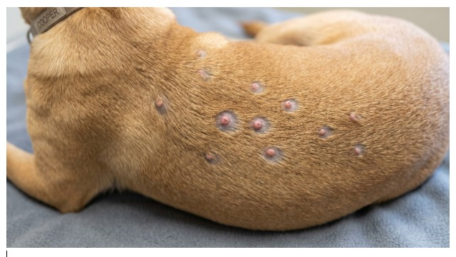
Session 3A
Significant Problem
A 3-year-old short-coated dog presents with roughly a dozen small, raised bumps scattered across the dorsal trunk, each about the size of a pinhead, most with a fine ring of hair loss immediately around them. On close exam, an intact hair shaft can be seen emerging from the center of several bumps. Gentle digital pressure on an intact bump produces a small amount of creamy material at the surface; there is no fluctuant swelling beneath the skin and no drainage tracts anywhere on the trunk. The dog does not flinch when the area is palpated.
Which lesion type and structural compartment best explain this presentation?
A. Bullae, reflecting fluid accumulation at the dermal-epidermal junction
B. Pustules, reflecting a follicle-centered process not yet extending into deeper tissue
C. Nodules, reflecting an inflammatory process already established in the deep dermis
D. Wheals, reflecting acute fluid shift into the superficial dermis
B. Correct.
Several findings have to be put together rather than read off individually. An intact hair shaft visibly emerging from the center of each bump tells you the follicle itself is still structurally intact and is the anatomic center of the lesion. Creamy material expressed with only gentle pressure tells you the contents are sitting at or near the surface, not walled off in a deeper compartment. The absence of fluctuant swelling and the dog's lack of pain on palpation both argue against the process having broken through the follicular wall into the surrounding dermis. Put together, this is a small, intact, surface-accessible, follicle-centered pustule, the earliest stage of a follicular inflammatory process, before any rupture has occurred
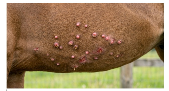
Session 3A
Significant Problem
A horse housed on pasture in northern Florida develops multiple small, intensely pruritic papules and crusts concentrated along the mane, tail base, and ventral midline during the warm months, recurring each year at the same time. The horse rubs the affected areas raw against fence posts but has no intact blisters anywhere, and the skin does not separate or peel when firmly rubbed at the lesion margins. A biopsy from an early papule is read as showing a cellular infiltrate dominated by eosinophils, most heavily concentrated immediately surrounding small blood vessels in the upper dermis; the basal cell layer of the epidermis is reported as intact and unremarkable.
Which dermatitis pattern is most consistent with this presentation, and what specifically supports that choice over the alternative?
A. Interface dermatitis, because the recurrent and intensely pruritic nature of the lesions points to chronic basal cell injury
B. Intraepidermal pustular dermatitis, because the lesions are described as crusted and the horse is rubbing them open
C. Perivascular dermatitis, because the infiltrate centers on dermal vessels and the basal epidermal layer is unaffected
D. Nodular dermatitis, because the lesions recur in the same anatomic sites every year
C. Correct.
Two findings have to be weighed against each other, not just located. The biopsy explicitly places the dominant infiltrate around dermal vessels, which is the structural definition of a perivascular pattern — but the case also has to rule out interface dermatitis, which is the pattern most likely to be confused with this presentation given the chronic, recurrent, intensely pruritic picture. The biopsy reports the basal cell layer as intact and unremarkable, which is the specific finding that excludes interface dermatitis, since that pattern requires basal keratinocyte injury at the junction. The absence of intact blisters and the lack of epidermal separation on rubbing further argue against a junctional process. Seasonal recurrence and rubbing behavior describe the clinical course, not the histologic pattern, and should not be used to select an answer on their own.

Session 3A
Significant Problem
A dairy cow develops a cluster of follicular pustules on the rear limb that, over one week, progress to firm, painful subcutaneous swelling with intermittent rupture and drainage of purulent material. The surrounding skin is now diffusely thickened, warm, and edematous beyond the original lesion borders.
Which progression of disease best describes the current presentation?
A. Folliculitis has progressed through furunculosis into early cellulitis
B.This remains simple folliculitis without further progression
C. This represents a primary interface dermatitis without follicular involvement
D. This represents a primary perivascular hypersensitivity reaction.
A. Correct.
The case describes a clear progression: follicular pustules (folliculitis) that ruptured into firm, painful subcutaneous swelling (furunculosis, where the follicle ruptures and releases its contents into the dermis), now accompanied by diffuse thickening, warmth, and edema extending beyond the original lesion borders (early cellulitis, where inflammation spreads through the surrounding soft tissue beyond the original follicular focus). Recognizing this sequence as a continuum, not three unrelated findings, is the central reasoning skill this case is built to test.
Session 3B
Immediately after a skin wound occurs, platelets aggregate at the site and a fibrin clot forms, creating a provisional matrix. Which phase of wound healing does this describe?
A. Hemostasis, the earliest phase involving platelet aggregation and clot formation
B. Inflammation, the phase involving neutrophil and macrophage debridement
C. Proliferation, the phase involving granulation tissue and new capillary formation
D. Remodeling, the phase involving collagen reorganization and scar maturation
A. Correct.
Platelet aggregation and fibrin clot formation are the defining events of hemostasis, the first phase of wound healing. This phase occurs immediately after injury and establishes the provisional matrix that subsequent phases build upon.
Session 3B
Which sequence correctly orders the four phases of integumentary wound healing from earliest to latest?
A. Inflammation, hemostasis, remodeling, proliferation
B. Proliferation, hemostasis, inflammation, remodeling
C. Remodeling, proliferation, inflammation, hemostasis
D. Hemostasis, inflammation, proliferation, remodeling
D. Correct.
Wound healing proceeds in this order: hemostasis establishes the initial clot and provisional matrix; inflammation follows, with neutrophils and macrophages debriding the wound; proliferation builds granulation tissue, new capillaries, and re-epithelialization; and remodeling reorganizes collagen and matures the scar over the longest time course. Each phase depends on the structures or signals established by the preceding phase.
Session 3B
A wound with closely apposed edges, sutured together with minimal tissue gap, is expected to heal with little granulation tissue formation. Which type of healing does this describe?
A. Second intention healing, defined by wide tissue gaps and reliance on granulation tissue to fill the defect
B. First intention healing, defined by apposed wound edges and minimal granulation tissue requirement
C. Remodeling, defined by collagen reorganization occurring independently of wound edge apposition
D. Contraction, defined by myofibroblast activity drawing wound margins together over time
B. Correct.
First intention healing is defined by closely apposed wound edges, typically achieved through suturing, which minimizes the tissue gap that needs to be filled. Because there is little gap to bridge, minimal granulation tissue is required and healing proceeds primarily through reepithelialization.
Session 3B
Second intention healing generally takes longer than first intention healing and produces a more visible scar. Which mechanistic reason best explains why?
A. Second intention wounds skip the inflammatory phase entirely, which delays the start of healing
B. Second intention wounds receive less blood supply by definition, regardless of location
C. Second intention wounds require more granulation tissue and contraction to close a larger gap, generating more new tissue and a larger scar
D. Second intention wounds are always contaminated, and contamination alone accounts for the slower time course
C. Correct.
Because second intention wounds have a larger tissue gap that is not surgically closed, the wound must generate more granulation tissue and rely more heavily on wound contraction to bring the margins together. Producing this additional tissue takes more time than simple reepithelialization across apposed edges, and the larger volume of new tissue is what produces a more visible scar.
Session 3B
Adequate blood supply delivers the oxygen and nutrients required for fibroblast activity and new capillary growth during wound healing. Which phase of healing would be most directly impaired by poor local perfusion?
A. Proliferation, since granulation tissue formation depends on oxygen and nutrient delivery to support new tissue growth
B. Hemostasis, since platelet aggregation requires more oxygen than any other phase of healing
C. Remodeling, since collagen reorganization occurs independently of blood supply at the wound site
D. Inflammation, since neutrophils and macrophages do not require oxygen to function at the wound site
A. Correct.
Proliferation is the phase most dependent on a robust blood supply, since fibroblasts require oxygen and nutrients to synthesize new collagen and matrix, and new capillary growth (angiogenesis) is part of building the vascular supply the tissue needs. Poor local perfusion directly limits the resources available for this tissue-building phase.

Session 3B
Significant Problem
A dog sustains a laceration on the lateral thigh after running through a fence. By the time the dog is seen six hours later, the wound edges are jagged and gape 2 centimeters apart. The surrounding tissue is moderately swollen, and the wound bed contains visible dirt and plant debris that cannot be fully flushed out in a single attempt. The owner elects conservative management rather than surgical closure.
Given how this wound will be allowed to heal, which trajectory should be expected over the following weeks?
A. Healing primarily through reepithelialization alone, with minimal granulation tissue and a thin, barely visible scar
B. Healing within 24 to 48 hours, since clean wound edges always heal faster regardless of contamination
C. Healing through granulation tissue formation and wound contraction over a longer time course, with a more visible scar than a closed wound
D. Healing with no inflammatory phase at all, since the wound was not surgically closed
C. Correct.
Several features here point to second-intention healing rather than first: the wound edges are gapped rather than apposed, the tissue is contaminated with debris that cannot be fully removed, and the decision was made not to surgically close it. When a wound is left open like this, it must fill the gap itself, which requires the formation of granulation tissue and wound contraction to draw the margins inward over time. This is a fundamentally slower process than a cleanly apposed, sutured wound, and it characteristically leaves a larger, more visible scar because more new tissue must be generated to fill the defect.

Session 3B
Significant Problem
A horse sustained a deep laceration on the distal aspect of the cannon bone region three weeks ago. The wound was left to heal without surgical closure. On recheck, the wound bed is filled with a mass of soft, dark red, bumpy tissue that now bulges above the surrounding skin surface and bleeds easily when touched. The margins of the surrounding normal skin show no evidence that reepithelialization is advancing over the raised tissue.
What is happening at the wound bed, and why is this particular location prone to this outcome?
A. Granulation tissue has formed and stopped appropriately; the bulging appearance is a normal feature of second-intention healing at any body location
B. Granulation tissue production has become excessive and is now physically blocking epithelial migration, an outcome distal limb wounds are prone to due to high motion and limited surrounding soft tissue
C. The wound has become infected with a fungal organism, which is why the tissue bleeds easily when touched
D. The wound is undergoing normal wound contraction, and the bulging tissue will flatten on its own within another week
B. Correct.
Normal granulation tissue should provide a surface over which epithelial cells migrate inward from the wound margins, eventually covering the defect. Here, the tissue has instead grown above the skin surface, and the margin shows no advancing reepithelialization, which means the granulation tissue itself has become the obstacle rather than the substrate for healing. The distal limb is specifically prone to this because it has constant motion, minimal overlying muscle, and comparatively limited blood supply and skin laxity compared to wounds on the trunk; the same biological drivers that make this location vulnerable to delayed healing also predispose it to this overgrowth response once granulation tissue begins forming.

Session 3B
Significant Problem
A dairy cow in late lactation, currently in a herd-wide period of restricted forage availability and documented low body condition score, sustains a moderate skin laceration on the flank from a fence post. Two weeks later, the wound shows minimal granulation tissue formation, little wound contraction, and wide margins with no clinical sign of active infection. A herd mate with normal body condition who sustained a similar wound at the same time has healed substantially further.
What is the most likely explanation for the difference in healing progress between these two animals?
A. The affected cow's wound has likely become infected, as infection is the most common cause of delayed healing in cattle.
B. Wound healing rate in cattle is determined almost entirely by wound location, so the difference reflects where each wound is positioned on the body
C. The herdmate's wound was smaller, and wound size alone explains essentially all of the difference in healing speed observed
D. The affected cow's poor nutritional and body condition status is limiting the protein and energy substrate needed for fibroplasia and collagen deposition, slowing the proliferative phase of healing.
D. Correct.
Granulation tissue formation and collagen deposition are metabolically demanding processes that depend on adequate protein and energy availability to support fibroblast activity and new tissue synthesis. A cow in a herd-wide period of restricted forage with a documented low body condition score is in a systemic state of nutritional deficit, which directly limits the substrate available for these proliferative-phase processes. This is offered as the explanation specifically because the case provides a same-timing, same-injury-type comparison animal in better body condition who is healing further along, isolating systemic nutritional status as the variable that differs between the two animals while ruling out timing or wound type as confounds.
Session 3C
A superficial skin lesion has lost its crust and now has an exposed, moist surface. The clinician wants to collect cells by placing a glass slide directly against the lesion. Which sampling method is being used?
A. Fine-needle aspiration
B. Impression smear
C. Tape preparation
D. Swab preparation
B. Correct.
An impression smear collects cells by pressing a glass slide directly onto an exposed, moist, or exudative lesion surface.
Session 3C
A dog has intact, lichenified skin, and superficial Malassezia overgrowth is suspected. Which cytology collection method is most appropriate for collecting surface debris and organisms?
A. Tape preparation
B. Fine-needle aspiration
C. Impression smear
D. Deep tissue biopsy
C. Correct.
Tape preparation is useful for collecting superficial epidermal debris and organisms such as Malassezia from intact skin.