Fertilization
 Call Kai
 Learn
Learn Practice Test
 Spaced Repetition
 Match
Match Flashcards
 Knowt Play
1/45

There's no tags or description
Looks like no tags are added yet.
Name | Mastery | Learn | Test | Matching | Spaced | Call with Kai | Chat |
|---|
No analytics yet
Send a link to your students to track their progress
46 Terms
day 0-1
fertilization —> zygote forms; sperm pierces the zona pellucida
day 2-3

end of day 3

day 4
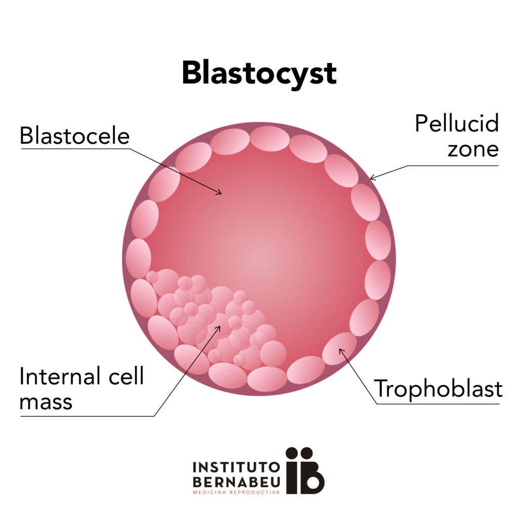
differentiation into trophoblast; blastocyst forms
day 7
hypoblast delaminates from inner cell mass
day 8
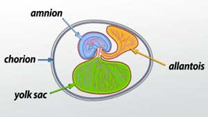
bilaminar disc (epiblast + hypoblast); amnion and primary yolk sac appear
day 8-12
growth
day 12
extraembryonic mesoderm and extraembryonic coelom form

day 13
connecting stalk and prochordal plate form; oval disc
day 15
primitive streak appears
day 17
cloacal membrane forms; gastrulation —> trilaminar embryo
day 17-21
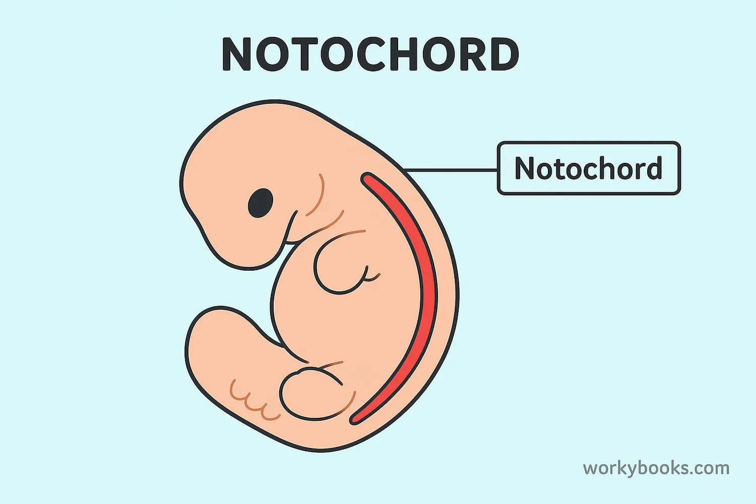
notochord forms and grows cranial
day 20-21
neurulation (neural folds) —> neural groove —> neural tube; nueral crest appears
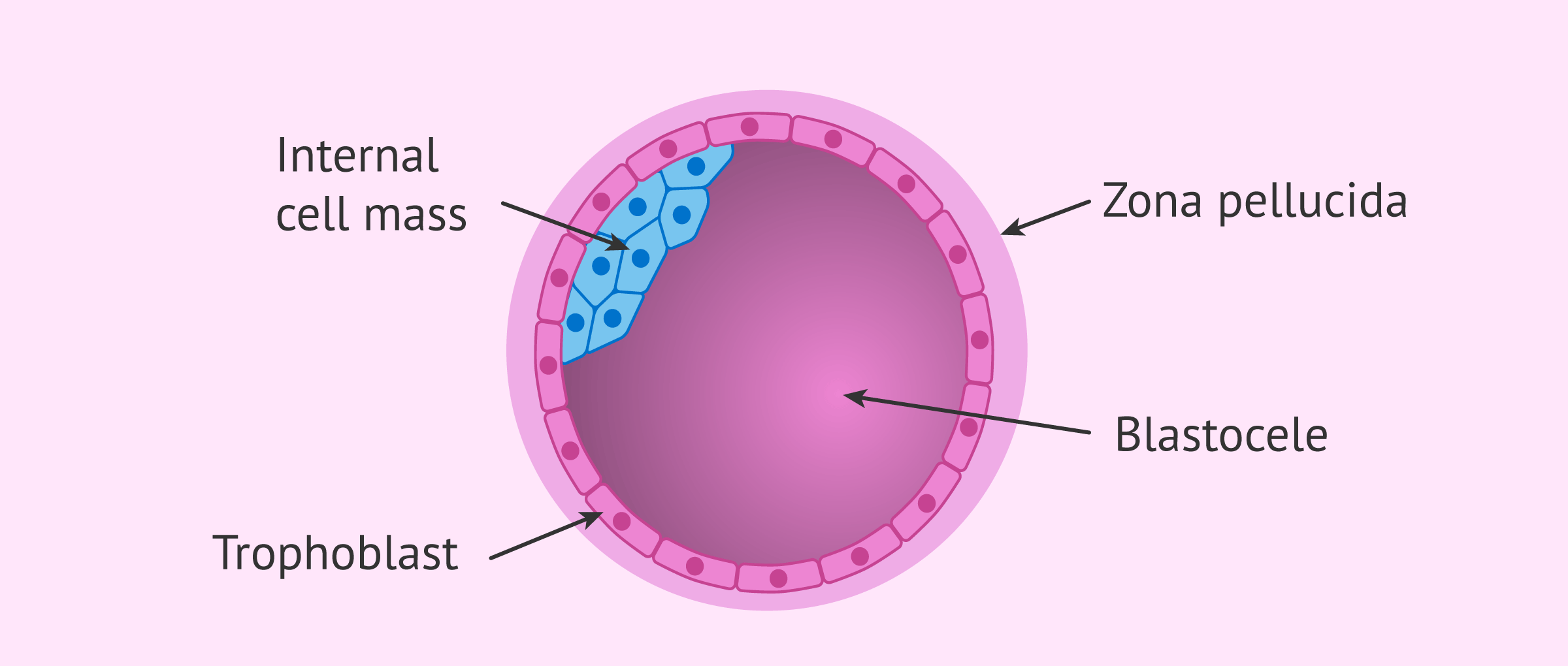
zona pellucida
pierced by the sperm at fertilization

blastomere
individual cells produced by cleavage

morula
solid ball of 16 blastomeres

trophoblast
outer layer of cells on the blastocyst; will contribute to the placenta, not the embryo itself
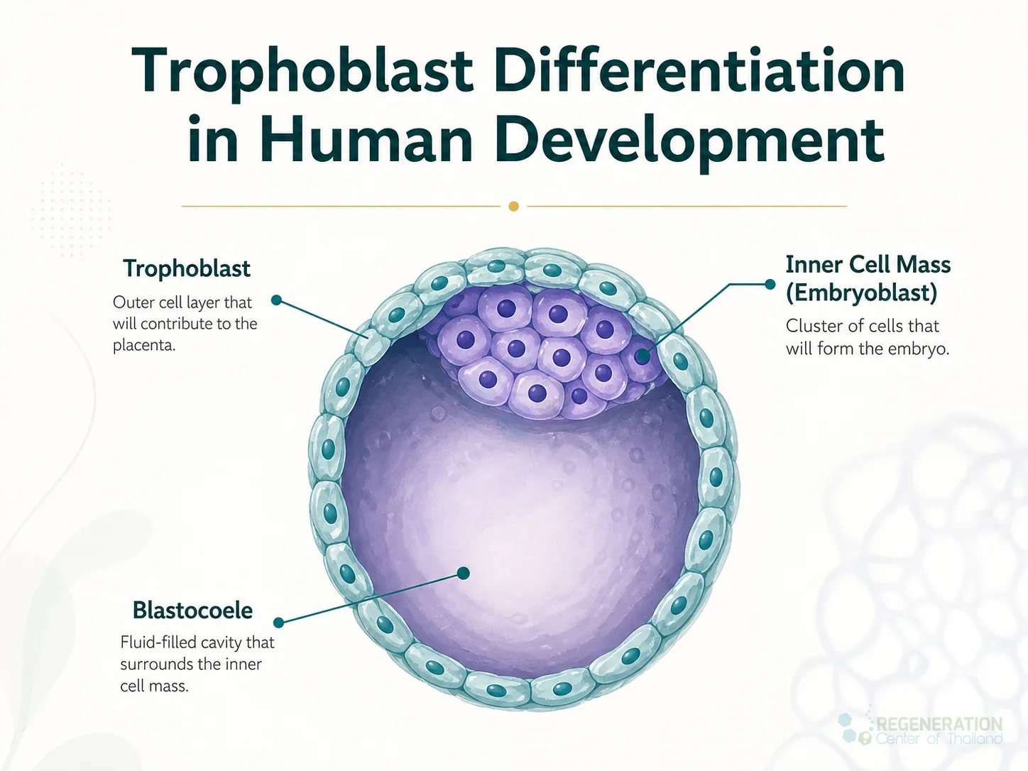
embryoblast (inner cell mass)
inner clump of cells inside the trophoblast sphere; develops into the embryo

blastocyst cavity
fluid-filled cavity inside the sphere of trophoblast cells

blastocyst
germ sac
hypoblast
the under layer of the bilaminar disc
contributes to the yolk sac lining and later turns into the endoderm

epiblast
upper layer of the bilaminar disc
gives rise to the amnion and later turns into the ectoderm

bilaminar embryo (embryonic disc)
2 layer epiblast
hypoblast disc present

amnion
membrane that closes the amniotic cavity
keeps the embryo from drying out
exocoelomic membrane (primary yolk sac)

extraembryonic mesoderm
goo that surrounds the amnion and yolk sac

extraembryonic coelom
small fluid spaces that form in the extraembryonic mesoderm and merge into a larger cavity

connecting stalk
stalk of the extraembryonic mesoderm linking the embryo to the mesoderm lining the trophoblast wall; forerunner of the umbilical cord
prochordal plate
marks for the mouth
primitive streak
site where the cells migrate inward to form the mesoderm during gastrulation
cloacal membrane
marks for the a*us
gastrulation
formation stage where the “gut” layer arrangement first appears

mesenchymal cells
migratory cells extruded from the primitive streak that invade the space between epiblast and hypoblast and replace the hypoblast
cardiogenic area
region where heart development begins
ectoderm
new name for the epiblast once gastrulation is complete
outer skin layer
endoderm
new name for hypoblast layer when the cells are replaced by mesenchymal cells; inner skin layer
intraembryonic mesoderm
new middle layer of tissue sitting between the ectoderm and endoderm
trilaminar embryo
has the ectoderm, mesoderm, and endoderm layer
notochord
establishes the embryo’s body axis and signals the ectoderm to form the neural plate
neural fold

neural tube
precursor for the central nervous system

neural groove

neural crest

median sagittal plane
plane that bisects the embryo into 2 symmetrical halves
cranial/caudal
head-ward/tail-ward directions
ectopic pregnancy
implantation outside the uterus