CB.8 Analysis of biological macromolecules. Nucleic acid hybridization and antibody labelling
 Call Kai
 Learn
Learn Practice Test
 Spaced Repetition
 Match
Match Flashcards
 Knowt Play
1/26

Earn XP
Description and Tags
Learning outcome: CB1.7 Outline the technical approaches available to analyse biological macromolecules.
Name | Mastery | Learn | Test | Matching | Spaced | Call with Kai | Chat |
|---|
No analytics yet
Send a link to your students to track their progress
27 Terms
In clinical practice why is specific detection of cellular components important?
Diagnosis/ Prognosis/ Informs treatment
General nucleic acid or protein stains are useful for identifying cell structures - How?
Detection of specific macromolecules is accomplished by: Antibodies – for proteins and Nucleic acid probes - for DNA/RNA
o Antibody → recognises a specific protein structure
o DNA probe → recognises a specific DNA/RNA sequence
Whilst proteins do not form strong covalent bonds how do they interact with each other to form specific/strong interactions?
Through forming several relatively weak but specific interactions e.g. Hydrogen bonds, Electrostatic attractions, Van der Waals interactions, Hydrophobic interactions.

What is an antibody?
Protein produced by b-cells/plasma cells. Binds to a specific antigens epitope.
When B cells detect a specific antigen they will be stimulated to secrete more antibodies specific to that antigen.
What is the basic structure of an antibody?
Four-chain structure of an antibody. The basic unit is composed of two identical light (L) chains and two identical heavy (H) chains, which are held together by disulfidebonds to form a flexible Y shape.
Each chain is composed of a variable (V) region and a constant (C) region.
—> The variable region allows the antibodies to have widely varying binding sites specific for variety of antigen epitopes.

How do antibodies/antigen interact?
What is the scientific testing relevance of this?
o Antibody binds to a specific region on a the antigen (epitope) with a complementary shape and chemical environment.
o This is the basis of antibody -based detection techniques: Protein → contains epitope → antibody recognises epitope. Scientists make antibodies that specifically recognise a particular protein within a sample.

The antibody/antigen binding in the lab is not visible therefore how to scientists detect the interaction?
To detect where the antibody has bound, we use a secondary antibody. This leads to primary and secondary antibodies.
Primary antibody = binds to protein.
Secondary antibody detects that interaction = as it is attached to something to allow us to see/measure an amplification signal e.g. an enzyme or a fluorophore
Summary: Protein → primary antibody → secondary antibody → detectable signal
What is a fluorophore?
Fluorophore emits a particular light wavelength when excited by an appropriate light. It is in example of a type of amplification signal = produced by the secondary antibody
There are three different forms scientific processes used to detect proteins (using antibody/protein principles) - what are they?
Immunohistochemistry (IHC) - is the protein IN the tissue? where IN the tissue is it?
ELISA = Enzyme-Linked Immunosorbent Assay - ESTIMATE amount of protein in the tissue.
Western blotting / immunoblotting - WHAT size is the blot/protein? has the blot been modified?
Which of the three antibody techniques would you use if you wanted to simply know if the protein was in a sample?
Is a protein present in a tissue/biopsy? Immunohistochemistry
Which of the three antibody techniques would you use if you wanted to know how much protein was in a sample?
How much of a specific protein is present? ELISA
Which of the three antibody techniques would you use if you wanted to know how where the protein was in a tissue sample or biopsy?
Specifically where a protein is present in a tissue/biopsy? Immunohistochemistry
Which of the three antibody techniques would you use if you wanted to know what size a protein was in a sample?
What size is a particular protein? Western blot
Which of the three antibody techniques would you use if you wanted to know if much protein was in a FLUID sample?
ELIZA or Western blot will tell if you there is a specific protein present in a fluid sample.
What is the purpose of immunohistochemistry? And what are the basic principles?
IHC can tells us IF the tissue has the protein in a tissue sample/ biopsy and WHERE it is. Detects a specific protein within a tissue
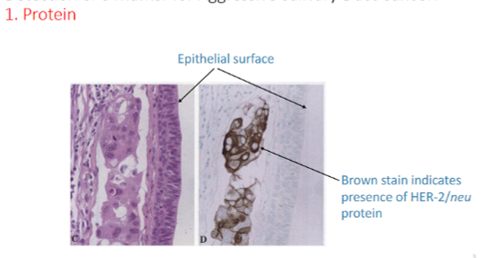
(E.g. when investigating the HER-2 gene related to certain cancers the first question is does this tumour contain HER2 protein? The protein is first through immunohistochemistry = Brown staining will detect if the HER2 protein is present)
Also tells us where the protein is located within a tissue e.g. cell membrane/cytoplasm/nucleus
What is the purpose of Enzyme-Linked Immunosorbent Assay? And what are the basic principles?
ELISA = Enzyme-Linked Immunosorbent Assay
Is a scientific test performed in the lab (also based on antibody detecting antigen/protein principle) that is able to QUANTIFY the number of proteins.
But instead of looking at where a protein is located in a tissue, ELISA can be used to measure how much of a particular antigen is present.
The basic concept is: Antigen → antibody → enzyme → colour
The amount of colour produced can be measured: More target antigen → more enzyme associated with the assay → stronger colour signal. Therefore ELISA can be used quantitatively.
E.g. suppose you're testing a patient's sample for a particular protein. You could compare the colour produced with standards of known concentration and determine the approximate concentration of the protein in the patient's sample.

What is the purpose of Western blotting / immunoblotting? And what are the basic principles?
Is a scientific test performed in the lab (also based on antibody detecting antigen/protein principle) that is able to tells us the SIZE of the protein and any MOFIFICATIONS (e.g proteins).
Protein sample → gel electrophoresis → proteins separated by size → transferred to membrane → antibody detection tells us the size.
Why separate the proteins first? Proteins have different molecular masses. During gel electrophoresis, they can be separated according to size. Then an antibody is used to identify the particular protein we're interested in. The position of the band gives information about the protein's approximate molecular weight (Mr).
+ A protein can be modified after it is produced. One important modification is the addition of a phosphate group: Protein → phosphorylated protein. You can use antibodies that specifically recognise the phosphorylated form. (This can therefore provide information about cell signalling)

In the detection of proteins antibodies are used. In the detection of nucleic acids what is used?
DNA probes! → they recognise a specific DNA/RNA sequence
How are restriction enzymes used?
Restriction enzymes are proteins that cut DNA at particular nucleotide sequences.
o Large DNA/chromosome → restriction enzyme → smaller DNA fragments
o These fragments can then be separated and analysed.
What are the general differences between DAPI vs nucleic acid hybridisation? (in terms of what they detect)
o DAPI: detects DNA generally
o Nucleic acid hybridisation detects specific DNA sequences.
How does DAPI detect DNA?
DAPI (stands for 4′,6-diamidino-2-phenylindole) is a fluorescent DNA stain/molecule used in microscopy and cell analysis.
The fluorescent molecule that binds to DNA – by binding in the minor groove of double-stranded DNA. When DAPI is bound to DNA, it fluoresces blue. So the DNA/nucleus is fluorescent blue.


What can you see on this slide?
DAPI has been combined with antibody/protein staining systems:
o DAPI → binds to DNA → blue
o Antibody → binds to actin → red
o Antibody → binds to tubulin → green
Nucleic acid hybridisation is the technique when we want to identify specific DNA sequences – when might this be needed clinically?
Look for specific gene mutations or gene amplifications in patients.
Identify bacteria in complex samples.
Detect chromosome deletions or rearrangements (e.g. to correlate with disease)
What is the purpose of DNA hybridisation? And what are the basic principles?
DNA hybridisation - technique used to locate specific DNA sequences on chromosomes.
Basic technique
Identifying oligonucleotide probe (short piece with a complementary sequence to the DNA sequence you want to find) is fluorescently labelled.
DNA is heated so two strands sperate (denaturation).
DNA is then cooled so the probe binds (hybridises) through complementary base pairing.
And the fluorescent signal can then be seen under a microscope.

What is FISH?
Allows us to detect where the DNA sequence is in the cell/chromosome: stands for FISH = Fluorescence In Situ Hybridisation
o Fluorescence → the probe can be detected using fluorescence
o In situ → detection occurs in the original cellular/chromosomal location
o Hybridisation → complementary nucleic acid sequences pair together

HER-2 in cancer investigation is a clinically important example. How does this utilize principles of immunohistochemistry and FISH?
HER-2, human epidermal growth factor receptor 2, (or HER2) = protein involved in cell growth signalling. Increased HER2 expression or amplification of the HER2 gene can be associated with certain cancers (e.g. salivary duct, breast/colorectal/gastric/endometrial).
When investigating tissue samples for cancer:
First through immunohistochemistry = Brown staining will detect if HER2 protein is present. (Anti-HER2 antibody binds to HER2 protein).
Then using Fluorescence in situ hybridisation (FISH) = labelled DNA probe will bind to HER2 DNA.
—> In the process nuclei-stained orange and HER2 gene is stained as yellow dots.
—> In cancer = gene amplification of the HER2 gene will have occurred (so cancer can produce more protein which helps cancer proliferate) = FISH will detect more yellow dots

How do you detect genetic Deletions/ Duplications/amplifications or Translocations/rearrangements?
FISH (Fluorescence In Situ Hybridisation)