A+P 2 Lab Review
 Call Kai
 Learn
 Practice Test
 Spaced Repetition
 Match
 Flashcards
 Knowt Play
1/95

There's no tags or description
Looks like no tags are added yet.
96 Terms
Lymphatic system returns leaked interstitial fluid and plasma back to the blood via the:
Lymphatic vessels
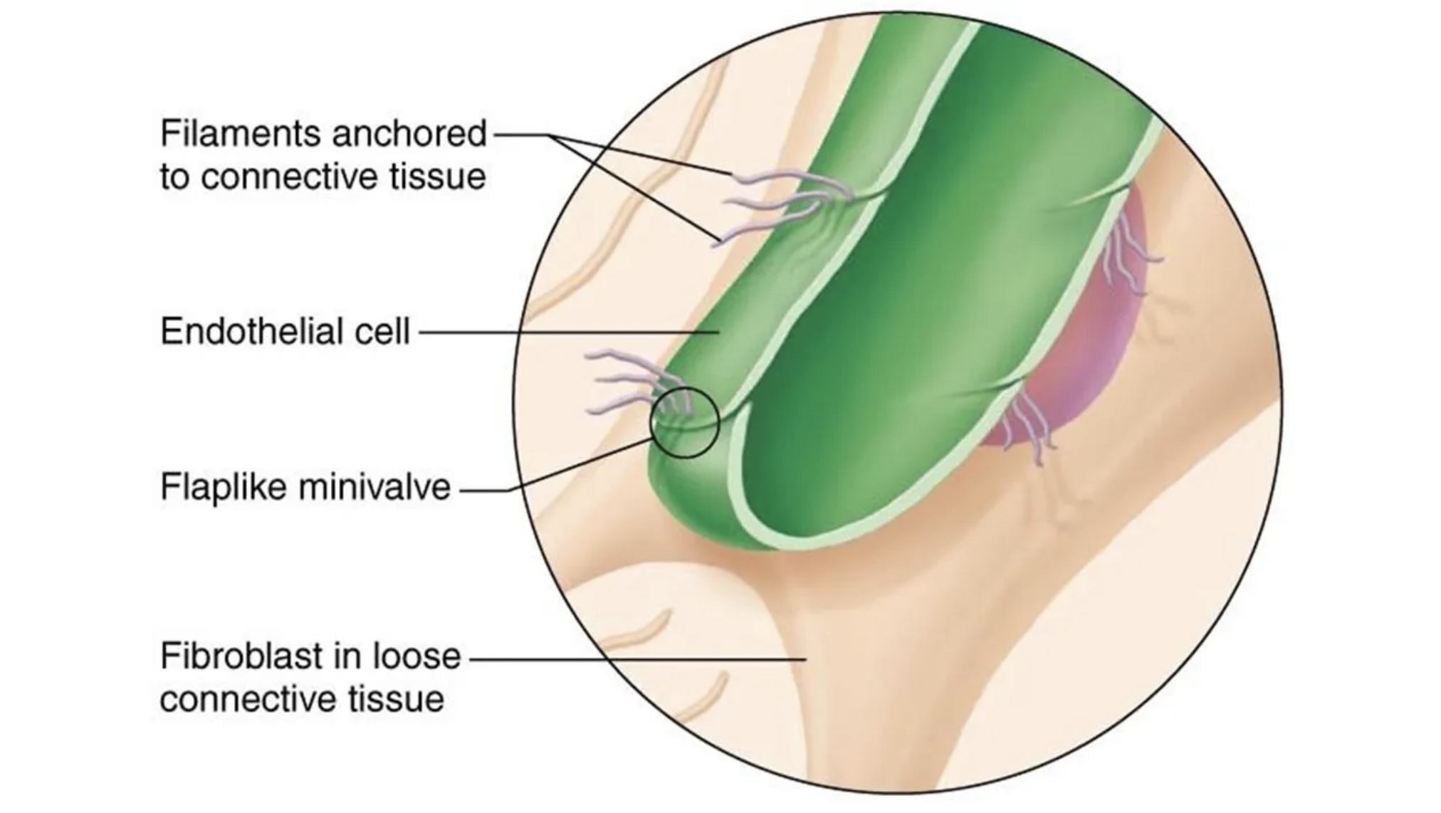
Blind ended tubes in which adjacent endothelial cells overlap each other to form flaplike mini-valves are called:
Lymphatic capillaries
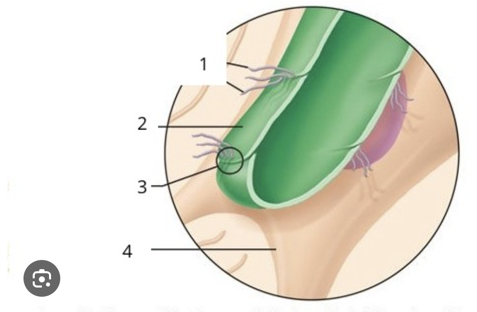
Label #1
Filaments anchored to connective tissue

Label #2
Endothelial cell

Label #3
Flaplike mini-valves
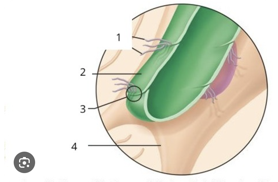
Label #4
Fibroblasts in connective tissue
Which of the following regions DO NOT drain into the right lymphatic duct?
Right lower limbs
The thoracic duct drains the lymph into which vein?
Left Subclavian Vein
The right lymphatic duct drains the lymph into which vein?
Right Subclavian Vein
Appendix + Peyer’s Patches + Tonsils are related with MALT (mucosa associated lymphatic tissue), which are key components of:
GALT (gut associated lymphatic tissue)

Lymphoid nodules that destroy bacteria and prevent them from breaching into intestinal wall are:
Peyer’s Patches
Lymphoid organ that is involved with the maturation of B-cells:
Red Bone Marrow
Lymphoid organ that is involved with the maturation of T-cells:
Thymus
Primary organs of the Lymphatic System are:
Thymus
Red Bone Marrow
Fetal Liver
Secondary organs of the Lymphatic System are:
Lymph nodes
Spleen
MALT (tonsils, appendix, and peyer’s patches)
Red Bone Marrow is also actively invovled in:
Hemopoiesis

Where to find this tissue?
Thymus
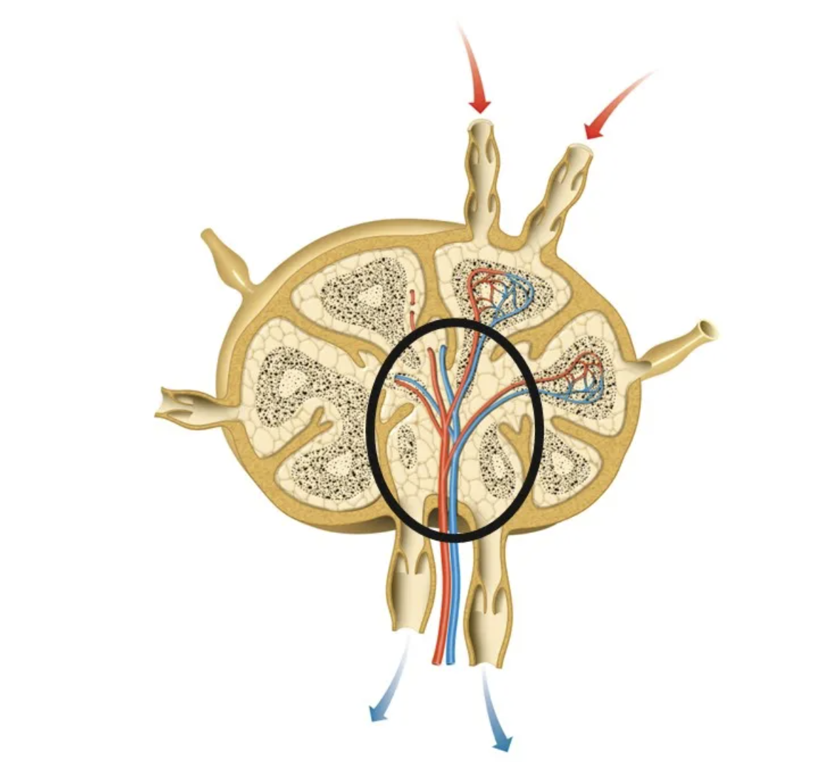
What is the circled section called?
Medullary sinus
What type of tissue does the Pharyngeal Tonsil have?
Ciliated Pseudostratified columnar epithelium or Respiratory System

What is the circle section called?
Tonsillar Crypt

Letter E is called:
Stratified squamous epithelium

Letter F is called?
Germinal center

What slide is this called?
Palatine Tonsil
Which is NOT a function of the respiratory System?
Returns leaked fluid into the blood
Conducting Zone of the Respiratory System ends at:
Terminal Bronchioles
Site fro gas exchange is:
Alveoli

Inferior boundary of the nasal cavity is called:
Vomer

Superior boundary of the nasal cavity is called:
Perpendicular plate
Posterior opening of the nasal cavity is called?
Choanae
What is the hair of the vestibule called?
Vibrissae
Region of the pharynx posterior to the oral cavity is called:
Oropharynx
Laryngopharynx has what type of tissue?
Stratified Squamous Epithelium
Oropharynx has what type of tissue?
Stratified Squamous Epithelium
Nasopharynx has what type of tissue?
Ciliated Pseudostratified Columnar Epithelium
Laryngeal Prominence is located in what cartilage of the larynx?
Thyroid Cartilage

What is the circled portion called?
Cardiac Impression/Notch
What type of tissue is known as the respiratory epithelium?
Ciliated Pseudostratified Columnar Epithelium
Cells that makeup 5% of the Alveoli and secretes surfactant are called:
Cuboidal Great (Type II) Alveolar Cells
Cells that makeup 95% of the Alveoli and form thin squamous barrier specialized for gas exchange are called:
Squamous (Type I) Alveolar Cells
Which statement is true when it comes tot he Boyle’s Law?
If volume increases then pressure decreases
Which muscles do you use other than the diaphragm during quiet expiration?
Internal Intercostal Muscles
Which muscles do you use other than the diaphragm during quiet inspiration?
External and Parasternal Intercostal Muscles
Inspiration Muscles - External Intercostal Muscles:
between ribs
contract to elevate rib cage upward/outward
increase the width of chest cavity
Inspiration Muscles - Parasternal Intercostal Muscles:
lift upper rib cage
work with other muscles to stabilize the chest wall
Expiration Muscles: Internal Intercostal Muscles
active process
no skeletal muscles are actively used

What’s the name of this image?
Spirometer

The wave that is encircled in the diagram is the respiratory volume exchanged during quiet breathing. It is called the __________.
Tidal Volume (TV)

This wave encircled in the image is called the __________.
Inspiratory Reserve Volume (IRV)
Formula for Inspiratory Capacity (IC):
IC = TV + IRV
Formula for Functional Residual Capacity (FRC):
FRC: ERV + RV
Formula for Vital Capacity (VC):
VC = TV + IRV + ERV
Formula for Total Lung Capacity (TLC):
TLC = TV + IRV + ERV + RV
TV/Tidal Volume:
500 mL
air exchanged during normal breathing/relaxed
IRV/Inspiratory Reserve Volume:
3100 mL
Extra air inhaled after inhalation
ERV/Expiratory Reserve Volume:
1200 mL
Extra air exhaled after expiration
RV/Residual Volume:
1200 mL
Remaining of air in lungs after full forceful expiration
When blood is too acidic =
Acidemia - Bicarbonate binds with excess H+ → carbonic acid → carbon dioxide + water → extra carbon dioxide is hyperventilated out by lungs
Acidemia:
High H+ and Low pH
When blood is too basic:
Alkalemia - carbonic acid dissociates/separate → releases more H+ → replenish/restore bicarbonate → leftover bicarbonate is managed by kidney and emptied into urine
Alkalemia:
Low H+ and High pH
Bicarbonate Buffer System: What is it + Formula?
Mechanical mechanism that maintains the normal blood pH in the human body
CO2 + H2O → H2CO3 → H+ + HCO3- OR
CO2 + H2O ← H2CO3 ← H+ + HCO3-
What enzyme catalyzes CO2 to HCO3- and reverse?
Carbonic anhydrase
Respiratory Volume can be used to asses:
Respiratory status
Respiratory Volume can be combined to calculate ____ which gives the respiratory status.
Respiratory capacities
Spirometer:
A tool used to measure respiratory volumes
Spirometry distinguishes between:
Chronic Obstructive Pulmonary Disease (COPD)
Restrictive Pulmonary Disease (RPD)
COPD/Chronic Obstructive Pulmonary Disease:
increases airway resistance
ex: Bronchitis
Hyperinflation of lungs (TLC, FRC, and RV may increase)
RPD/Restrictive Pulmonary Disease:
TLC decreases due to disease/exposure to environmental agents
ex: tuberculosis (disease)
ex: fibrosis (exposure to environmental agents)
Lung expansion compromised (TLC, FRC, RV, and VC decrease)
Gas Exchange involves what?
Oxygen
Carbon Dioxide
Where does Gas Exchange across?
Respiratory Epithelium/Ciliated Pseudostratified Columnar Epithelium
Gas Exchange is influenced by:
Pressure gradient
Thickness/surface area of respiratory epithelium
Ventilation-perfusion coupling
Ventilation-Perfusion Coupling:
Coordination between air + blood flow in alveoli to ensure efficient gas exchange
Gas Exchange: Movement of O2
Alveoli → Capillaries/Blood
Gas Exchange: Movement of CO2
Capillaries/Blood → Alveoli
Gas Exchange: Venous pressure of O2
40 mmHg
Gas Exchange: Alveolar pressure of O2
104 mmHg
Gas Exchange: Venous pressure of CO2
45 mmHg
Gas Exchange: Alveolar pressure of CO2
40 mmHg
Driven force of gas exchange:
Simple Diffusion (Partial Pressure + Pressure Gradient)
CO2 diffuses → Alveoli because
Partial pressure in blood is higher than in the alveoli
O2 diffuses → Capillaries/Blood because
Partial pressure in inhaled air inside alveoli is higher than oxygen-depleted blood
Oxygen is transported in 2 ways:
2% is dissolved in plasma
98% is bound to hemoglobin (Hb) in RBCs; 4 polypeptide chains, each with iron containing heme group, transporting 4 O2 molecules each
Carbon Dioxide is transported in 3 forms:
7 - 10% dissolved in plasma
20% bound to globin part of hemoglobin aka carbaminohemoglobin
70% transported as bicarbonate ions (HCO3-) in plasma
2 Divisions of the Respiratory System:
1). Conducting Division - Nostrils → Terminal Bronchioles
Upper Respiratory Tract: Nose → Superior of the Larynx
Lower Respiratory Tract: Larynx → Lungs
2). Respiratory Division - Alveoli + Gas Exchange
What’s the Lung airflow?
Bronchi → Bronchioles → Alveoli
Functions of the external part of the nose:
Warming/Cleansing/Humidifying
Odor
Amplification of voice
Septal Cartilage
Cartilage that separates the right and left nasal passage of the nose
What cartilage attaches the Larynx → Trachea?
Cricoid cartilage
Trachea:
12 cm long
16-20 C-shaped rings of hyaline cartilage
anterior to the esophagus
lined with pseudostratified columnar epithelium (ciliated cells/goblet)
Carina
The inferior most fork of the trachea
Glottis
Vocal cords + openings
Elastic Arteries:
thick walled
large resistance lumen
Aorta/major branches or Conducting Arteries - conduct blood from heart to medium sized vessels
Muscular Arteries:
also called “Distributing Arteries” - deliver blood to body organs
thickest tunica media w/ more smooth muscle + active vasoconstriction
Arterioles:
smallest of all arteries
control flow into blood capillaries via vasodilation + vasoconstriction of smooth muscle
also called “resistance arteries” - changing diameter changes resistance to blood flow
Lymphatic Vessels (Lymphatics):
one way system → heart
includes lymphatic capillaries + larges lymphatic vessels
Lymphatic capillaries:
Found in (limited) in areas of the meninges → help drain interstitial fluid from CSF
Similar to blood capillaries but more permeable
take up larger molecules/particles
proteins, cell debris, cancer cells, pathogens