Genetics Exam 3
 Call Kai
 Learn
 Practice Test
 Spaced Repetition
 Match
 Flashcards
 Knowt Play
1/125

There's no tags or description
Looks like no tags are added yet.
126 Terms
chromosome condensation
2 meters (6.5 ft) of DNA in every cell in our body
Large DNA molecules must be tightly condensed to fit within the nucleus of a cell
condensation occurs in stages:
nucleosomes
solenoids
chromatin loops
condensed chromatin
chromatin folded around protein scaffold
intimately associated with the cell cycle and gene expression
chromatin
a complex of DNA and protein found in eukaryotic cells
primary function: to package long DNA molecules into more compact, denser structures
euchromatin
lightly packed chromatin rich in gene concentration and is most often under active transcription
heterochromatin
tightly packed chromatin consisting mainly of genetically inactive sequences
faculative
constitutive
faculative heterochromatin
type of heterochromatin that, under the right conditions, can change to euchromatin
gene silencing, barr bodies
constitutive heterochromatin
type of heterochromatin
very gene poor, centromeres and telomeres
ALWAYS closed, can never be accessed
folded fiber model
1965 E.J. dupraw
whole mounts of human white blood cells
found few or no free fiber ends and concluded each chromatid must consist of a single fiber
.2nm double helix of DNA
Type A fiber- 1-10nm (ratio of ~6:1)
Type B fiber- 20-25 nm (ratio of ~10:1)
extensive folding of Type B forms a chromatid
replication form each end toward centromere
Condensation occurs through extensive, irregular looping and folding both longitudinally and transversely
Describes each chromatid as a single continuous, unbranched 20 nm20nm to 30 nm30nm nucleoprotein fiber
nucleosome model
1974, R.D. Kornberg; 1975 P. Oudet
most commonly accepted model for DNA packaging
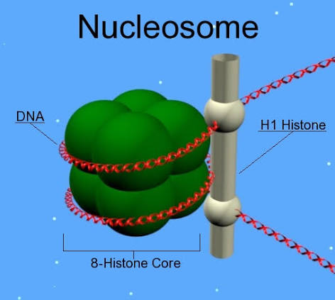
nucleosome
Fundamental repeating structural unit of eukaryotic chromatin, producing the "beads-on-a-string" appearance (10 nm fiber)
nucleosome
simplest packaging structure of all eukaryotic chromatin
localized areas of transcription; better fit for protein biosynthesis

core histones
H2A, H2B, H3, H4
consist of approximately 120 amino acids each
highly conserved during evolution
in combination, form the core particle (ocatmer)
VERY BASIC in charge (approx 25% lysine and arginine)

linker histones
H1
consist of approximately 200 amino acids
tissue specific expression and NOT highly conserved
loosely associated with core particle
fairly basic
not considered a part of the core

10 nm fiber
nucleosome
200 bp DNA
146 bp wrap core
54 bp link to next nucleosome- LINKER DNA
reduce length by 7 times
linear arrangement
primary packaging of the chromatin
“beads on a string”
30 nm fiber
type of condensation of chromatin
H1 histone responsible for packaging
solenoid
supercoiling- reduces length of DNA by ~7 times

solenoid
type of condensation of chromatin
helical coiling of 10nm fibers consisting of 6 nucleosomes

H1 histone
works by binding 2 distinct regions of DNA
linker DNA
portion of the 146 bp core
indues tighter DNA wrapping around histones= ~40x
involved with creation of solenoid
zig-zag model
alternative model
DNA backbone not flexible enough to bend between nucleosomes
straight linker DNA connect opposite nucleosomes
this model allows for more compaction
middle of “tube” has DNA
experimental results show that both model topologies may both simultaneously be present in chromatin fiber

300nm fiber
type of higher order coiling of chromatin
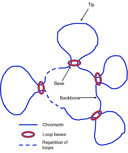
“chromatin loops”
built around a scaffold of topoisomerase 2
compaction level of euchromatin
700nm fiber
final condensation of chromatin
DNA diameter is ~700nm
spiral scaffold composed of topoisomerase 2 and about 15 non-histone proteins
compaction level of heterochromatin
metaphase chomosome
chromatin formation
cell cycle
I: interphase: DNA is replicated in S phase
P: Prophase:
early: chromosomes condense
late: nuclear envelope breaks down, mitotic spindle assembles
M; metaphase: mitotic spindles arrange chromosomes at cell equator
A: Anaphase: sister chromatids separate
T: Telophase: cell plate forms
biological complexity
the result of the hierarchical organization of nested levels of cells, tissues, and higher order part types interacting together
C value paradox
excess DNA is present in the genome that does not seem to be essential for the development or evolutionary divergence of an organism
genome size does not correlate with organismal complexity
the onion test
T. Ryan Gregory
Onion
diploid (2n= 16)
haploid= 17pg
17×10^6bp (has 5x more DNA)
Human
diploid (2n= 46)
haploid= 3.5 pg
3.5×10^6bp
G-value paradox
the number of genes does not correlate with organismal complexity
ex: watercress and humans ~ 20,000 genes
coding sequences: 2-5% of the human genome
classes of nucleotide sequence
highly repetitive DNA sequence (HR)
moderately repetitive DNA sequence (MR)
single copy DNA sequence (unique)
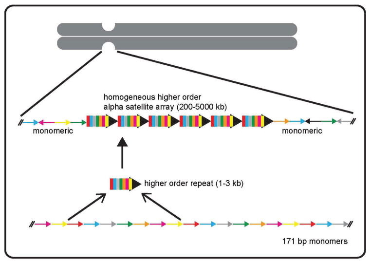
highly repetitive DNA sequence
comprises about 10% of the human genome
most is located in heterochromatic regions around the centromere/telomere
“non-coding DNA regions”
occurs as variable length modifs (5-100 bp), in long tracts of up to 100 Mb
present at >10^6 copies per genome
postulated functions include structural and organization role (centromeres and telomeres) to nothing more than junk
e.g. alpha satellite DNA- from 2 to more than 30 repeats of 171 bp tandem repeats
heterochromatin (tightly coiled)
moderately repetitive DNA sequence
comprises about 30% of the human genome
found mostly throughout euchromatin (faculative heterochromatin)
average 300bp in size
present at 10-10^5 copies per genome
function in length mutability, modulation of transcription factor binding, spacing between promoter elements, cytosine methylation, alternative splicing, mRNA stability, transcription start and termination sites, etc.
also includes “redundant” genes for histones, and ribosomal RNA and proteins (gene-products present in cell in large numbers)
e.g. microsatellite DNA (repeating sequences of 2-30 bp of DNA); interspersed repetitive DNA (transposable elements)
scattered throughout euchromatin

microsatellites
variable number of tandem repeats typically occurring in non-coding regions of the genome
occurs through a mutation process known as “slippage recognition”
type of moderately repetitive DNA sequence
useful genetic markers as they tend to be highly polymorphic
used to sequence the human genome
markers for certain disease conditions
primary markers for DNA testing in forensic cases

single copy DNA sequence
unique
comprises about 1-5% of the human genome
found throughout the euchromatin
“coding DNA regions = GENES”
~20,000 protein coding genes
present at single or low copy number per genome
euchromatin- GENES
gene
sequence of unique nucleotides (genotype) that carry the genetic information which is to be expressed (phenotype)
“instruction manuals for our bodies”
every person has 1 copy from mother and 1 from father
>99.5% of the DNA, number of genes, types of genes, protein products, are the same in ALL people
<1% of the total number of genes contribute to each person’s unique physical features
the basic physical and functional unit of heredity
transcriptional unit
molecular level “gene” - DNA sequence that gives rise to an RNA molecule
contains:
promoter
transcribed region
regulatory sequence
terminator

transcribed region
contains exons and introns
flanking regions
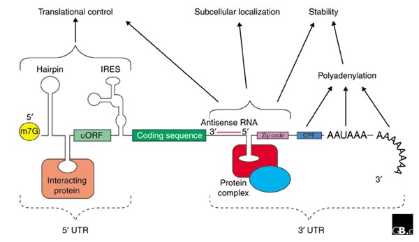
5’ untranslated region
3’ untranslated region
exon
“coding sequences” = phenotype
make it into mature mRNA
part of the transcribed region of a gene
intron
“intervening sequences” = areas of genes that generally dont code for phenotype
taken out of mature mRNA
part of the transcribed region of a gene
5’ untranslated region
part of the transcribed region of a gene
mRNA that is directly upstream from the initiation codon
differs in prokaryotes and eukaryotes
mostly involved in regulation of translation
recruitment of ribosomal subunits
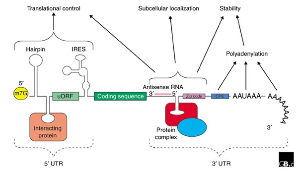
3’ untranslated region
part of the transcribed region of a gene
section of mRNA that immediately follows the translation termination codon
influences the localization, stability, export, and translation efficiency of an mRNA
post-transcriptional influences on gene expression
promoter
part of the structural view of a gene
DNA sequence onto which the transcription machinery binds and initiates transcription
ex: TATA box

TATA box
type of core promoter
highly conserved sequence in DNA serving as the binding site for transcription factor binding
core DNA sequence= TATAAA
ON/OFF for transcription
Basal level of transcription
regulatory sequences
enhancer/silencer regions
CAAT box
GC Box
RE1
“ramped up level of transcription or silenced transcription”
CAAT box
type of regulatory sequence
GGCCAATCT- consensus sequence that occurs upstream by 60-100 bases to the initial transcription site
typically required for inducible genes to be produced in sufficient amounts

GC box
type of regulatory sequence (proximal region)
region of DNA that can be bound with proteins (activators) to activate transcription of a gene or genes
ramps up or silences

RE1
type of regulatory sequence (proximal region)
region of DNA that binds repressor proteins preventing neuronal genes from begin expressed in non-neuronal tissues

terminator
section of nucleic acid sequence that marks the end of a gene or operon in genomic DNA during transcription

types of genes
solitary genes
duplicated genes
multigene families
psuedogenes
repeated genes
solitary genes
(unique) type of gene
a single copy of a gene (haploid situation) or two copies in diploid
comprises the bulk of euchromatin
duplicated genes
type of gene
process by which a portion of a chromosome is duplicated resulting in an additional copy of a gene
results in a copy of the original gene called a paralog gene
either of the 2 genes may mutate and change the original function of the gene
usually occurs due to an error during meiosis
mulitgene families
type of gene
set of several similar genes, formed by duplication of a single original gene, and generally with similar biochemical functions
most often located in similar region of the chromosome
most often used or synthesized at different times
pseudogenes
type of gene
dysfunctional relatives of genes that have lost their protein-coding ability
often the result of multiple mutations within a gene
most likely used to be functional (through evolution lost function)
repeated genes
type of gene
multiple copies of small genes clustered throughout the genome at specific sites
present in high copy numbers
many times present in a “heat to tail” configuration
ex; genes for tRNA and rRNA
cell division
process where a parental cell divides into 2 daughter cells
cell size is limited due to the ability of the cell to transport food and oxygen from the cell membrane to the interior of the cell
maximizes SA:V ratio
outside of the cell unable to keep up with the inside because the inside grows at a faster rate
DNA replication
process by which genetic information is duplicated by the use of an existing strand of DNA as a template for the synthesis of a new, identical strand
ensures that each cell in an organism has a complete and correct copy of the organism’s genome or genetic data
G0: resting phase
G1: growth phase; cell increases size for DNA synthesis
S- DNA REPLICATION
G2: growth phase; cell increases size for mitosis
M- cell growth sops and cells divide
Fredrick Griffith
1928- “Transforming principle”
suggested that bacteria could transfer information from one to another
first suggestion of DNA at heritable material
Avery-Macleod and McCarty
1994: experimental demonstration that DNA is the substance causing bacterial transformation
basically confirmed Griffith’s work and isolated DNA as the heritable material
Hershey-Chase
1952- confirmed that DNA was the genetic material
responsible for inheritance using radioactively labeled bacteriophage to infect bacteria
Franklin and Wilkins
1952: used X ray crystallography to study the structure of DNA
image B51: X ray diffraction image of DNA
Watson and Crick
1952: used data from image B51 to propose the double helical structure of DNA
proposed a possible copying mechanism (due to the specific pairing of bases)
Predictions
the DNA helix is “unwound” during replication
the sequence of the “parental strand” can serve as a template for synthesis of “new strand” of DNA
each nucleotide of the DNA strand is copied precisely due to the complimentary base-pairing.
process occurs semi-conservatively
semi-conservative model
the double stranded DNA contains one parental and one daughter strand following replication

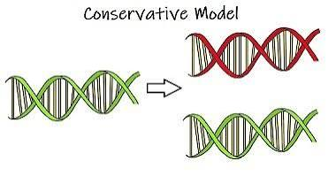
conservative model
both parental strands stay together after DNA replication and the daughter molecules contain all new nucleotides
dispersive model
parental and daughter DNA are interspersed in both strands; consist of old and new strands

Meselson-Stahl Experiment
Cultured E. coli in medium containing heavy nitrogen (15N), then switched cells to medium containing light nitrogen (14N)
Separated DNA by density using CsCl (cesium chloride) equilibrium density gradient centrifugation:
Generation 0: All heavy DNA (15N/15N15N/15N)
Generation 1: 100%100% intermediate/hybrid density DNA (15N/14N) — ruled out the conservative model
Generation 2: 50%50% hybrid DNA (15N/14N) and 50%50% light DNA (14N/14N) — ruled out the dispersive model
Supported Watson and Crick's proposal that each replicated double helix contains one conserved parental strand and one newly synthesized daughter strand
replication stages
initiation
unwinding
primer synthesis
elongation
replication initiation
origin of replication (OriC in bacteria)
~250 base pairs AT-rich region (DnaA boxes)
GATC methylation sites (precise timing of replication)
initiator proteins (origin binding proteins) DNaA protein
replication bubbles
replication occurs bi-directionally
replication forks

unwinding of dsDNA strand
step 2 of replication
topoisomerase
helicase
single strand binding proteins

topoisomerase
involved in step 2 of unwinding dsDNA
aka: gyrase
reduce torsional strain causing unwinding of double helix
helicase
involved in step 2 of unwinding dsDNA
breaks hydrogen bonds between complimentary nucleotides
single strand binding proteins
involved in step 2 of unwinding dsDNA
stabilize ssDNA until elongation beings
prevent ssDNA from reannealing
priming of ssDNA strand
step 3 of replication
primase
primers are typically 10-12 bases in length
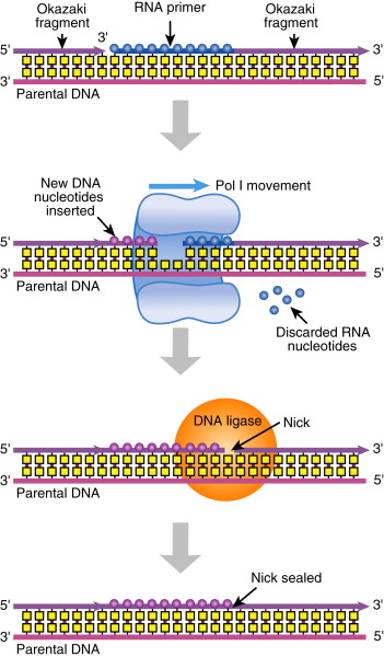
primers are removed following elongation and replaced with DNA nucleotides

primase
involved in the 3rd step of transcription initiation
RNA polymerase that adds a ribonucleotide primer to ssDNA

transcription elongation
4th step of replication (DNA synthesis)
DNA polymerase (prok) 1,2,3,4,5
DNA polymerase (euk) a,d,e
limitations:
DNA polymerase cannot initiate DNA synthesis by itself; requires addition of RNA primer
DNA polymerase can only synthesize DNA in 5’-3’ direction
replication is semi-conservative
replication is bi-directional

DNA polymerase
enzymes that catalyze attachment of nucleotides to make new DNA during replication

DNA polymerase 1
prokaryotic DNA polymerase
removes RNA primer and starts synthesis
DNA polymerase 3
prokaryotic DNA polymerase
responsible for most of replication process
DNA polymerase 2,4,5
prokaryotic DNA polymerase
synthesize lagging strand
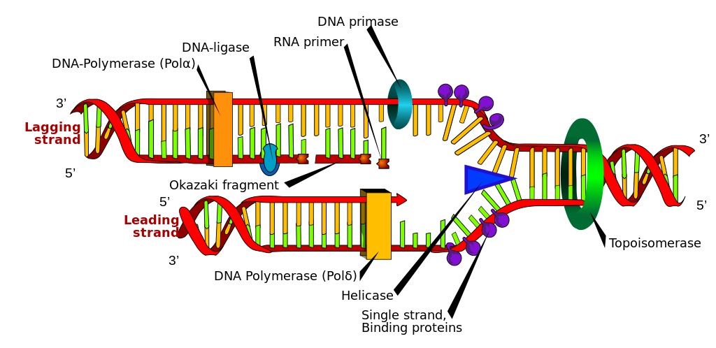
DNA polymerase alpha
eukaryotic DNA polymerase
synthesizes the primer

DNA polymerase delta
eukaryotic DNA polymerase
synthesize leading strand

DNA polymerase eplison
eukaryotic DNA polymerase
synthesize lagging strand

Watson and Crick DNA model
two strands of DNA
run anti-parallel to each other
sugar phosphate backbone
hydrophilic sugar and phosphate backbone on the outside with nitrogenous base inside
nucleotides joined together by phosphodiester bonds (5’C-3’C)
Base pairing A=T C- - - G

DNA elongation steps
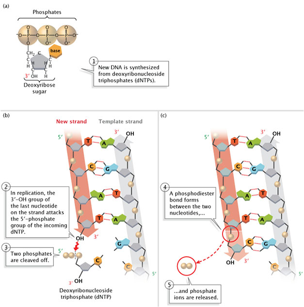
new DNA is synthesized from deoxyribonucleotide triphosphates (dNTP’s)
In replication, the 3’OH group of the last nucleotide on teh strand interacts with the 5’PO4 group of the incoming dNTP
two phosphates are cleaved off the incoming dNTP
a phosphodiester bond forms between the two nucleotides
and phosphate ions are released
leading strand synthesis
continuous synthesis
goes INTO replication fork
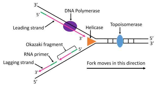
lagging strand synthesis
discontinuous synthesis → Okazaki fragments
goes away from the replication fork
DNA ligase

DNA ligase
joins together newly synthesized DNA strands by creating a covalent phosphodiester bond

high fidelity
exonuclease activity
mistakes are very rare (1 × 10^6 bases)
instability of mismatched bas e pairs
configuration of DNA polymerase 3 active site
proofreading/exonuclease activity of DNA polymerase 3 (backs up in the 3’-5’ direction to fix mistakes

transcription
DNA to mRNA (specifically hnRNA- heteronuclear RNA)
Where: cell nucleus (eukaryotes) and sometimes mitochondria
When: either G1/S or G2/M (waves of transcription that coincides with different transition points during the cell cycle
immature mRNa (hnRNA) consists of both introns and exons
mature mRNA, introns have been removed (splicing)
RNA polymerase makes the new mRNA strand

rules of transcription
only one of the two DNA strands serve as a template
template strand (DNA strand that mRNA is built from)
coding strand (matches mRNA except U’s are T’s in coding strand)
selective process- each gene has its own transcription protocol
always in 5’ to 3’ direction (nucleotides added at 3’ growing tip)
results in an RNA compliment (anti-parallel and unidirectional)
steps of transcription
Pre-recognition: accessibility to DNA
recognition: pre-initiation complex formation
initiation: binding of RNA polymerase complex
elongation: movement of RNA polymerase 2 and formation of mRNA
termination: cleavage of new transcript
pre-recognition
DNA packaged in the nucleus as heterochromatin
facultative
constitutive
Lys (K) acetylation
histone acetyl transferase (HAT)→ allows DNA to be read
histone deacetylase (HDAC) → clamps tail back down
DNA must be converted to euchromatin prior to transcription
part of gene expression regulation
decreases positive charge of the histone core and loosens the DNA strand
transcription recognition
pre-initiation complex formation at the promoter
formation of large complex of ~100 proteins (PIC) required for RNA polymerase 2 to bind
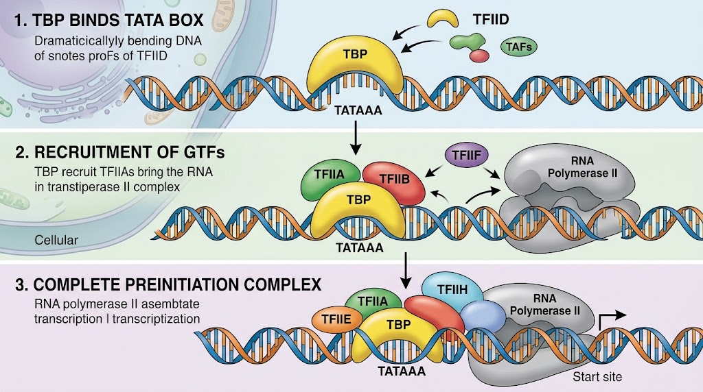
TATA binding protein (TBP) binds to TATA box
general transcription factors (e.g. TF IID) recruited by TBP
TBP binds to TATA → recruits other proteins and TF → formation of pre-initiation complex
trasnscription initiation
binding of RNA polymerase complex (i.e. mediator)
other transcription factors (b,f,h) and RNA polymerase 2 recruited to complex
Mediator complex (~20 proteins)
ATPase and Helicase activity unwinds DNA
~1 turn of DNA unwinds and forms transcription bubble
RNA polymerase 2 binds to the template strand via the polymerase active site
RNA polymerase 2 is unphosphorylated at its carboxyl end (CTD)

elongation
movement of RNA polymerase 2 and formation of mRNA
RNA polymerase 2 is phosphorylated at CTD by mediator complex
RNA polymerase 2 traverses the template strand and creates an RNA copy
RNA polymerase reads template (3’-5’) and writes (5’-3)
exact copy of coding strand (except thymines are replaced with uracils, and the nucleotides are composed of a ribose (5C sugar)

termination
cleavage of new transcript
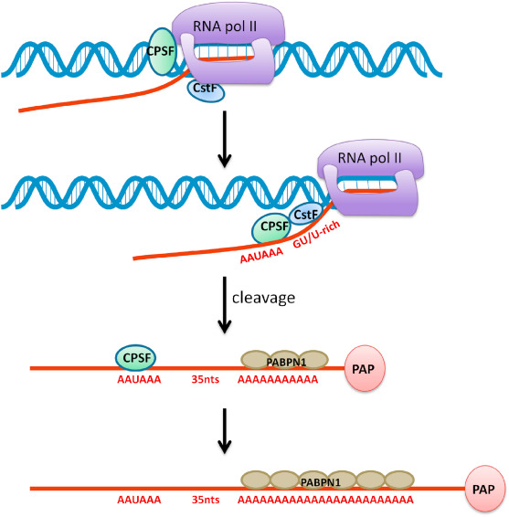
two protein complexes carried by CTD recognize poly A signal (AAUAA)
CPSF (cleavage and polyadenylation specificity factor)
CSTF (clevage stimulation factor
post transcriptional processing can now occur
5’ capping
co-transcriptional regulation
when a growing RNA chain is ~30 nucleotides long, a guanine cap (7-methyl guanine) is added to the 5’ end by capping enzymes
functions:
regulation of nuclear export- CBC recognized at nuclear pore
promotes translation- helps ribosome recognize the message
prevention of mRNA degradation in the cytoplasm
involved in intron splicing

3’ polyadenylation
post transcriptional regulation
nascent RNA is cleaved by ribonuclease downstream of conserved AAUAAA site
Poly (A) polymerase adds adenine ribonucleotides to the 3’ end of the RNA molecule—up to 200 bases
not dependent on template
functions;
enhances the stability of the RNA molecule
regulates transport to the cytoplasm
RNA processing
co-transcriptionally and post-transcriptionally
splicing
introns
exons
spliceosome
splicing
the mechanism by which introns are removed
introns
intervening sequences of RNA not expressed in proteins
GU nucleotide sequence at the 5’ splice site (donor site)
AG nucleotide sequence at the 3’ splice site (acceptor site)
polypyrimidine tract (PPT) just upstream of the 3’ splice site
promotes the assembly of the spliceosome
branch point sequence (UACUAAC)- binding site for snRNp-U2
exons
retained in mature mRNA and are the expressing sequences
spliceosome
large complex of RNA and proteins responsible for removal of introsn from transcribed mRNA (splicing)
responsible for both cleavage of the intron from the RNA and ligation of the remaining exons
1977: sharp and Roberts lab revealed that genes of highe rorgansimsa re “split” or present in several distnct segmetns along the DNA molecule
hnRNA
snRNP
mRNA
hnRNA
pre-mRNA
immature single stranded mRNA present in the nucleus (contains introns)
snRNP
snurps
small nuclear RNA molecules associated with specific ribonuclear proteins playing essential role in splicing (U1,U2,U4,U5,U6)
mRNA
mature mRNA
RNA that is translated to become a polypeptide chain
hnRNA + snRNP
written in linear form
comma-less (no pauses)
coded in triplets (codon)
unambiguous (each codon only codes for 1 amino acid)
degenerate (1 amino acid can have more than 1 codon—ex: val has 4 diffferent codons)
specific start and stop codons
non overlapping
universal

spliceosome assembly
U1 binds to 5’ splice site
U2 binds to BPS
trimer of U4, U5, and U6 from hnRNA leaving U5 and U6 bound
U1 and U4 dissociate from hnRNA leaving U5 and U6 bound
U2 and U6 associate
“lariat” of hnRNA is formed from the intron
5’ splice site is cleaved by U5 and U5 binds to 3’ splice site
3’ splice site is celaved and U5 ligates exons togeher via ATP hydrolysis
snRNP’s are released along with spliced intron