Neuro Exam 1
 Call Kai
 Learn
Learn Practice Test
 Spaced Repetition
 Match
Match Flashcards
 Knowt Play
1/234

There's no tags or description
Looks like no tags are added yet.
Name | Mastery | Learn | Test | Matching | Spaced | Call with Kai | Chat |
|---|
No analytics yet
Send a link to your students to track their progress
235 Terms
In the visual pathway, do nasal RGC fibers cross contralateral, or stay ipsilateral?
Contralateral
In the visual pathway, do tempoal RGC fibers cross contralateral, or stay ipsilateral?
Ipsilateral
If you have central vision loss in one eye and temporal visual field oss in the other eye, you most likely have
Junctional scotoma
A junctional scotoma is:
A lesion that's localized in the optic chiasm region
What is part of the CNS?
Brain - Spinal chord - optic nerve (CN2)
What is part of the PNS?
Cranial Nerves - Spinal nerves
How many cranial nerves and spinal nerves are there?
12/31
cranial-12
spinal-31
What can be found in the anterior cranial fossa?
Inferior surface of frontal lobe: - Olfactory bullb/tract
What can be found in the middle cranial fossa?
Optic chiasm - Pituitary gland - Temporal lobe
What can be found in the posterior cranial fossa?
Brainstem - Cerebellum
What parts of the brain are inferatentorial?
Brainstem and cerebellum
What parts of the brain are supratentorial?
Everything except for brainstem and cerebellum
Which dura fold seperates the cerebellum and the cerebrum?
Tentorium cerebelli
Which dura folds seperate the 2 cranial hemispheres?
Falx cerebri
The brainstem is composed of what structures?
Midbrain - Pons - Medulla
What is found in the telencephalon?
Cerebral cortex - Subcortical white matter - Basal ganglia
What is found in the diencephalon?
Hypothalmus - Midbrain - LGN
What is found in the mesencephalon?
Midbrain
What is found in the metencephalon?
Pons - Cerebellum
What is found in the myelencephalon?
Medulla
When does neurelation occur?
3rd and 4th week (day 18-28) post-fertilization
What occurs during neurulation?
Development and closure of the neural tube
How is gestational age calculated?
Post-fertilization age + 2 weeks
The CNS is derived from what?
The neural tube
Where are ventricles developed from?
The hollow part of the neural tube
Wher is the PNS derived from?
Neural crest cells
What causes spinal bifida?
Incomplete closure of the neural tube
What's the best way to prevent spinal bifida?
Follic acid
The optic nerve and retina are derived from what brain section during development?
The diencephalon
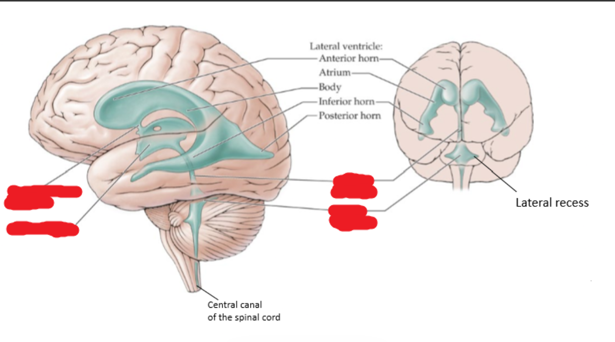
Name the covered parts of the ventricles.
-What structures were ventricles developed from?
from the hollow part of the neural tube

Most neurons in the CNS are:
Multipolar
The olfactory receptor neurons in the olfactory epithelium are:
Bipolar
The dorsal root ganglion neurons are:
pseudounipolar
What type of cells make myelin in the CNS?
Oligodendrocytes
What type of cells make myelin in the PNS?
Schwann cells
What is the difference between nuclei and ganglia?
Nuclei - Cell bodies in the CNS (central-nuclei)
Ganglia - Cell bodies in the PNS (P-GANG)
Bundles of fibers in the brain are called
Fasciculi - Brachium - Column - Peduncles - Lemiscus
Bundles of axons in the PNS are called
Nerves - Nerve roots - Nerve trunk - Nerve chord - Ramus
Why doesn't the basal ganglia fit the definition of a ganglia?
"The basal ganglia is in the CNS while any other ""ganglia"" is in the PNS"
basal=the og base.
Basal ganglia in CNS= grey matter
What is white matter?
Areas of the CNS that contain myelinated axons (fibers in the inner brain)
-messengers
What is grey matter?
Areas of the CNS that mainly consists of neuron cell bodies- (outer layer of cortex)
-thinking/sends orders
Includes: thalamus, basal ganglia, cortex
How are grey matter and white matter organized in the cerebral cortex?
Grey matter on the outside, white matter on the inside

How are grey matter and white matter organized in the brainstem?
White matter on the outside and between sections of the inside
Grey matter more concentrated in chunks on the inside

How are grey matter and white matter organized in the spinal cord?
White matter all around the outside
Grey matter on the inside

How many cervial spinal nerves are there?
8
C1- C8
(think of your daily schedule; starts at 8)
How many thoracic spinal nerves are there?
12
T1- T12
(lunch at 12)
How many lumbar spinal nerves are there?
5
L1- L5 spinal nerves
How do you remember the parts of the spine in order?
Come To Learn Some Chemistry
At what vertebral bones does the spinal chord end?
L1-L2 BONES
Where are lumbar punctures (spinal taps) performed and why?
Below L2 usually between L3 or L4 because they aren't part of the spinal cord
Dorsal root ganglion are part of the
PNS
(aka spinal ganglion), dorsal=on your back
The dorsal root of the spinal nerve is sensory or motor?
Sensory
(dorsal/behind)
The ventral root of the spinal nerve is sensory or motor:
Motor
(ventral=frontal)
Why is the spinal nerve considered a mixed nerve?
It has sensory, motor, and autonomic functional components
Functional components of spinal nerves:
General somatic afferent (GSA)
sensory fibers from skin, skeletal muscles, and joints to the CNS
-cell bodies in the dorsal root ganglia
Functional components of spinal nerves:
General viseral afferent (GVA)
sensory fibers from visceral organs to the CNS
-cell bodies in dorsal root ganglia
Functional components of spinal nerves:
General somatic efferent (GSE)
motor fibers to skeletal muscles
-cell bodies in the anterior horn of the spinal cord
Functional components of spinal nerves:
General visceral efferent (GVE)
autonomic motor fibers to smooth muscle, cardiac muscle, and glands
Sympathetic: T1-L2/L3;
-preganglionic neurons in the intermediolateral cell column
Parasympathetic: S2-S4;
-preganglionic neurons in sacral parasym. nucleus of the intermediate grey matter
What is referred pain?
Pain that is felt at a different location from the site of origin
Why are heart attacks considered an example of referred pain?
Because the pain from the heart attack can be felt at the left shoulder or arm as opposed to the heart itself
because of referred pain, intracranial or cervical pathological issues can present themselves as:
Eye pain
"What does ""somatic efferent"" refer to?"
Ventral horn cells/cranial nerve motor nuclei that project directly from the CNS to skeletal muscles

"What does ""autonomic efferent"" refer to?"
a 2-neuron chain that synapses in the peripheral ganglia before reaching its target organ

What are the common sympathetic functions?
Fight or flight
- Bronchodilation
- Pupil dilation(cats)
- Increased heart rate
- Digestion inhibition
- Piloerection
What part of the spinal cord is responsible for the sympathetic divisions
Thoracic (T1-12); sympathetic ganglia: prevertebral, paravertebral, adrenals
Lumbar (L1-3); Norepi & Ach

What are the common parasympathetic functions?
What a SLUD! (rest/digest=gut stuff )
Salivation
Lacrimation
Urination
Defication
- Pupil constriction
- Bronco-constriction
- Slowed heart rate
What part of the autonomic nervous system is responsible for the parasympathetic division
Cranial
CN 3- oculomotor
CN 7- Facial
CN 9- Glossopharyngeal
CN 10- Vagus nerve; (parasympathetic ganglia in/near end organs)
Sacral
S2-4
*S4- Ach, parasym. ganglia in/near end organs

How do you find the central sulcus in MRI scans?
"Look for the superior frontal sulcus (yellow/long) and follow the ""letter T"" to the precentral sulcus (blue/short).
The sulcus immediately following the ""T"" is your central sulcus (red)

Somatosensory deficits are associated with the:
Post-central gyrus (behind=sensory)
Motor deficits are associated with the:
Pre-central gyrus (frontal=motor)
What seperates the frontal lobe and parietal lobe?
The central sulcus
What seperates the parietal lobe and the occipital lobe?
The parietal-occipital sulcus
What seperates the parietal lobe from the temporal lobe?
The lateral fissure (sylvian fissure)
What are the common motor defects?
Paralysis/-plegia - Paresis - Palsey - Para - Quadri- or teta-
What does paralysis/-plegia mean?
No movement
What does paresis mean?
Weakness (partial paralysis)
What does palsey mean?
Weakness or no movement
"What does ""para"" mean?"
Both legs
"What does ""quadri"" or ""tetra"" mean?"
All four limbs
What are common sensory pathway deficits?
Sensory loss - Paresthesias
What are paresthesias?
False feelings of pain, burning, tingling, etc.
What brodmann area is the primary visual cortex?
Brodmann Area 17 (aka striate cortex)
(17= “ see-teen”)
Why is the primary visual cortex also called the striate cortex?
The presence of the line of gennari
Summary:
(Brodmann Area 17 = Primary visual cortex = Striate cortex)
“At age 17, I got my FIRST pair of Striate glasses to VISUALIZE the world."
What is the line of Gennari?
The dense axonal input from the thalamus to layer 4 of the visual cortex
Broadmans areas 18 and 19 are known as:
The extrastriate cortex
What are the Main Long Tracts and what are they responsible for
Posterior column-medial Lemiscus Pathway (sensory): vibration, joint position, fine tactile discrimination (poke w/2 needles; what’s the diff?)
Anterolateral Pathway (sensory): pain, temp, crude touch (non-discriminative; something is touching me but idk what is it)
Lateral corticospinal Tract (motor): voluntary movement of the extremities
If you have a cortical lension, it'll present itself as
A contralateral deficit
What type of pathway is the medial lemniscus pathway? What is it for?
Sensory - Vibration - Joint position (proprioception) - Fine tactile descrimination
What type of pathway is the anterolateral pathway? What is it for?
Sensory - Pain - Temperature - Crude touch
What type of pathway is the lateral corticospinal pathway? What is it for?
Motor - Voluntary movement of extremities
Recite the medial lemniscus pathway
Primary sensory neuron
- Input goes to the spinal cord via the dorsal root ganglion
- Dorsal root ganglion -> Posterior (dorsal) columns - Up the spine via posterior (dorsal) columns -> medulla
Secondary sensory neuron (Medial lemniscus)
- *Contralateral crossover in medulla via internal arcuate fibers
- Up the medulla -> VPL in Thalmus
Tertiary sensory neuron
- VPL in thamlus -> Primary somatosensory cortex

Recite the anterolateral (spinothalmic) pathway
Primary Sensory Neuron
- Input enters spinal chord via dorsal root ganglion - Dorsal root ganglion -> Dorsal/posterior horn (synapse)
Secondary Sensory Neuron
- Crossover to contralateral side in spinal chord
- Up the spinal chord via the anterolateral pathway (spinothalmic tract)
- Synapse in the VPL of thalamus
Tertiary Sensory Neuron
- VPL -> primary somatosensory cortex

Recite the corticospinal pathway (pyramidal tract)
Upper motor neuron
- Precentral gyrus in brain -> Medulla
- Crossover to contralateral side via pyramidal decussation
- Medulla -> Spine via lateral corticospinal tract (synapse)
Lower Motor Neuron
- Spine -> Skeletal muscle
If you have a hemi-chord lesion, what do you expect to happen?
Ipsilateral loss of motor control segments below the lesion
- Ipsilateral loss of medial lemniscus senses below the lesion
- Contralateral loss of spinothalamic senses below the lesion
- No loss of function above the lesion
cortical lesions cause what type of deficits?
contralateral
The precentral gyrus is also known as the:
Primary motor cortex
The postcentral gyrus is also known as the:
Primary somatosensory cortex
The pre/postcentral gyri (homunculus) dictate the _____ side of the somatosensory and motor functions
Contralateral
Somatotopic organization in primary somatosensory & primary motor cortex using homunculi:
FAT
Face (lateral bottom)
Arm (middle)
Trunk (top & into the (butt) crack)

The medial inter-hemispheric region of the homunculus controls what area of the body?
Legs (trunk)
