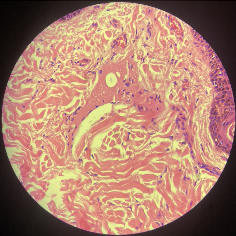
VPTH 122 1st Lab Exam Review (Exercise 1-5)
 Call Kai
 Learn
Learn Practice Test
 Spaced Repetition
 Match
Match Flashcards
 Knowt Play
1/29

There's no tags or description
Looks like no tags are added yet.
Name | Mastery | Learn | Test | Matching | Spaced | Call with Kai | Chat |
|---|
No analytics yet
Send a link to your students to track their progress
30 Terms
Lesions: pin point to pin head hemorrhage in cortical surface, pale
Morphological Diagnosis: Kidney: Hemorrhage, multifocal, miliary, cortical.
Identify
Lesions:
Morphological Diagnosis:

Lesions: pale, radially oriented streaks extending from the cortex toward the medulla
Morphological Diagnosis: Kidney: Fibrosis, interstitial, multifocal, chronic
Identify
Lesions:
Morphological Diagnosis:

Lesions: Numerous multifocal nodules distributed along the surface which varied in size round to irregular, tan to yellow. some appear grey to black, suggestive of hemorrhage or necrosis
Morphological Diagnosis: Kidney: Nodules, multifocal to coalescing, cortical and capsular
Identify
Lesions:
Morphological Diagnosis:

Lesions: spindle cell proliferation expanding the submucosa,
neoplastic cells have elongated to tapering nuclei with fine stippled chromatin, inconspicuous nucleoli, and scant, indistinct eosinophilic cytoplasm
Morphological Diagnosis: Urinary bladder: Suggestive of Spindle cell neoplasm
Identify
Lesions:
Morphological Diagnosis:

Lesions: irregular nests, cords, anastomosing trabeculae and variably sized tubules and gland-like structures supported by a prominent fibrocollagenous (desmoplastic) stroma.
Neoplastic cells are cuboidal to polygonal with moderate amounts of lightly eosinophilic cytoplasm and distinct cell borders.
Morphological Diagnosis: Urinary bladder: Invasive urothelial carcinoma
Identify
Lesions:
Morphological Diagnosis:

Lesions: extensive interstitial fibrosis, tubules are markedly atrophic, irregular and occasionally dilated,
with occasional intraluminal eosinophilic proteinaceous casts
Morphological Diagnosis: Kidney: Chronic tubulointerstitial nephropathy with marked interstitial fibrosis and tubular atrophy, multifocal glomerulosclerosis and chronic interstitial inflammation
Identify
Lesions:
Morphological Diagnosis:

Lesions: marked thickening of transitional epithelial cells lining the epithelium
Morphological Diagnosis: Urinary bladder: Invasive urothelial carcinoma
Identify
Lesions:
Morphological Diagnosis:

Lesions: dark red to reddish-purple and consolidated, with irregular pale areas
Morphological Diagnosis: Lung: Consolidation, multifocal to coalescing, cranioventral, with pulmonary hemorrhage
Identify
Lesions:
Morphological Diagnosis:

Lesions: mucosal surface appears diffusely dark red
Morphological Diagnosis: Trachea: Hemorrhage, diffuse, mucosal
Identify
Lesions:
Morphological Diagnosis:
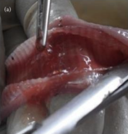
Lesions: diffusely dark red to reddish-purple, with a heavy, consolidated appearance
Morphological Diagnosis: Lung: Hemorrhage, diffuse, severe, with multifocal to coalescing areas of consolidation
Identify
Lesions:
Morphological Diagnosis:

Lesions: thickening of the interstitium due to infiltration of inflammatory cells predominantly lymphocytes.
Eosinophilic materials inside parabronchial lumen/atria.
Morphological Diagnosis: Lung: Pneumonia, interstitial, multifocal and spreading, moderate with pulmonary edema.
Identify
Lesions:
Morphological Diagnosis:

Lesions: multifocal epithelial disruption and sloughing,
with irregular accumulation of eosinophilic material and cellular debris along the mucosal surface
Morphological Diagnosis: Trachea: Tracheitis, necrotizing and degenerative, multifocal, moderate to severe
Identify
Lesions:
Morphological Diagnosis:

Lesions: irregular areas of red discoloration and prominent epicardial superficial blood vessels,
pale tan to whitish patchy discoloration on the atrium and ventricle
Morphological Diagnosis: Heart: Fibrosis, multifocal
Heart: Epicardial congestion and multifocal hemorrhage.
Identify
Lesions:
Morphological Diagnosis:

Lesions: The pericardial sac is filled with a large volume of clear, amber to yellowish-tinted fluid.
Epicardial surface is markedly pale, slightly irregular, and partially obscured by thin strands of translucent to white fibrin.
Morphological Diagnosis: Heart: Hydropericardium
Heart: Epicarditis, fibrinous
Identify
Lesions:
Morphological Diagnosis:

Lesions: widespread, diffuse loss of normal myocardial architecture.
Cardiomyocytes show varying degrees of degeneration and atrophy.
Cells are fragmented, poorly defined, and exhibit loss of normal cross-striations with faded, pyknotic, or completely absent nuclei.
Morphological Diagnosis: Snake Heart: Cardiomyopathy, necrotizing and degenerative, moderate to severe, diffuse, chronic-active
Identify
Lesions:
Morphological Diagnosis:

Lesions: diffuse, widespread disruption of the normal cardiac muscle architecture.
Cardiomyocytes exhibit profound degeneration, vacuolation, and loss of fiber detail.
Individual myofibers are fragmented, poorly defined, and show marked loss of distinct cross-striations.
Multiple areas demonstrate fading or absent nuclei (karyolysis/necrosis)
Morphological Diagnosis: Avian Heart: Cardiomyopathy, necrotizing and degenerative, severe, diffuse, chronic-active
Identify
Lesions:
Morphological Diagnosis:

Lesions: diffuse loss of normal myocardial architecture.
The cardiomyocytes exhibit advanced degeneration, severe fragmentation, and liquefactive-type necrosis.
numerous large, coalescing, irregular clear spaces and round vacuoles.
Morphological Diagnosis: Heart: Cardiomyopathy, necrotizing and degenerative, severe, diffuse, chronic-active
Heart: Myocardial edema and vacuolar degeneration, severe, multifocal to diffuse
Identify
Lesions:
Morphological Diagnosis:

Lesions: moderately enlarged with rounded, blunt capsular margins
mosaic of dark red to reddish-brown zones intermixed with lighter, pale tan to yellowish zones.
Morphological Diagnosis: Liver: Chronic passive hepatic congestion.
Identify
Lesions:
Morphological Diagnosis:

Lesions: lobes are diffusely enlarged;
uniform pale tan-orange to light yellowish-brown discoloration,
capsular surface appears smooth, glistening, and somewhat greasy or friable in texture.
Morphological Diagnosis: Liver: Hepatic steatosis/lipidosis
Identify
Lesions:
Morphological Diagnosis:

Lesions: moderately enlarged with slightly rounded, blunt lobar edges;
parenchyma is mottled dark red to tan with variable-sized, discrete, round, pale cream-to-white nodules
Morphological Diagnosis: Liver: Hepatitis, granulomatous
Identify
Lesions:
Morphological Diagnosis:
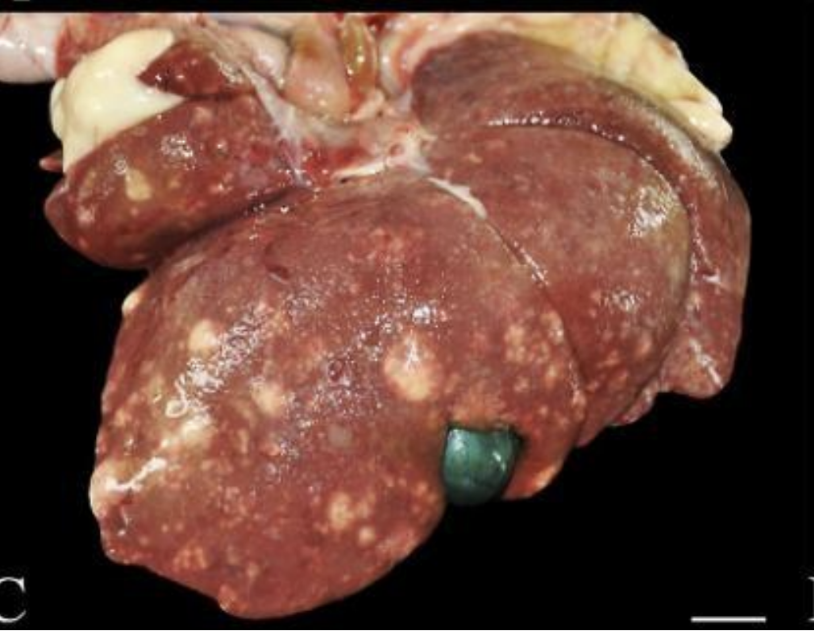
Lesions: loss of hepatocellular architecture,
hypereosinophilic cytoplasm,
nuclear pyknosis and karyorrhexis, and areas of karyolysis.
The necrotic hepatocytes are separated by pale, fragmented cellular debris.
The lesions are predominantly centered around central veins, with variable extension into the surrounding hepatic parenchyma.
Morphological Diagnosis: Hepatocellular necrosis, centrilobular, diffuse, severe
Identify
Lesions:
Morphological Diagnosis:

Lesions: hepatocytes show numerous cytoplasmic vacuoles, with affected centrilobular regions exhibiting hypereosinophilic cytoplasm, nuclear pyknosis, karyorrhexis, and karyolysis.
Extensive loss of hepatocellular detail and disruption of the hepatic parenchymal architecture are evident in the necrotic areas.
Morphological Diagnosis: Liver: Hepatocellular necrosis, centrilobular, diffuse, severe, with widespread cytoplasmic vacuolation
Identify
Lesions:
Morphological Diagnosis:

Lesions: increased eosinophilia of hepatocytes, loss of nuclear detail, and disruption of hepatic cords in areas adjacent to portal tracts
The affected hepatocytes show features consistent with cellular degeneration and necrosis, with extension of the lesions into the surrounding hepatic parenchyma.
The remaining hepatocytes exhibit relatively preserved architecture.
Morphological Diagnosis: Liver: Hepatocellular necrosis, periportal, multifocal to diffuse, severe
Identify
Lesions:
Morphological Diagnosis:

Lesions: widespread hepatocellular cytoplasmic vacuolation, characterized by numerous variably sized, optically clear vacuoles within the hepatocyte cytoplasm.
The vacuoles are prominent and, in some areas, coalesce, imparting a foamy appearance to the hepatic parenchyma.
Scattered hepatocytes exhibit enlarged, rounded, and occasionally hypereosinophilic cytoplasm, with some cells showing nuclear pyknosis or loss of discernible nuclear detail.
Multifocal areas of hepatocellular degeneration and possible necrosis are present.
Morphological Diagnosis: Liver: Hepatocellular vacuolar degeneration, diffuse, severe, with suspected multifocal hepatocellular necrosis.
Identify
Lesions:
Morphological Diagnosis:

Lesions: The center of the patch is well-demarcated raw, bright red, with round margin, and appear moist or oozing. Surrounding it is a ring of darker, skin with scabbing, crusting, and matted hair. There is complete hair loss, alopecia, directly over the affected area.
Morphological Diagnosis: Skin: Dermatitis, necrotizing and ulcerative, localized, moderate to severe with crust, scales
Identify
Lesions:
Morphological Diagnosis:

Lesions: Large, multi-lobulated and multinodular exophytic mass arising from the oral mucosa and gingiva, characterized by dark grey to black coloration with a smooth to irregular glistening texture.
Morphological Diagnosis: Gingiva: Oral Melanoma
Identify
Lesions:
Morphological Diagnosis:

Lesions: Elongated, well-demarcated by a dark red ring and highly erythematous lesion within an area of loss of hair suggestive of alopecia. The center is pale pink, exhibiting moist, glistening surface.
Morphological Diagnosis: Skin: Dermatitis, ulcerative + exudative with crust formation
Identify
Lesions:
Morphological Diagnosis:

Lesions: There are round basophilic cells supported by fibrous stroma infiltration displacing surrounding connective tissue
Morphological Diagnosis: Maxilla/Gingiva: Oral Mast Cell tumor
Identify
Lesions:
Morphological Diagnosis:

Lesions: Proliferation of spindle shaped cells
Morphological Diagnosis: Hindleg: Suggestive of fibrosarcoma
Identify
Lesions:
Morphological Diagnosis:
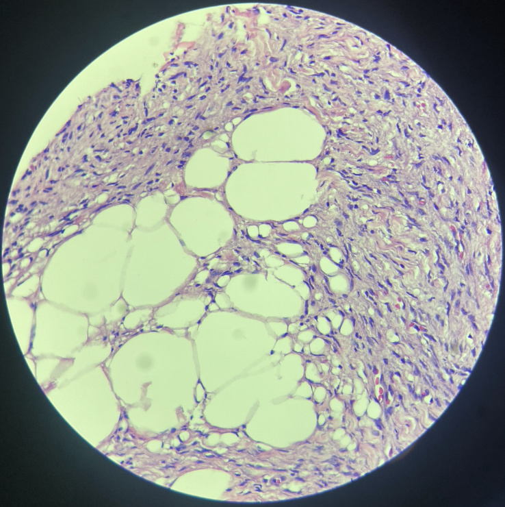
Lesions: Scattered fibroblasts and cell infiltrate with eosinophilic material displacing connective tissue
Morphological Diagnosis: Skin: Dermatitis, eosinophilic, multifocal to coalescing, moderate with collagenolysis
Identify
Lesions:
Morphological Diagnosis: