12. Nervous System: Brainstem, Cerebellum, Spinal Cord, Sensory & Motor Pathways
 Call Kai
 Learn
 Practice Test
 Spaced Repetition
 Match
 Flashcards
 Knowt Play
1/88

There's no tags or description
Looks like no tags are added yet.
89 Terms
brainstem
housed within posterior cranial fossa
3 components
midbrain
pons
medulla oblongata
consists of white matter tracts interspersed w/ multiple nuclei
integration centres for primitive reflexes
regulation of consciousness
cranial nerves III-XII (except CN CI) → projections
each cranial nerve pair originates from pair of nuclei embedded within this structure

midbrain
part of brainstem
medial to temporal lobes
connected to cerebellum via superior cerebellar peduncles
contains
left + right cerebral peduncles = crus cerebri
interpeduncular fossa
substantia nigra
tegmentum
tectum

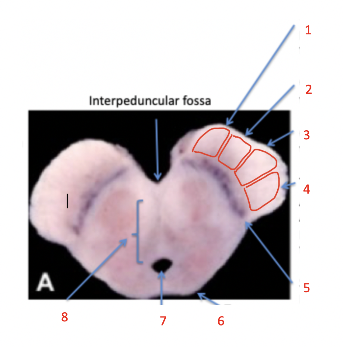
crus cerebri
(1-4) part of midbrain → left + right cerebral peduncles
composed of white matter tracts
different parts - medial → lateral:
(1) frontopontine fibers
starts at frontal cortex → goes down to pons
(2) corticobulbar tract
somatic motor to head, face, neck
(3) corticospinal tract
somatic motor to rest of body = major motor output
from cortex to spine
(4) -pontine fibers
temporo-
parieto-
occipito-

substantia nigra
(5) part of midbrain - component of basal nuclei
presence of neuromelanin pigment
less pigmentation in Parkinson’s patients
tegmentum
(8) part of midbrain - white matter tracts (lemnisci)
extends through midbrain, pons + medulla oblongata
sensory info coming through lemnisci

tectum
(6) part of midbrain - posterior 4 bumps
2 superior colliculi
vision reflex center
2 inferior colliculi
hearing reflex center

pons
part of brainstem
anterior to 4th ventricle + cerebellum
connected via middle cerebellar peduncles
structures
basilar groove for basilar artery
trigeminal nerve = CN V
sensory from face + mouth
motor to muscles of mastication
fibers
fronto-/temporo-/parieto-/occipito-pontine fibers synapse in pontine nuclei
pontocerebellar fibers from pontine nuclei decussate
project to cerebellum via middle cerebellar peduncles
corticospinal fibers run through → interspersed w/ pontocerebellar fibers
contains autonomic respiratory centres
nuclei supporting medullary respiratory centre = breathing control

medulla oblongata
part of brainstem
anterior to cerebellum
connected via inferior cerebellar peduncle
continuous w/ spinal cord
contains
medullary pyramids
olives
nucleus gracilis + cuneatus
other nuclei within → controls vital body functions
cardiovascular
heartbeat rate + force
vasoconstriction + vasodilation
respiratory
breathing rhythm
vomiting
deglutition
coughing, sneezing, hiccuping

medullary pyramids
part of medulla oblongata
formed by corticospinal tracts
somatic motor to body
decussation
on each side → 90% of corticospinal tracts decussates to contralateral side

olives
part of medulla oblongata
bumps lateral to medullary pyramids
inferior nuclei connected to cerebellum via inferior cerebellar peduncles

nucleus gracilis
part of medulla oblongata
component of posterior column sensory pathway for
fine touch
vibration
pressure
proprioception
lower trunk + extremity

nucleus cuneatus
part of medulla oblongata
component of posterior column sensory pathway for
fine touch
vibration
pressure
proprioception
upper trunk + extremity

cerebellum
posterior to pons + medulla oblongata
connected to brainstem by pairs of superior, middle, inferior cerebellar peduncles
function
receives sensory info
proprioception = body position, muscle length + tension
receives descending info from cerebral cortex
compares + integrates incoming info
makes minute adjustments → fine-tune motor response
regulate movement + posture
motor learning
features
left + right cerebellar hemispheres
vermis → “worm”
cerebellar cortex → gray matter = folia
cerebellar white matter → arbor vitae

cerebellum peduncles
bands of axons in cerebellum
superior
middle
inferior

superior cerebellar peduncles
afferent connections from body
proprioception
efferent connections to midbrain + thalamus
modify thalamic output to primary motor cortex
cerebellar neurons run through → cross to contralateral side → side + synapse in ventral lateral thalamus nucleus
convey up to primary motor cortex = fine-tuning

middle cerebellar peduncles
afferent connections from pons
pontocerebellar fibers
delivers info from cerebral cortex
inferior cerebellar peduncles
afferent connections from body + inferior olivary nucleus
proprioception
spinal cord
conduction pathway info transmitted to and from brain + contains somatic reflex arcs
exits skull via foramen magnum → within vertebral column
31 pairs of spinal nerves transmitting sensory + motor info (mixed nerves)
branch off spinal cord at every vertebral level
exit vertebral column via intervertebral foramina

vertebral column
bony protection for spinal cord
33 vertebrae
7 cervical
12 thoracic
5 lumbar
5 sacral (fused)
4 coccygeal (fused)

vertebra features
stacking of vertebrae
vertebral arches form vertebral canal
vertebral bodies connect via intervertebral discs
spinal cord within vertebral canal
spinal nerves exit column via intervertebral foramina

vertebral joints
intervertebral discs
b/w contiguous vertebral bodies
solid symphysis joint → fibrocartilage
2 zygapophyseal joints
b/w superior articular processes of vertebra + inferior articular processes of above vertebra
synovial plane joint

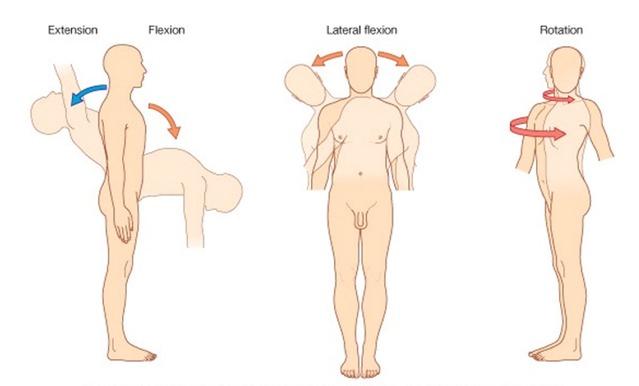
vertebral column movements
extension + flexion
lateral flexion
rotation
spine curvatures
redistributes weight of ventral cavity
primary
derived from embryonic curve
secondary
forms overtime due to musculature
cervical curve = hold head up
lumbar curve = prevent falling forward from ventral cavity weight

spinal nerve pairs
exit at each spinal cord
8 cervical
8th lies b/w vertebrae C7 + T1
*7 cervical vertebrae BUT 8 cervical pairs
12 thoracic
5 lumbar
5 sacral
1 coccygeal
spinal cord internal anatomy
consists of ascending sensory + descending motor tracts
internal = gray matter
neuron cell bodies of interneurons + lower motor neurons (CNS)
surrounds central canal → filled w/ CSF
external = white matter
tracts of myelinated axons (CNS)
each tract transmits similar sensory/motor info
features:
dorsal horn
ventral horn
lateral horn

dorsal horn
feature of spinal cord
cell bodies of interneurons
synapse w/ somatic + visceral sensory neurons that enter spinal cord via dorsal root

ventral horn
feature of spinal cord
cell bodies of somatic lower motor neurons
axons exit spinal cord via ventral root → project to skeletal muscles

lateral horn
feature of spinal cord - only in thoracic + lumbar regions
cell bodies of sympathetic motor neurons
axons exit spinal cord via ventral root → project to internal organs (visceral motor innervation)

dorsal root
white matter = axons → sensory info entering dorsal horn
delivers sensory info to CNS
joins ventral root to form mixed spinal nerve
ventral root
white matter = axon → motor info exiting ventral horn
delivers motor info to muscles
joins vedorsal root to form mixed spinal nerve
somatic sensory info transmission
somatic sensory info enters spinal cord via dorsal root → enters dorsal horn
2 ways
synapse w/ lower motor neuron = reflex arc
transmit towards primary somatosensory cortex via
ascending tract
synapse w/ interneuron
sensory info transmits along chain of 3 neurons to reach primary somatosensory cortex
in most cases
tracts decussate = cross to contralateral side
sensory input sorted at thalamus → VPL nucleus
main pathways:
anterolateral (spinothalamic) tract
posterior (dorsal) column tract
somatic motor info transmission
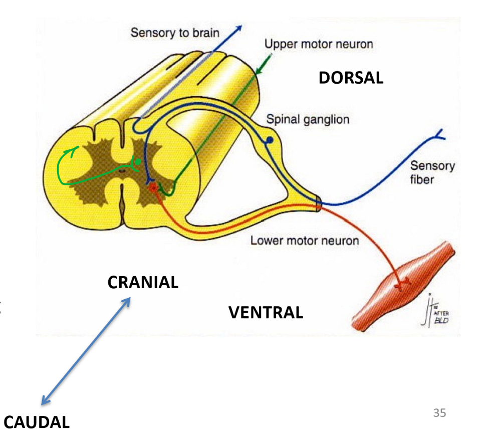
2 neuron chain from primary motor area to effector skeletal muscle
upper motor neuron
lower motor neuron
motor info transmitted from primary motor cortex via upper motor neuron in descending motor tract to given vertebral level
synapse w/ lower motor neuron in ventral horn
axon leaves via ventral root to target skeletal muscle
motor signals further modified by basal nuclei
main pathway:
lateral + anterior corticospinal tract

anterolateral tract (spinothalamic tract)
somatic sensory transmission pathway
lateral → pain + temperature
anterior → crude touch
1st order sensory neurons synapse in dorsal horn
2nd order neurons decussate to contralateral side at lumbar/cervical cord
ascend as spinal lemniscus in brainstem
lemniscus: axons of 2nd order neurons - specific to brainstem + upwards to thalamus
synapse in ventral posterolateral (VPL) nucleus of thalamus
3rd order neurons project to primary somatosensory cortex

posterior column tract (dorsal column tract)
somatic sensory transmission pathway
fine touch, pressure, vibration, proprioception
1st order sensory neurons enter spinal cord
ascend in ipsilateral posterior column to dorsal column nuclei in medulla oblongata
nucleus gracilis: lower trunk + lower extremity
nucleus cuneatus: upper trunk + upper extremity
2nd order neurons decussate to contralateral side at medulla
ascends as medial lemniscus
synapse in ventral posterior lateral (VPL) nucleus of thalamus
3rd order neurons project to primary somatosensory cortex

upper motor neuron
descends from primary motor cortex to ventral horn at given vertebral level
lower motor neuron
extends from ventral horn project through ventral root to target skeletal muscle
lateral and anterior corticospinal tract
axons of upper motor neurons descend from primary motor cortex
form left + right cerebral peduncles of midbrain
lateral corticospinal tract = 90% corticospinal tract
decussates to contralateral side at pyramids of medulla oblongata
descends to given vertebral level
synapses w/ lower motor neurons
anterior corticospinal tract
remains on ipsilateral side
decussates at given vertebral level
synapses w/ lower motor neurons

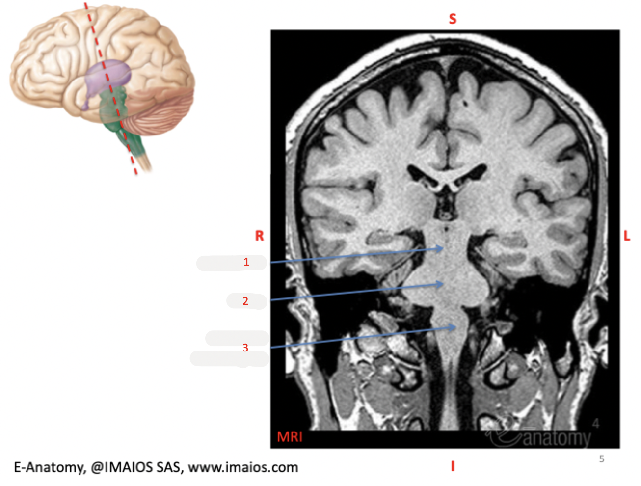
midbrain
(1)
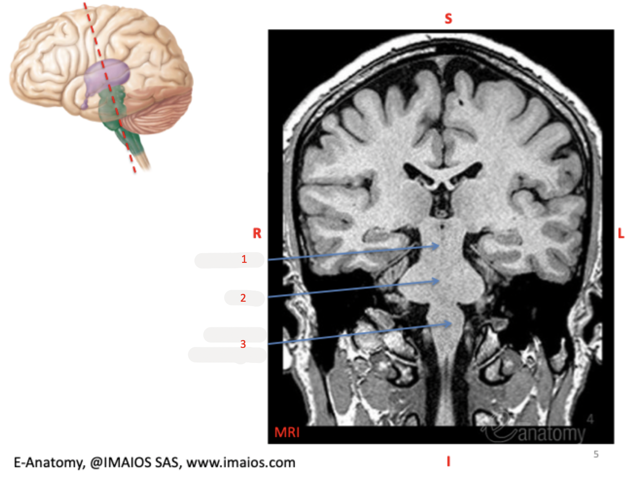
pons
(2)

medulla oblongata
(3)
infundibulum
(1)
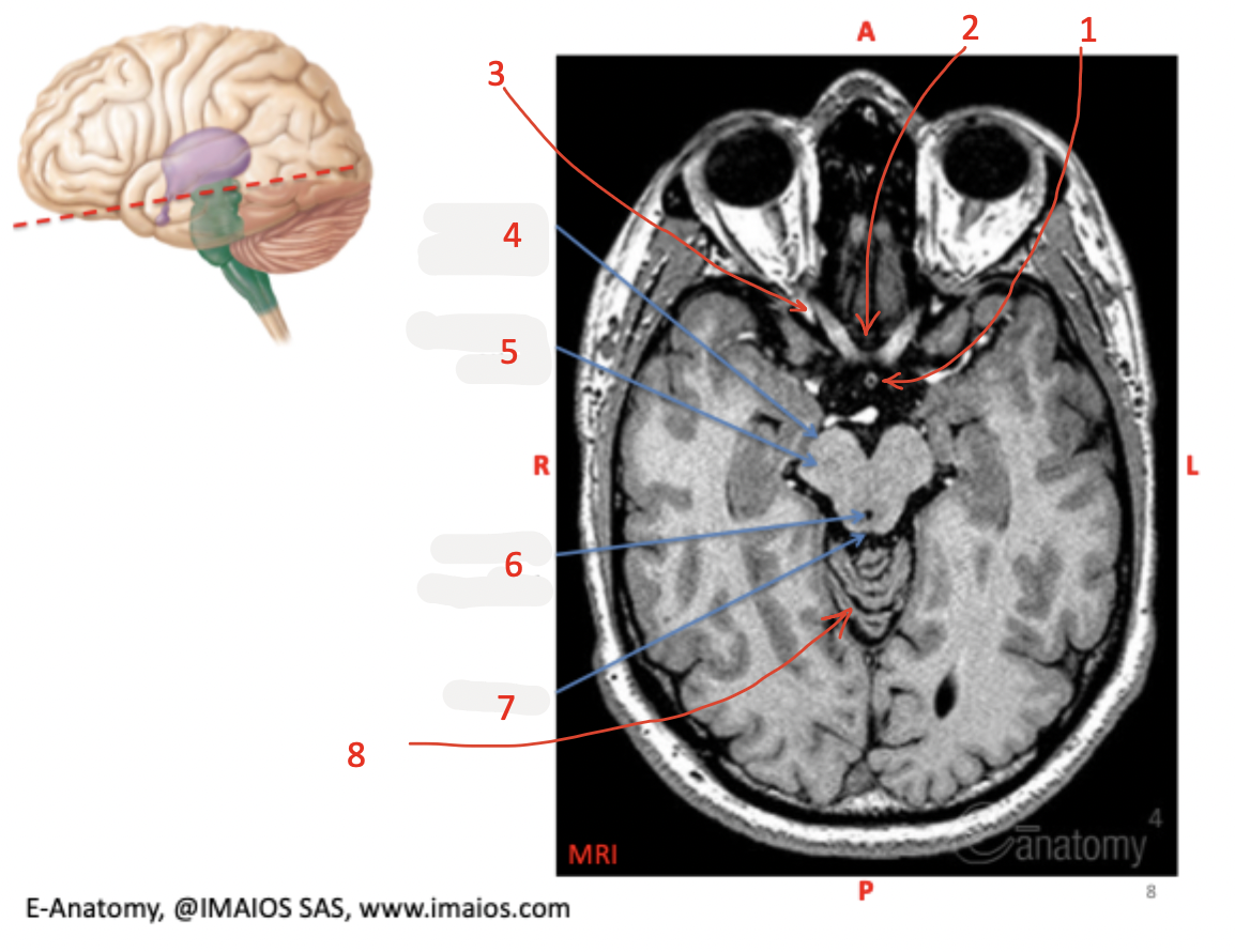
optic chiasm
(2)
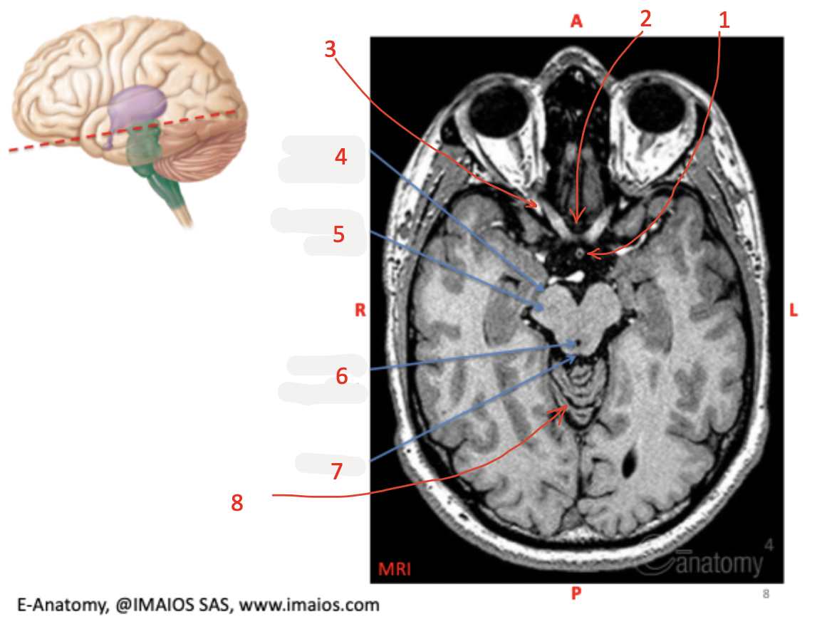
optic nerve (CN II)
(3)

cerebral peduncle
(4)

substantia nigra
(5)

cerebral aqueduct
(6)
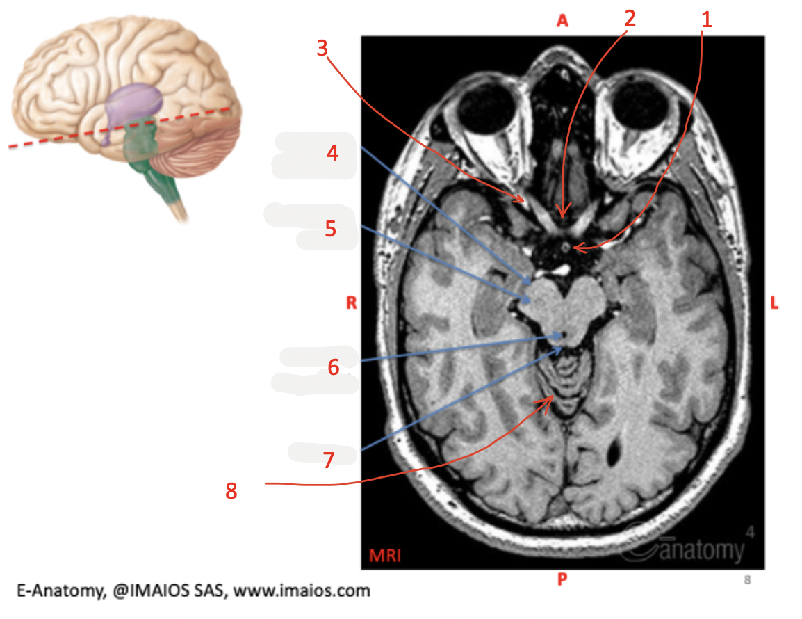
tectum
(7)

cerebellum
(8) posterior to midbrain

thalamus
(1)

hypothalamus
(2)

optic nerve
(3)

infundibulum
(4)

pituitary gland
(5)

pons
(6)

medulla oblongata
(7)

cerebellar tonsil
(8)

cerebellum
(9)

cerebral aqueduct
(10)

inferior colliculi
(11)

superior colliculi
(12)

pineal gland
(13)

cerebral peduncle of midbrain
(14)

fornix
(15)

basilar artery
(1)

basilar groove
(2)
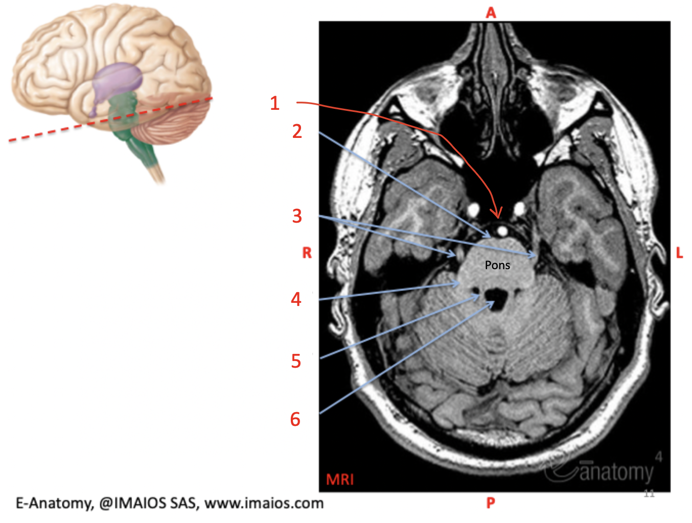
CN V (trigeminal nerve)
(3)

middle cerebellar peduncle
(4)

superior cerebellar peduncle
(5)

4th ventricle (fourth ventricle)
(6) contains CSF
pyramid
(1)

olive
(2)

medulla oblongata
(3)

cerebellar hemisphere
(4)
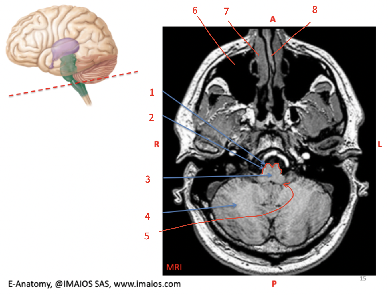
inferior cerebellar peduncle
(5)

maxillary sinus
(6)
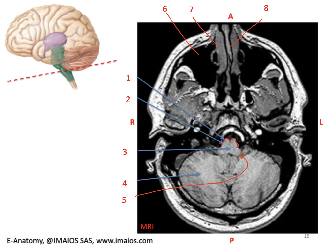
nasal concha
(7)

perpendicular plate
(8)

frontal lobe
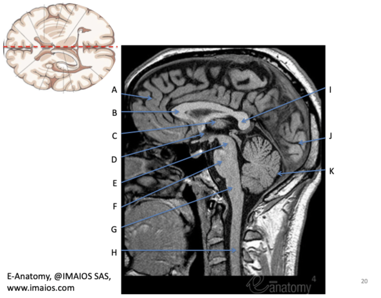
(A)

corpus callosum genu
(B)

thalamus
(C)

hypothalamus
(D)

midbrain
(E)

pons
(F)

medulla oblongata
(G)

spinal cord
(H)

corpus callosum selenium
(I)

occipital lobe
(J)

cerebellum
(K)