NEUR305 Exam 2
 Call Kai
 Learn
Learn Practice Test
 Spaced Repetition
 Match
Match Flashcards
 Knowt Play
1/92

There's no tags or description
Looks like no tags are added yet.
Name | Mastery | Learn | Test | Matching | Spaced | Call with Kai | Chat |
|---|
No analytics yet
Send a link to your students to track their progress
93 Terms
Recall properties common to all sensory systems
-mediated by distinct classes of receptor neurons in specific sense organs
-stimulus → electrical signals encoded as action potentials
Sensation vs perception vs percept
-sensation: the initial activation of the nervous system in response to a stimulus in the environment (sensitive to changes!)
-a percept: the mental representation of the sensed stimulus
-perception: the construction of the percept (how you interpret the environmental stimuli sensed by your nervous system)
modality-specific
sensory receptors are responsive to a single type of stimulus energy
sensory transduction
process of converting sensory signal to electrical signal - sometimes the receptor will only respond to energies with specific temporal or spatial pattern
sensory receptors don’t all generate APs by themselves - some communicate by changing their membrane potential and neurotransmitter release
receptive fields
a delimited medium where some physiological stimuli can evoke a sensory neuronal response
-think of as the spatial resolution of a neuron (better spatial resolution = smaller receptive fields)
base pathway for all sensory systems (except olfaction)
receptor → first order neuron → second order neuron → thalamus → primary sensory cortex
*olfaction doesn’t go through thalamus
most ancient/underdeveloped sensory system
olfactory
describe structurally and functionally why you associate memories with a particular smell
olfactory receptors → olfactory bulb → (pyriform cortex → orbitofrontal cortex)/(olfactory tubercle → orbitofrontal cortex, thalamus, hypothalamus) / (entorhinal cortex → hippocampal formation)
Unlike most other sensory systems, olfactory information can reach the primary olfactory cortex without first passing through the thalamus. Its connections with emotion (amygdala)- and memory-related (hippocampal) regions help explain why a smell can suddenly bring back a vivid memory or feeling. The specific pathway is bulb → entorhinal cortex → hippocampus (which is important for episodic memory)
sensory adaptation vs. habituation
-sensory adaptation is reduced sensory response, habituation is reduced behavioral response
-sensory involves changes in sensory receptor responsiveness while habituation involves changes in neural processing of a repeated stimulus
explain/identify the function of the vomeronasal organ
detects pheremones
valence
the classification of odors as attractive or repellant
olfactory receptors embedded in olfactory epithelium (smell odorants, convergence and divergence)
-detect odorants and identifies concentration & quality
olfactory receptor neurons
-bipolar
-small, unmyelinated axons
-are turned over
-olfactory microvilli protrude into mucus
-sensitive to subsets of stimuli
contained in the olfactory epithelium, not inside the bulb
individual olfactory neurons generally express only one olfactory receptor gene, but each receptor can recognize more than one odor
olfactory bulb
-the first major processing structure receiving info from ORNs
-located on underside of frontal lobes above the nasal cavity
-formed by glomeruli axons (1 bipolar receptor can activate 8000 glomeruli and 1 glomeruli can receive info from up to 750 bipolar receptors)
olfactory coding is combinatorial meaning that
instead of dedicating an individual olfactory receptor to a specific odor, the olfactory system uses combinations of receptor types to greatly reduce the number of receptors required to convey a broad range of odors
olfactory pyriform cortex
-primary olfactory cortex
-key brain area involved in processing/coding of olfactory info
-receives highly converged inputs from distributed glomeruli of the main olfactory bulb
-also receives inputs from cortical and limbic systems
what kind of neural coding in the olfactory system
temporal coding: timing and sequence of glomeruli activation encodes the odor identity
vomeronasal organ - pheremones vs odorants
-pheremones: a chemical substance produced and released into the environment by an animal, affecting the behavior or physiology of others of its species
-MANY odorants, only a few pheromones
what three human sensory systems involve chemical sensing
olfactory (smell), vomeronasal (pheremone sensing), gustatory
taste receptors
organized into taste buds, many of which are associated with papillae on the tongue. the three types of taste-bearing papillae are fungiform, foliate, and circumvallate
tastant produces change in cell’s electrical activity to communicate with sensory nerve fibers
ventral posterior medial (VPM) nucleus of the thalamus
where the cranial nerves lead to. thalamus acts as relay/processing center
primary gustatory cortex - gustotopic maps and TRPA1
input from the VPM. primarily associated with regions of the insula and frontal operculum.
-gustotopic map: spatial organization of neural activity associated with different taste qualities. NOT A TONGUE MAP - refers to how taste information is organized in the BRAIN. investigated using extracellular eletrophysiological recording & anterograde tracing
-TRPA1 is a component of the nociceptive response to CO2
gustatory pathway
tastant molecules → taste receptor cells → cranial nerves → brainstem → VPM nucleus of thalamus → primary gustatory cortex
what cranial nerves do receptor cell axons travel along
facial, glossopharyngeal, and vagus
how can taste affect our behavior? what about other sensations like pain or temperature
the experiment of mice having their sweet vs bitter regions stimulated and how that affected how much time they spent in the chamber (positive vs negative valence). this showed that taste-related neural circuits can influence whether an animal approaches or avoids and environment.
not every sensation you experience in your mouth is a taste. nocireceptors, carbon dioxide, in carbonated drinks, acetic acid in vinegar, trigeminal nerve are all in your mouth and related to pain & temperature
compare and contrast rods and cones in the retina
rods = dim-light vision, cones = color vision & high spatial acuity
primary projection pathways of the visual system
light → retina → retinal ganglion cells → lateral geniculate nucleus (LGN) → primary visual cortex (V1) → higher-order visual areas
identify the potential role of ganglion cells based on primary projection
output signals from the retina toward the brain
vision: bipolar vs horizontal vs amacrine vs ganglion cells
-bipolar: transmit signals between photoreceptors and ganglion cells
-horizontal: lateral processing involving photoreceptor pathways
-amacrine: processing involving bipolar and ganglion cell pathways
-ganglion: output signals from retina toward the brain
on-center and off-center ganglion cells
center-surround receptive fields are divided into two regions: a central region and a surrounding ring.
on-center ganglion cells increases its activity when light illuminates the center of its receptive field but inhibits activity during illumination of the surrounding region.
off-center ganglion cells increase activity when illumination in the center decreases and excites when there is light in the surround.
explain the opponent-process theory of color vision
proposes that color information is processed through opposing channels. major opponent pairs are red v. green and blue v. yellow
compare and contrast receptive fields of neurons in the lateral geniuculate nucleus (LGN) and the primary visual cortex (V1)
increasing receptive-field complexity: LGN→V1→V2→V4→inferotemporal cortex
cortical magnification in V1
a disproportionately large area of visual cortex is devoted to processing information from the center of the visual field because central vision provides much more detailed information
Identify and state inputs and outputs of V1
retinal ganglion cells → LGN (thalamus) → layer 4 of V1 → further cortical processing
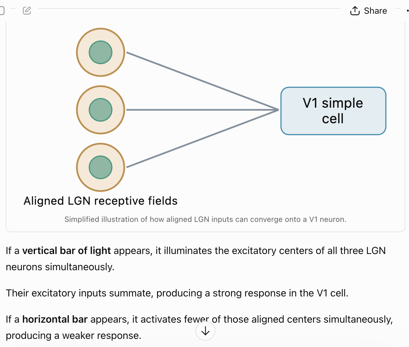
Explain how receptive fields of V1 neurons are formed
V1 simple cells are neurons that have spatially organized excitatory and inhibitory subregions in its receptive field
Identify and state inputs, outputs, and functions of V2
more detail compared to the V1 - more color continuation (gestalt principle - brain groups visual features into organizes shapes/objects) and details.
-organized into stripes: thin stripes (color - projected from blobs), pale stripes (spatial form - projected from interblobs), thick stripes (motion - projected from layer 4b)
Compare and contrast receptive fields of neurons in the lateral geniculate nucleus
(LGN), the primary visual cortex (V1), V4, inferotemporal area (TEO)
retinal ganglion cells and many LGN have circular center-surround receptive fields. many v1 neurons respond preferentially to edges or lines at particular angles (ex. V1 neuron might respond strongly to a vertical line but weakly to a horizontal line. this is because inputs from several LGN neurons can converge onto a V1 neuron so if their receptive fields are arranged along a particular orientation, a line matching that orientation can activate multiple inputs together)
the iris & pupil of the eye (especially dilation)
iris is the muscular structure that regulates pupil size. pupil is the opening through which light enters. pupil dilation is associated with arousal or cognitive effort.
path of light entering the retina
light entering → ganglion cell side (light passes through inner retinal layers) → interneuronal layers → photoreceptors → retinal pigment epithelium
retina of the eye: midget (parvocellular) vs. parasol (magnocellular) vs koniocellular
-midget ganglion cells: parvocellular pathway (layers 3-6): small receptive fields, high spatial acuity, important for red-green color processing,
-parasol ganglion cells: magnocellular pathway (layers 1-2): larger receptive fields, lower spatial acuity, important for detecting motion & changes in visual stimulation
-koniocellular: blue and yellow
lateral geniculate nucleus, pretectum, and superior colliculus are projected to ipRGCs. what makes ipRGCs special?
-express photopigment called melanopsin (so ipRGCs can respond directly to light)
-project to hypothalamus (circadian rhthyms)
-respond to light even without rod and cone input
LGN pathway: tonic vs burst mode
-tonic: alert: regular, individual action potentions
-burst: lost focus: LGN neuron becomes hyperpolarized, neuron receives arousing input and responds with a burst of action potentials, meaning the burst has a better chance of activating neurons in V1
LGN pathway: V1 (pinwheel, stereopsis, blobs vs interblobs vs layer 4b)
-retinotopic organization: neighboring locations in the visual field are represented by neighboring regions of visual cortex. allows visual cortex to maintain information about where objects are located in the visual field.
-orientation columns in V1 (groups of V1 neurons that share a similar preferred stimulus orientation) are organized into pinwheel structures. pinwheels form because as you move across the surface of V1, preferred orientations often change gradually. around certain points, all orientations are represented, creating a pinwheel-like pattern. orientation pinwheel centers are located near the centers of ocular dominance columns.
-ocular dominance columns: regions of V1 where neurons preferentially respond to input from one eye
-orientation columns and ocular dominance columns intersect at approximately right angles
-blobs vs interblobs: blobs are color-sensitive regions and interblobs are orientation-sensitive regions
-layer 4b extends across the cortical columns which is important because the hypercolumn model combines vertical and horizontal aspects of cortical organization
LGN pathway: V4
object constancy & more complex (ex. illusory contours)
LGN pathway: V5/MT
motion processing
visual processing: dorsal stream: where?
projects from visual cortex toward parietal lobe
-spatial location
-motion
-spatiotemporal info
-visual info used to guide actions
visual processing: ventral stream: what?
projects towards temporal cortex
-object identification
-color
-facial recognition
-object recognition
visual processing: object constancy
ability to recognize an object despite changes in how it appears on the retina (ex. different viewing positions, illumination, contexts)
visual processing: sensory adaptation (saccades)
saccades are rapid eye movements that shifts your gaze from one location to another. saccades direct important visual information towards the fovea. eye movements change the stimulation reaching retinal neurons.
-if an image is artificially stabilized on the retina, portions of the image can begin to fade perceptually because the neurons responding to the constant visual stimulus can adapt
why are saccades crucial to vision
saccades rapidly shift gaze, allowing different parts of the visual scene to be examined with high acuity. they also change retinal stimulation, helping counteract perceptual fading associated with sensory adaptation.
what is sensory adaptation and how does it apply to vision
sensory adaptation = a gradual decrease over time in the responsiveness of a sensory system to a constant stimulus. prevents sensory response from remaining saturated during strong stimulation.
do rods and cones have action potentials? why or why not?
no, they have graded potentials (likely do to their very short axons) so the magnitudes of the changes in membrane voltage can vary.
how do horizontal bipolar cells and ganglion cells create a surround
horizontal cells integrate information laterally between neighboring photoreceptor pathways. their inhibitory interactions help create the antagonistic surround that influences bipolar and ganglion cell responses. the ganglion cell’s receptive field therefore reflects more than its direct vertical photoreceptor input. it also reflects lateral interactions across the retinal network (lateral inhibition is when a neuron’s response to stimulation is inhibited by activity in neighboring neurons)
*the point of center-surround receptive fields
what are the functions of each of the different pathways retinal ganglion cells project to (ex. retinal ganglion - pretectum pathway)?
-retina→LGN: visual perception
-retina→superior colliculus: eye/head orientation
-retina→pretectum: pupillary light reflex
-retina→hypothalamus: circadian rhythms
auditory pathway
auditory (cochlear) nerve → cochlear nuclei → superior olivary nucleus → inferior colliculus (IC) → medial geniculate nucleus (MGN) → primary auditory cortex (A1)
Summarize the roles of the outer, middle, and inner ear in auditory processing
-outer and middle: collect and mechanically transmit sound
-inner (cochlea): converts mechanical motion into neural signaling
Describe how inter-aural processing aids sound localization and compare and contrast when interaural time versus interaural level differences would be used
Identify the brain region associated with a particular auditory task (interaural localization, etc)
Summarize models of auditory processing in the barn owl
barn owls have asymmetrically positioned ears vertically. helps to determine not only whether prey is to its left or right but also whether the prey is above or below it.
-azimuth: horizontal: left vs. right: ITD (time)
-elevation: vertical: above vs. below: ILD (intensity)
Distinguish between the roles of the primary (core) and secondary (belt) auditory cortices
-A1 (core) processes sound features, including frequency.
properties of sound: frequency vs. amplitude vs. phase
-frequency = pitch: the number of oscillations per second
-amplitude = loudness: the magnitude of pressure changes in a sound wave
-phase: timing: position of wave within its repeating cycle
anatomy: outer ear (pinna, ear canal)
-pinna: visible external ear: ridges and folds modify sound differently depending on its direction
-ear canal: the passage that directs sound toward the tympanic membrane
anatomy: middle ear: ossicles (stapes, incus, malleus, tensor tympani)
contains ossicles (three small bones that transmit vibrations)
primary function is impedance matching, which means that the middle ear helps transfer sound energy efficiently from air into fluid-filled cochlea
anatomy: inner ear: cochlea: hair cells, spiral ganglion cell axons
-scala vestibulu vs scala tympani: vestibuli is when sound vibrations enter the cochlea when the stapes pushes on the oval window. tympani is when the pressure wave ultimately travels through the cochlear fluid system and is relieved at the round window
-the organ of corti: has auditory hair cells that feed into spiral ganglion cells
auditory pathway: auditory nerve
auditory pathway: spiral ganglion neurons
spiral ganglion neurons are what generate action potentials that travel through the cochlear nerve. the hair cells detect the mechanical vibrations but these are what fire. they connect to the cochlear nerve to carry the signals.
auditory pathway: dorsal cochlear nuclei
brain stem’s first synapse for hearing
-where the neural processing of auditory info begins
-maintains tonotopic map of the cochlea, where inputs from the apex are low frequencies and inputs from base are high frequencies. helps with spectral analysis that helps with processing the timbre of the sound stimulus.
auditory pathway: superior olivary nucleus contains
-medial superior olive: interaural time difference (ITDs): compares when sound reaches each ear: primarily low frequencies
-lateral superior olive: interaural level differences (ILDs): compared how loud sound is at each ear: primarily high frequencies
auditory pathway: inferior colliculus
hub in the midbrain that pulls hearing cues together. keeps tonotopic map of the frequencies of sound being brought into the pathway and mixes inputs from the MSO and LSO. from here, sends a clean and more prioritizes signal to the rest of the pathway - goes directly to MGN.
auditory pathway: medial geniculate nucleus
gate between the inferior colliculus and the primary auditory cortex. shapes and filters incoming spectro-temporal signals, which are representations of a signal over time and frequency. It’s also modified by attention and links sound to the reward system, so task relevant cues are boosted before they hit the cortex. It also performs filtering from complex sounds.
auditory pathway: A1
primary auditory cortical processing. builds precise and fine tonotopic map using input from medial geniculate nucleus of the thalamus
auditory pathway: A2
combines A1 outputs into complex sounds, timbre, pitch, etc. makes meaningful sound objects that are context dependent
konishi’s model
coincidence detection is a mechanism for computing interaural time differences
-a coincidence detector is a neuron that responds especially strongly when inputs from the two ears arrive at approximately the same time. (associated with ITDs NOT ILDs)
why do ILDs work best at high frequencies
because when sound travels toward your head from one side, your head can partially block the sound from reaching the opposite ear. this creates an auditory shadow. but the strength of this shadow depends on sound frequency. high frequencies produce stronger auditory shadows (larger intensity differences between the ears)
*ILDs are represented through comparisons of firing activity. LSO neurons receive ipsilateral excitation and contralateral inhibition via the MNTB.
how does tonotopic mapping work
this is the systemic spatial organization of sound frequencies along the cochlea. different frequencies produce maximal vibration at different locations along the basilar membrane.
base is high frequency. apex is low frequency.
where does frequency by coding take place
in the cochlea of the inner ear
-place coding in the basilar membrane inside the cochlea
-temporal coding in the auditory nerve fibers connected to hair cells
State the main touch receptors and compare receptive fields
-meissner’s: touch: rapid adaptation, small receptive field
-merkel cells: pressure: slow adaptation, small receptive field
-ruffini: stretch: slow adaptation, large receptive field
-pacinian: vibration: very rapid adaptation, large receptive field
Describe or explain the significance of differential myelination of somatosensory axons in terms of behavior and sensation
-large, myelinated axons = fast touch signals
-lightly/unmyelinated axons = slower pain signals
dorsal column-medial lemniscus pathway
-transmits touch, vibration, and pressure
receptor → dorsal spinal cord (ipsilateral) → medulla (crosses midline) → VPL nucleus of thalamus → primary somatosensory cortex (S1)
anterolateral/spinothalamic pathway
transmits temperature and pain
receptor → spinal cord (crosses midline) → ascends on contralateral side → VPL thalamus → S1
Compare the roles of the primary and secondary somatosensory cortex
-S1: located in postcentral gyrus, organized as a somatotopic map (homunculus), receives input from VPL nucleus of the thalamus. is the initial cortical processing
-S2: receives input from S1 and both hemispheres. integrates bilateral sensory info and processes texture/object size
touch: homunculus
body parts are drawn larger or smaller depending on sensory sensitivity, not physical size. somatotopic map is where different body regions map to specific cortical areas, with neighboring body parts being represented in neighboring brain regions.
pain: nocireceptors
-no capsule so that axons are free and open to minute changes in chemistry and temperature of skin
-free nerve endings are free to respond to changes in pH, ATP, & other signaling molecules
pain types: nociceptive vs inflammatory vs neuropathic pain
-nociceptive: direct response to tissue damage
-inflammatory: immune response following injury
-neuropathic: caused by nerve damage
pain types: first and second pain
-first pain: lightly myelinated fiber: sharp, fast, localized experience
-second pain: unmyelinated fiber: slow, dull, lingering (punishment/learning)
pain ascending pain pathways: gate control theory of pain
pain perception can be reduced when non-pain sensory input is activated (ex. rubbing an injured area reduces pain)
because touch fibers activate inhibitory interneurons in the spinal cord that “close the gate” to pain signals
descending pain modulation
brain can actively suppress pain signals through descending pathways. activate endogenous opioids (endorphins, enkephalins) that inhibit pain transmission in the spinal cord
involves Periaqueductal Gray (PAG), nucleus raphe magnus (NRM) and locus coeruleus (LC) to activate the opioids
ascending pain pathways (anterolateral system)
first pain (sensory-discriminative so determines location and intensity of pain): sharp, fast: spinal cord → VPL thalamus → S1/S2
second pain (affective-motivational so controls the emotional and motivational responses to pain): slow, dull, emotional: projects to anterior cingulate cortex, insula, amygdala, hypothalamus
neural coding: encoding vs decoding
-encoding: how the brain maps external sensory stimuli into patterns of neural activity
-decoding: how the brain uses neural response to infer the original stimuli
neural coding spikes: rate vs sparse vs population
-rate: the rate/frequency of action potentials is the data
-sparse: Information is encoded by a small subset of active neurons out of a much larger group
-population: Information is encoded by the combined activity of many neurons.
neural coding: firing rate: raster plot vs PSTH
-raster plots: dots are active potentials and each row is a trial
-PSTH measures given neuron’s activity over time
neural coding: rate coding vs temporal coding
-rate coding: the rate or frequency of action potentials represent data
-temporal coding: the timing of and between spikes represents data
neural coding: labeled line vs place vs population
-labeled line: identity of active neural pathway carries info
-place: location of activity along an organized sensory map carries info
-population: combined activity of multiple neurons carries info