MICRB Midrterm 2 Cram
 Call Kai
 Learn
 Practice Test
 Spaced Repetition
 Match
 Flashcards
 Knowt Play
1/69

There's no tags or description
Looks like no tags are added yet.
70 Terms
First Complete Genome Sequenced
Published in 1995 (“Science” was the journal)
Subect was Haemophilus influenzae (pathogenic, causes meningitis and pneumonia but noo the flu)
Done by Craig Venter using “shotgun sequencing” (sequence random bits of DNA then let computers figure out how it all fits together) which was more efficient + faster than the other approaches of the time
Now, complete genomes aren’t interesting enough to get a whole article in a journal beause they’re extremely accessible compared to when it first came out
Sanger Sequencing
o Developed by Fredrick Sanger in 1970s – Nobel prize (one of his two!)
o Based on DNA polymerase building a complementary strand using: (i) mostly normal dNTPs and (ii) rare special dNTPs that lack a 3’OH and therefore cannot be elongated further
o Special “ddNTPs” each labelled a different way (different fluorophores)
o Build DNAs of different lengths, each terminated with a labelled ddNTP
o Determine sequence based on identity of terminating residues (e.g. – 26 nt sequence terminated with a “T”, 27 nt sequence terminated with a “G”, etc.)

Next Generation Sequencing or Massively Parallel Sequencing
o Different variations - Illumina sequencing popular
o Chips generate millions of “clusters”, each represents (many copies of) a different DNA molecule being sequenced
Reversibly terminated dNTPs used:
o Insert one labelled residue, take an image (A, C, G, T each a different colour of
fluorophore)
o Unblock the 3’ end so you can add another residue. Repeat.
o Each round, you get an image of what residue is at each cluster for that position
(1st nt, 2nd, 3rd…)
What can genome sequencing tell us?
o Metabolic capabilities of an organism
o Virulence genes, antibiotic resistance genes, etc
o Unusual mutations that account for unusual phenotypes
o Discover new genes that might be of medical/industrial interest
o etc…
Provides DNA blueprint required for many studies/analyses
o Genetics approaches (e.g. making mutations to genes)
o Transcriptomics, qPCR, etc – studies of RNA expression
o Proteomics – studies of proteins
o INseq (Tn-seq)
o Etc.
Metagenomics
o Metagenomics is the study of the complete genetic content of an
environmental sample
o Massive sequencing of DNA purified from environmental samples can
provide genomic information (sometimes even complete genomes) for
organisms that cannot be cultured in the lab
o Can tell us about the composition of
microbial communities & how that
composition changes
o Can also look at gene level. E.g. –
how does the frequency of antibiotic
resistance genes compare in the
microbiomes of animals from different
farms?
Transcriptomics: RNA-seq
o RNA can be converted to DNA using a process called reverse
transcription, which can then be sequenced via next-generation methods
o Can get a complete picture of the relative abundance of all of the
transcripts in the cell under a given set of conditions
o One way this is used is comparing expression under different conditions.
E.g. – which genes does a pathogen specifically express during infection?
Proteomics
o Another important “omics” technology is proteomics.
o Often relies on knowing the genomic DNA sequence, but doesn’t use
DNA sequencing.
o Instead, uses mass spectrometry to identify proteins/protein levels
o Like RNA-seq, can tell you what proteins are present under which
conditions. Can also be used in many other creative ways. E.g. which
proteins interact with a protein of interest
The Phases of Transcription (and Actors)
Initiation
Promoters
Sigma factor
Transcription ( residues numbered based on +1 which is the first nucleotide transcribed)
RNA polymerase
Termination
Terminator
Transcriptional Promoters
Transcriptional initiation is guided by DNA sequences called
promoters - DNA sequences bound by factors that promote
transcriptional initiation. Reside upstream of (before) genes.
Whether or not a sequence acts as a promotor & if promoter is active
is dictated by binding of sigma factors (next slide) & regulatory
proteins (future lectures) to the promoter region.
Sigma Factors
Transcription uses an enzyme called RNA polymerase (see future
slides)
A special subunit of RNA polymerase called a sigma factor binds DNA
as an essential step in initiating transcription
Bacteria encode multiple different sigma factors that are produced under
different conditions. They recognize different sequences (promoters) –
see next slide.
The housekeeping (most commonly used/most important) sigma factor
is called or 𝛔70 (or RpoD) – it recognizes two sequences upstream of the
transcriptional start site. See next slide.
Sequences are degenerate – don’t usually match exactly. It can be
difficult to find an RpoD promoter by looking at raw sequence
RNA Polymerase
RNA polymerase binds to DNA template and makes an RNA copy of
one of two strands
Copied strand = coding strand, other strand = template strand
Only builds RNAs 5’ - 3’ – Because transcription goes in only one
direction
Because DNA strands run opposite (5’-3’), transcription from two
DNA strands goes in opposite directions
RNA polymerase core enzyme made up of 5
subunits: ⍺ (2 copies), β, β’, ω. Holoenzyme
also includes 𝛔 (sigma) subunit (sigma factor)
Sigma factor binds promoter region, then
dissociates from core enzyme
Core enzyme unwinds DNA to expose template
- forms transcription bubble
Using NTPs (ATP, CTP, GTP, UTP) as
substrates and the template strand as guide, the
RNA chain is built one nucleotide at a time
Ultimately, RNA polymerase will encounter a
transcriptional terminator (see next slide) and
will dissociate from the template & release the
RNA
Terminator
Transcription will (generally) continue until RNA polymerase (RNAP)
encounters a transcriptional terminator. RNAP then dissociates from
DNA, stops making RNA & releases transcript.
Intrinsic (rho-independent) terminators form when RNA hairpin
structures form, followed by a string of “U” residues. U residues act as
pause signal for RNAP – formation of hairpin forces RNAP off
template.
Rho-dependent terminators: A
protein called Rho binds RNA as it is
being transcribed and causes RNA
polymerase to dissociate after it
encounters certain sequences
What are some of the products of transcription, and what’s the lingo?
o There are 3 major classes of RNAs (PLUS: other non-coding RNAs
with a range of functions – often regulatory):
o Messenger RNA (mRNA) – converted to protein via translation
o Transfer RNA (tRNA) – functional RNAs, used in translation process
o Ribosomal RNA (rRNA) - functional RNAs, used in translation process
o ~95% of RNA in cell is rRNA and tRNA!!!!
o mRNAs contain both Open reading frames (ORFs) and
untranslated regions (UTRs). ORFs are translated to protein, UTRs
are parts of the mRNA transcript that are not translated into protein
o mRNAs that encode multiple ORFs are polycistronic. Such genes are
arranged in an operon. Genes in an operon are cotranscribed.
Organization of bacterial mRNAs
In a simple mRNA encoding a single open reading frame (ORF):
o 5’UTR – everything from first transcribed residue (+1) through the start
codon of gene. Contains ribosome binding site (RBS), more?
o ORF – Start codon (e.g. AUG) through stop codon (e.g. UAA)
o 3’ UTR – everything from the stop codon of the gene through the final
transcribed residue. Often contains transcriptional terminator sequences
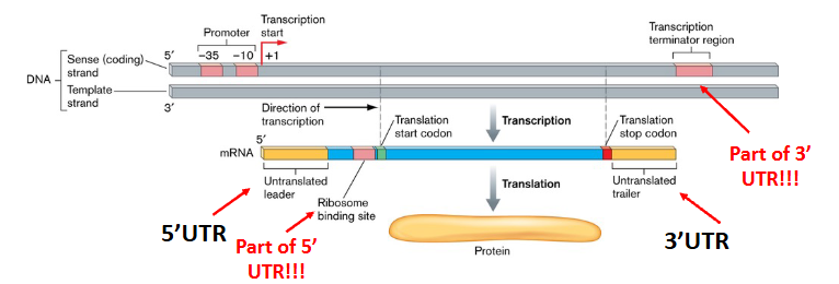
How’s transcription different in Eukaryotes?
o Eukaryotes have 3 RNA polymerases. RNA polymerase II produces mRNA
(encode proteins). Others encode RNA transcripts that are not translated – e.g.
rRNA, tRNA, many other RNAs
o Eukaryotic RNA polymerases are more complex – 12+ subunits
o Eukaryotic RNA polymerase requires transcription factors to recognize
promoters – they bind specific DNA sequences & recruit RNA polymerase
o An example is the TATA box (promoter sequence) bound by TATA-binding
transcription factors (complex of multiple proteins).
o Eukaryotes do not use operons – each gene has its own promoter
o Eukaryotic primary mRNA transcripts contain protein-coding exons and non-
coding introns. mRNAs are processed (spliced) to remove introns
o Eukaryotic mRNAs are polyadenylated at the end (3’) of transcript and capped
at 5’ end using a modified G residues attached using an unusual linkage
o Eukaryotic mRNAs transcribed/spliced in nucleus, translated in cytoplasm
Transcript processing & transport in eukaryotic cells

Transcription in Archaea
o Core aspects of archaeal transcription are very similar to eukaryotic
transcription.
o The archaeal RNA polymerase resembles RNA polymerase II from
eukaryotes and has 11-13 subunits. Is also recruited to promoters using
transcription factors – many of which are related to eukaryotic proteins.
o Archaea also uses TATA boxes & transcription factors that bind these
elements & recruit RNA polymerase
o Others aspects of transcription in archaea more like bacteria…mostly in
terms of “simplicity”
o Archaea transcription (like bacteria):
o No 5’ cap, no poly A tail, mRNAs do not have introns & are not
spliced
o No nucleus – transcription and translation often coupled (like in
bacteria)
o Archaea also use operons, like bacteria - multiple genes encoded by one
RNA (single promoter controls expression of several genes)
Proteins (basic info)
o Proteins are polypeptides. They are comprised of polymers of amino
acids connected by peptide bonds.
o Amino acids have an amino group on one side, an ⍺–carbon in the
middle (has “R group”), and a carboxyl group on the other side.
o Peptide bonds are between the carboxylic acid group of one amino
acid and the amino group of the next amino acid.
o Like DNA or RNA, proteins are directional. Instead of 5’-3’, they
run from N-terminus (free amino terminus) to C-terminus (free
carboxylic acid terminus)
The 20 amino acids
o There are 20 different amino acids that make up proteins. They have
the same backbone, but different R groups. (* 2 rare amino acids,
selenocysteine/pyrrolysine, are rarely used and only in certain organisms)
o They can be grouped by the properties of their R groups: Non-polar
(hydrophobic), polar, positively charged, negatively charged.
Protein Structure
o The chain (sequence) of amino acids in a protein is referred to as its
primary structure
o Small segments of protein adopt simple local structures (local in 3D
space, not necessarily in sequence) called secondary structure. Most
common secondary structure elements = ⍺-helices & β-sheets. Formed
by hydrogen bonding in peptide backbone (amide H & carbonyl O)
o The full 3D structure of a protein called its tertiary structure. Will
typically include multiple secondary structure elements arranged in
different ways & other structural features as well
o Quaternary structure is the result of multiple different polypeptides
coming together – multimeric proteins (or protein complexes).
o The individual polypeptide chains in a multimeric protein are subunits.
Subunits can be identical (homomeric) or different (heteromeric)
tRNA
o The ribosome uses tRNAs to convert (“translate”) the mRNA sequence
into a protein sequence
o tRNAs are ~70-95 bases long, extensively structured, and often contain
modified bases (residues that are chemically altered after transcription)
o Each tRNA has a specific anticodon that
binds a particular three base codon.
o At other end, tRNAs carry the specific
amino acid (cognate amino acid) that
corresponds to that codon.
o tRNA synthetases are the enzymes that
“charge” tRNAs – add the amino
acid to the CCA at the 3’ end.

The gneetic code
o Each codon corresponds to a specific amino acid, or to a stop codon.
o There are 64 possible codons and 20 amino acids -- multiple different
codons can encode a particular amino acid
o Sometimes there is a distinct tRNA for each codon that encodes a given
amino acid
o In other cases, certain tRNAs can work even if there is a single
mismatch in 3rd position “wobble” – same tRNA for 2 different codons
Start codons
o The start codon encodes the first amino acid of a particular ORF (N-
terminal amino acid of a protein). Where translation begins. It is
typically an AUG (ATG in DNA sequence) for all three domains of life
o Alternative start codons also get used – mostly GUG, UUG. In E. coli:
83% AUG, 14% GUG, 3% UUG.
o In bacteria, start codon is translated to N-formylmethionine (fMet)
(chemically modified version of methionine) using a special tRNA.
o AUG encountered during normal translation encodes a standard
methionine. fMet often removed from proteins after translation.
o In Archaea/Eukarya, methionine (unmodified) is used
The ribosome
o Prokaryotic ribosome is the 70S ribosome -- is made up of two subunits
– 30S (small subunit) and 50S (large subunit). Each subunit is
comprised of rRNA and ribosomal proteins.
o E. coli 30S ribosome contains 16S rRNA & 21 proteins. 50S contains
5S/23S rRNA and 31 proteins. Ribosomes are large, complex machines.
o tRNAs and several other proteins interact with
ribosome transiently – perform important roles in
translation
o rRNA carries out much of the main function of
ribosome – including catalyzing peptide bond
formation. The first ribosomes comprised of
only RNA?
Initiation of translation
o RNA has many “AUG”s. To locate bona fide start codons, bacterial mRNAs
use a ribosome binding site (RBS, or “Shine-Dalgarno sequence”).
o RBS is an RNA sequence (some variation in exact sequence)that is recognized
by ribosome - must be appropriately spaced from a start codon (e.g. AUG).
o 16S rRNA component of a free 30S ribosomal subunit (not bound to 50S)
binds to RBS/start codon to initiate translation.
o Initiator (fMet) tRNA then binds the start codon. Once bound, energy from
GTP is used to recruit 50S subunit & full 70S ribosome forms.
Elongation (Translation)
The ribosome has 3 tRNA binding sites:
• A (aminoacyl) site: Where new charged tRNAs enter and recognize the
codon being translated. Once in place, the growing peptide from P site is
transferred its amino acid & peptide bond forms.
• P (peptidyl) site: After peptide bond formation, translocation occurs –
RNA moves 3 bases (one codon). The tRNA from A site moves to P site.
This tRNA transfers growing amino acid chain to the new charged tRNA
that has entered the A site. P site tRNA now lacks amino acid (uncharged)
• E (exit) site: Uncharged tRNAs exit here
During translation, this process repeats until ribosome encounters a stop
codon
Translation Diagram

Terminating translation
o Translation will continue until the ribosome hits one of the three
stop codons: UAA, UGA & UAG
o Once this occurs, a protein called a release factor binds
o This results in ribosome releasing the mRNA & the completed
polypeptide
o 30S/50S subunits dissociate. Ribosome is free to begin a new round
of translation
Polysomes & coupling of transcription/translation
o The same mRNA can be simultaneously translated by multiple different
ribosomes – multiple ribosomes on a single transcript called polysomes
o As 5’ end of RNA in an ORF is translated & freed up, a new ribosome
can bind and begin making protein before previous ribosome is finished
- each ribosome on a polysome builds a complete protein.
o In addition, transcription and translation are often coupled in
prokaryotes – RNA being translated while transcription still going
on (RNA polymerase and ribosomes both attached to transcript)
Eukaryotic translation
o In eukaryotes, translation and transcription are not coupled and occur
in different compartments (transcription in nucleus, translation in
cytoplasm) – separated by both time and location.
o Each eukaryotic mRNA encodes one gene – many prokaryotic
transcripts encode 2+ ORFs (ribosomes re-initiate on same transcript)
o Main mechanistic differences in the initiation step. 5’ cap recognized
by initiation complex in eukaryotes (ribosome binding site). More
regulation/complexity at this step.
o Eukaryotic ribosomes are larger and a bit more complex - 40S (small
subunit)/60S (large subunit). 80S complete ribosome
Chaperones
o Some proteins require chaperones to fold into correct conformation.
Others can fold alone in most situations…but any protein can misfold
(randomly, or due to conditions, such as high/low temperature).
o Chaperones are proteins that help other proteins adopt their properly
folded and fully active state - all 3 domains in life require chaperones.
o Functions: initial folding, re-folding denatured proteins, helping
subunits in multimeric proteins come together, etc.
o There are RNA chaperones that help RNAs adopt the correct
structure.
o Special chaperones are activated in response to high or low temperatures
(heat shock / cold shock proteins) to assist with protein/RNA folding
o In E. coli DnaJ/DnaK & GroEL/GroES are major chaperones that assist
in protein folding – essential proteins for the cell to survive. Amongst the
most abundant proteins in the cell.
Use energy from ATP hydrolysis to enable unfolded or unstable conformations to re-fold in a controlled fashion (precise mechanisms complex & debated)
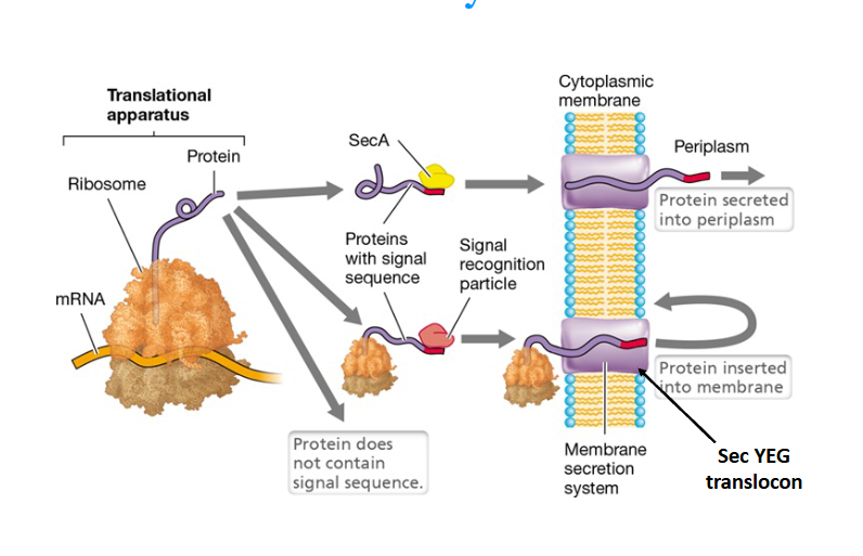
Protein secretion in prokaryotes
o All proteins are synthesized by ribosomes in the cytoplasm, but many
proteins are required in other locations (e.g. cytoplasmic membrane,
periplasm, outer membrane, outside of cell)
o Use translocase systems that transport proteins across (and into) the
cytoplasmic membrane.
o Most translocated proteins contain a signal sequence at the N-terminus
(within first ~25 amino acids) that targets the protein to a particular
secretion system – often removed after translocation.
o The Sec secretion system (see next slide) & twin arginine translocase
(Tat) are ubiquitous in prokaryotes – others are more specialized
o Eukaryotic protein folding, processing/modification and transport
systems are complex and not covered in this course
Sec system
o Tat pathway (not shown) secretes pre-folded proteins across membrane
o The Sec secretion system recognizes a signal sequence in the first ~20
amino acids of protein - translocates unfolded protein before it folds
In Sec pathway, proteins are either:
1) Passed across cytoplasmic membrane (SecA pathway)
2) Recognized by RNA/protein complex – signal recognition particle - and
inserted into the cytoplasmic membrane (SRP pathway).
Both pathways:
1) Pass the unfolded protein through a membrane channel - Sec YEG
translocon
2) Require ATP for energy
3) Signal sequence is cleaved following translocation
Transcription factors
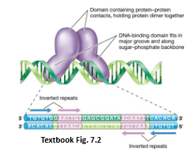
o Many regulatory proteins are DNA-binding proteins. Have DNA-
binding domains that usually bind the major groove of DNA helix.
o Also have other domains with activities like: dimerization, interacting
with other proteins (such as RNA polymerase), regulatory domains...
o Most common scenario: homodimer binds direct or inverted repeats –
one monomer binds each repeat. Dimerization required.
o DNA binding proteins usually
recognize a consensus sequence
…usually somewhat flexible.
o Specificity aided by need for dimer
to recognize both repeat sequences
o DNA structure can play a role too.
Activators and repressors
o Transcription factors that promote transcription = activators
o Transcription factors that inhibit transcription = repressors.
o Some transcription factors are both (activator of one gene, repressor of
another)
o Activators often work by binding DNA at promoter & recruiting RNA polymerase (sigma factor) to begin transcription.
o Repressors bind DNA & (often) prevent RNAP DNA binding or transcriptional initiation after it
binds. Sequence bound by repressor often called operator
Allosteric regulatory proteins
o Some transcription factors are regulated allosterically – binding of an
effector - usually a small molecule –activates or inactivates the protein.
Often works by altering whether it binds DNA.
o Inducers = effectors that “turn on” activator proteins or that inactivate
repressors
o Corepressors = effectors that activate repressor proteins
o An inducible system is one that is off by default, but can be turned on. A repressible system is one that is on by default, but can be turned off.
o A single gene is often controlled by both inducible and repressible systems
Arginine biosynthesis
o Example of a repressible system
o ArgR is an allosterically-controlled repressor protein that regulates the
expression of an arginine biosynthesis operon
o Regulates multiple co-transcribed genes that produce arginine from
precursor molecules in the cell
o When arginine is present, it acts as a co-repressor.
o When arginine levels are low, ArgR isn’t bound by arginine, doesn’t
bind DNA – genes are expressed and arginine is synthesized by cell
o When arginine levels are high, it binds ArgR, enabling ArgR to bindthe operator & prevent
transcription of this operon

Lac operon
o Inducible system
o Lactose is a good energy source for many bacteria, including E. coli.
o Machinery for breaking down lactose is encoded by the lac operon.
o Expressing these genes in absence of lactose not useful – to prevent
this, LacI repressor protein binds lac Operator, prevents transcription
o When lactose is available, a lactose isomer called allolactose binds LacI & inactivates it
(allolactose = inducer)
o Inducible system - catabolic systems are often inducible
o E. coli doesn’t want to use lactose if a better energy source is available -- Catabolite repression
o In the presence of glucose, production of cAMP is inhibited. Low cAMP levels in cell
o For lac operon to be expressed, also requires CRP (cAMP receptor protein) to bind cAMP. cAMP-bound CRP binds promoter region & recruits RNA polymerase
o Lac operon expression requires both: lactose AND low glucose levels.
o Glucose regulation is indirect (cAMP is direct inducer)

Cyclic AMP (and ppGpp)
o Cyclic AMP (cAMP) is an example of a signaling
molecule or second messenger
o Small molecules whose levels are controlled to serve
as regulatory molecules (send message around cell).
o In bacteria, nucleotide-based signaling molecules
common: cAMP, (p)ppGpp, cyclic-di-GMP, more!
o Produced in response to some condition –regulate
different processes in the cell to mount a
coordinated response to that condition.
o E.g. ppGpp produced in response to amino acid
starvation. ppGpp shuts down protein synthesis &
induces amino acid biosynthesis in a process that is
called stringent response
Quorum sensing
o Quorum sensing involves signaling molecules called autoinducers
o Quorum sensing involves sensing the local density of cells through
secreting/detecting specific molecules – regulating based on that info.
o Used to coordinate group behaviours like biofilm formation, virulence,
etc. Only useful to carry out these activities at high cell densities
o Basic idea: Produce autoinducer (small molecule), it diffuses away.
Doesn’t accumulate except at high density. Detect high density – know
you’re in a group – activate group behaviours
o Different autoinducers exist. Acyl homoserine lactones (AHL) common
in Gram -ve bacteria – different versions of AHL in different species.
o Quorum sensing is a form of chemical communication. It is used by
bacteria, archaea and eukaryotic microbes
o Numerous genes induced/repressed in response to the concentration of AHLs (sensed by the cell).
o Different species produce/detect their own specific versions of AHL (different R groups)
o Quorum sensing discovered in Vibrio fischeri, a symbiont of the Hawaiian Bobtail Squid.
Produces an enzyme that carries out a luminescent reaction, but only when present at high concentrations in a specialized light-producing organ of the squid

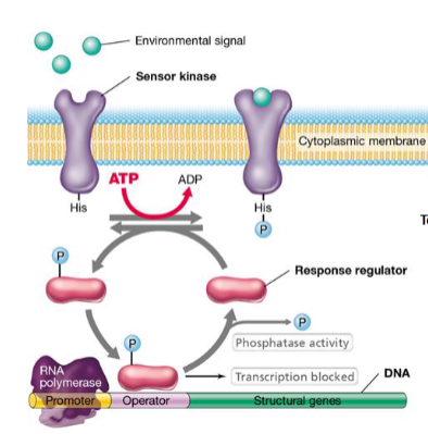
Two-component regulatory systems
o A common form of gene regulation in bacteria – form of signal
transduction. Sometimes they sit atop large regulatory networks –
control major biological programs.
Use two proteins: (sometimes more…but we’ll ignore this ☺)
Sensor kinase: Usually in cytoplasmic membrane. Senses signal(s), which
activates kinase activity - adds phosphate to response regulator. In absence of
signal, dephosphorylates response regulator.
Response regulator: When phosphorylated, becomes active. Many work as
DNA-binding transcription factors → Phosphorylated version binds DNA to
regulate expression of target genes (activator and/or repressor)
o One of largest gene families in bacteria ~30 different two component
systems/genome. Some species have > 200 different systems
o Expression of two-component systems often regulated…layers of
regulation (regulating the regulator).
Transcriptional regulation in Archaea
o Despite differences in transcription mechanisms (see lecture 4-1),
transcriptional regulatory systems in Archaea often analogous with bacterial
systems
o Activators/repressors bind DNA to affect recruitment of RNA polymerase
o Two-component regulatory systems also present in Archaea…but not as
common as in bacteria
o More complex regulation is observed in Eukarya (not covered in this course)
Regulation at the level of RNA
o There is a lot of regulation that happens after transcription starts, but
before the protein is produced
o Transcriptional attenuation is regulation that involves terminating
mRNA synthesis before gene(s) are transcribed
o The stability of mRNAs can be controlled – how long before they are
degraded (longer lifetime, more translation, more protein produced)
o Translation efficiency can be controlled – usually whether or not
RBS is free to be bound by the ribosome → RBS structure
o Much of this regulation is executed by RNA regulatory elements
mRNA lifetimes and protein levels
.
o Bacterial transcripts do not last long –half-life spans from seconds to
~an hour (often ~a few minutes). Different mRNAs can have very
different half-lives
o All cells contain multiple ribonucleases (RNases) that degrade mRNAs. Recognize different sequence/structural features, degrade RNA in different ways
o Why some mRNAs are targeted more/less by RNAses is complex and often unclear
o Regulatory proteins/RNAs can bind to mRNA and impact its half-life.
RNA structures that “shut off ” gene expression
1. For translation to initiate, the ribosome binding site (RBS) needs to
be “free” to bind ribosome, not involved in base-pairing
2. During transcription, the formation of intrinsic transcriptional
terminators before a gene’s transcription is complete (usually in
5’UTR) leads to transcriptional attenuation
Formation of stem-loop structures in mRNA can shut off gene
expression if they: (1) involve the RBS, or (2) involving the formation of a
transcriptional terminator upstream of a gene. This can be regulated.
Small RNAs
o All cells encode regulatory RNAs – noncoding RNA whose function is to
regulate gene expression
o Bacterial sRNAs (~50-300 nt long) generally exert effects by base-pairing
to target mRNA(s). Base-pairing with sRNA affects RBS availability
and/or RNase targeting, thereby controlling how much protein gets made
o One sRNA can sometimes regulate many different target mRNAs

sRNAs ribonuclease mechanism diagram

Hfq
o Many sRNAs have limited sequence complementarity to target mRNAs
(perhaps only a ~ 5-11 nt stretch base pairs)
o RNA chaperones such as Hfq are essential for mediating this regulation
o Hfq binds to both RNAs to stabilize their interaction - in some cases Hfq
also appears to recruit RNases to mediate degradation
Riboswitches
o Riboswitches are ligand-binding segments of mRNA found in 5’ UTRs
that regulate the expression of gene(s) within that mRNA.
o Include receptors (“aptamers”) that specifically bind a particular small
molecule. This binding allows riboswitches to sense their environment.
o Ligand binding changes RNA structure in 5’UTR – this change induces or
represses expression of downstream genes (next slide). Ligand binding
changes expression of genes from “on” to “off ”, or vice versa.
o Riboswitches have been found in all 3 domains of life, but mostly identified/
characterized in bacteria
o Very different from sRNAs
o Ligand binding changes base-pairing in the 5’UTR to affect formation of
either: (i) A stem-loop that sequesters the RBS (blocks translation) or (ii) a
transcriptional terminator that prevents transcription of the genes
o Riboswitches have been hypothesized to be “relics from the RNA world”
o Riboswitches bind a wide range of central metabolites – structurally
conserved aptamer domains in divergent lineages (ancient ancestry)
o Binding of such metabolites would have been essential in an RNA world in
order to sense the environment

Post-translational regulation (regulating protein levels/activity)
o An enzyme within a given biosynthetic pathway is sometimes inhibited by
the end product of that pathway -- feedback inhibition.
o Activity of proteins can also be regulated by protein-protein interactions
(one protein binds another to control its activity)
o Protein activity can be changed via post-translational modifications – a
chemical moiety such as a phosphate group is added to a specific amino
acid residue of a protein (changes activity, or turns it “on”/“off ”)
o Protein sometimes degraded
Protein degradation/turnover
o Like RNA, protein can also be degraded and their building blocks recycled.
This is accomplished by enzymes called proteases.
o Proteases are essential for clearing away and recycling misfolded proteins
o In other cases, proteolysis can be a very specific and regulated mechanism
to clear away specific proteins in the cell to regulate their abundance
o Some proteins require proteolytic activity (e.g. cleave a single peptide
linkage) in order to be activated
Enzymes
o Enzymes work by lowering the activation energy of a reaction
o They do NOT change the energetics (ΔG) or the equilibrium of the reaction
o By lowering the activation energy, enzymes increase the rate of a reaction – in some cases by many, many orders of magnitude
How do they work?
o Local concentrations of substrates are increased at the active site of the enzyme & are oriented properly for the reaction to take place
o Many enzymes alter the electronic distribution of the substrate(s), which enhances reactivity.
o Stabilization of transition states of the reaction
o Some enzymes use of coenzymes or prosthetic groups – some of which employ metallic elements (E.g. - Fe, Mg, Zn, Co…) that can help with challenging chemistry
ex. Lysozyme cleaveg of Beta-1,4 glycosdic bond in peptidoglycam

How do enzymes influence metabolism?
o Most biological reactions will not occur at a significant rate
in the absence of an enzyme
o Enzymes catalyze specific reactions, allowing the for accumulation
of those specific products
o In metabolic pathways, products from one reaction are the
reactants in the next – prevents accumulation of products of the
individual reactions (accumulation of products promotes the
reverse reaction)
o Enzyme activities can be controlled by regulating the amounts of
the enzymes or by controlling their activity. This controls which
metabolic reactions do/don’t take place in the cell.
Competitive inhibitors
o Competitive inhibitors of an enzyme “fit” in the same active site as
the substrates – inhibit substrate binding (and thus the reaction)
o This is the way many drugs work. Example below shows a sulfa drug,
which is an antibiotic that inhibits folate biosynthesis.

Allosteric activators/inhibitors for Enzymes
o Allosteric regulation of an enzyme: Catalytic activity of an enzyme controlled by a molecule (“effector”) that binds the enzyme at a location other than the active site.
o Allosteric activators bind the (inactive) enzyme & change the conformation of the active site to activate the enzyme

Feedback inhibition
o Strategy to prevent too much of product from being made
o End product acts as inhibitor of an early step in pathway (usually first committed step)
o Will evolve binding affinity for enzyme such that it binds/inhibits only when levels are high
o Once end product used up, levels drop, no longer inhibits, pathway resumes
Metabolic requirements for all life
1. Liquid water
2. Nutrients (e.g. sources of carbon, nitrogen, etc)
3. A source of energy to do work
4. A source of electrons for biochemical reactions
Nutrients for Metabolism
o All cells need the all the basic atoms/molecules required to build the
materials your cell needs (DNA, RNA, proteins, essential cofactors, cell
wall, membranes, etc).
o All cells need a usable source of the basic elements (E.g. - C, N, P, S)
o Cells also need other elements that serve essential cell functions, (E.g.
Fe, Mg) – some elements are only required in trace amounts
o Many microbes have evolved in environments where certain essential
molecules (e.g. amino acids) are available and they have lost (or never
had) the ability to make them. These would be essential nutrients for
those organisms
Classifying microbes by where they get their carbon
Autotrophs:
o Use CO2 (inorganic carbon) to build cell materials
o Takes a lot of energy
o Most chemolithotrophs and phototrophs
o Primary producers – very important – synthesize the organic
molecules that heterotrophs (like us) use
Heterotrophs:
o Obtain carbon from organic compounds
o Nearly all chemoorganotrophs
ATP
o Hydrolysis of ATP to ADP used to power synthesis of cell components
(proteins, membranes, cell wall, etc) and to power other cellular functions
(e.g. transport)
o Many ATPase enzymes in the cell – couple energy of ATP hydrolysis to
perform other reactions
o Energy can be conserved in other energy-rich bonds as well (see below)
Catabolism and Anabolism
Both types of metabolism
Catabolism
Reactions to obtain energy &
break down complex
molecules
Anabolism
Reactions used to synthesize
cellular material (use energy)
Standard Gibbs free energy
ΔG0’ is the free energy change of a reaction under standard conditions.
Can be calculated based on nature of reactants/products.
Negative value: Reactants have more energy than products
- Exergonic (releases energy)
- Spontaneous
Positive value: Reactants have less energy than products
- Endergonic (requires energy)
- Not spontaneous
Actual ΔG is effected by:
o Temperature
o Concentration of substrates
o Concentration of products
High K values - Relatively high [products] -
increase ΔG values.
Makes reactions less energetically favourable (in that direction)

Aerobic respiration of glucose
o This releases 2895 kJ of energy per mole
o ATP production requires at least 31.8 kJ/mole
o Could produce up to 91 moles of ATP per mole of glucose
o Actual number less (e.g. ~38 moles). Why?
o Not under “standard conditions” & reactions not 100% efficient -
energy lost as heat
o This is an example of a reaction used by a cell to get energy…but this
net reaction is carried out over many steps

Redox reactions
o Source of electrons
o Some chemical reactions are redox reactions
(reduction/oxidation)
o Electrons transferred from high energy state to lower energy
state – can capture and use this energy
o Basis for much cell metabolism – the source of energy for
chemotrophs
o Even phototrophic bacteria need a source of electrons to carry
out anabolic reactions in the cell
o Electrons are at a lower energy state when they are associated
with more electronegative atoms
o Electronegativity decreases with higher period and increases with higher group
The Glucose Redox Example (Catabolism of Glucose)
o For many chemoorganotrophs, sugars like glucose are a preferred
energy source. They carry out the net reaction above in a
series of oxidation reactions (not one big explosive reaction)
o This lecture uses this as a useful starting point for understanding
catabolism, but many other organic compounds can also be used
to generate energy, often using overlapping pathways
o Many other sugars (disaccharides, polymers, monosaccharides
other than glucose) can be converted to glucose or into an
intermediate in glycolysis or the citric acid cycle
Glucose to CO2 is one half reaction, O2 to H2O is the other half
reaction
• Glucose is the electron donor here. O2 is the electron acceptor.
• Glucose loses electrons, is oxidized. O2 gains electrons, is reduced
- Glucose is a great source of high energy electrons (C-H bonds,
neither element especially electronegative).
- Oxygen is a great electron sink – leaves electrons at low energy state.
- Energy released by electrons moving to lower energy state. This is
captured by the cell through a series of redox reactions

Redox couples, redox tower
o The are many, many possible redox couples.
o Glucose & CO2 are an example of a redox couple (half reaction)
o Glucose is the reduced form and CO2 is the oxidized form
o By convention, redox pairs written as (oxidized form/reduced form). So: CO2/glucose
o These redox pairs can be arranged in a redox tower
o Different half reactions can be combined into different redox reactions. Redox tower tell you which redox couple will be the electron donor, and how much energy will be released.
o The reduction potential (E0’ ) of a redox couple indicates its propensity to act as an electron donor or acceptor
o Redox couples with more negative values (top of table) have a stronger tendency to act as electron donors (to be oxidized)
o Redox couples with more positive values (bottom of table) have a strong tendency to act as electron acceptors (to be reduced)
o Glucose is a great electron donor, O2 is phenomenal electron acceptor
o Redox reactions between couples that have
bigger differences in redox potential produce
more energy:

Electron carriers
o In metabolism, electrons often transferred from an initial electron
donor to an ultimate acceptor over many biochemical reactions
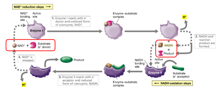
o Soluble electron carriers like Nicotinamide adenine dinucleotide (NAD+ / NADH) used to shuttle electrons around the cell
o NAD+ is oxidizing agent (gets reduced to NADH). NADH is reducing agent (Gets oxidized to NAD+)
o Electron carriers such as NAD+/NADH are enzymatic cofactors
o Electron carriers allow electrons donated in one reaction – stored in NADH – and accepted in a different reaction.
o They are soluble – transfer electrons around the cell
LEO says GER

How Biology generates ATP
i) Substrate level phosphorylation: ATP generated as a product of a metabolic reaction. Exergonic rxn –use excess energy to make ATP
ii) Oxidative phosphorylation: Energy from electron transfer reactions generate a proton motive force, which is used to generate ATP
iii) Photophosphorylation: Energy captured from light is used to generate a proton motive, which is used to generate ATP
Glycolysis