4 - The Pathophysiology of Dry & Neovascular AMD
 Call Kai
 Learn
Learn Practice Test
 Spaced Repetition
 Match
Match Flashcards
 Knowt Play
1/88

Earn XP
Description and Tags
Block 2
Name | Mastery | Learn | Test | Matching | Spaced | Call with Kai | Chat |
|---|
No analytics yet
Send a link to your students to track their progress
89 Terms
Describe the Beckman grading scale

How does vision typically differ between early/intermediate and late AMD?
VA is often unaffected and patients may be asymptomatic earlier
Late AMD causes central visual loss that is gradual in GA or rapid in nAMD
Which structures are primarily affected by AMD?
Choroidal circulation
Bruch’s membrane
RPE
Photoreceptors
What are drusen?
Localised deposits between the basement membrane of the RPE and Bruch’s membrane

What are hard drusen/druplets?
Tiny yellow-white lesions ≤63 µm
Small numbers are part of normal ageing
Numerous hard drusen increase the risk of soft drusen and AMD

What are soft drusen?
A hallmark of AMD
Larger drusen can be distinct or indistinct and may coalesce to form confluent drusen
Associated with diffuse thickening of Bruch’s membrane

How do drusen appear on OCT?
They cause elevation of the RPE

What can numerous drusen cause?
Drusenoid pigment epithelial detachment (PED)
What is the annual risk of CNV or foveal GA with bilateral drusen and good VA?
~3% per eye per year
How many patients with drusen developed nAMD over 4 years?
~10%
What features increase the risk of progression to late AMD?
Larger drusen
Greater number of drusen
Greater confluence drusen
Pigmentary changes - disruption of the overlying RPE causing hypo- or hyperpigmentation
What other features increase the risk of progression to late AMD?
Focal hyperpigmentation /RPE atrophy
Slow choroidal filling
Late AMD in the fellow eye
How do drusen change over time?
Growth is undulating; over 1 year
~50% increase in volume
~10% regress
~40% remain stable
Why is regression of drusen clinically important?
Drusen regression may precede development of GA or CNV

What is basal laminar deposit (BLamD)?
Material between the RPE plasma membrane and RPE basement membrane

Is BLamD specific to AMD?
No → it occurs in normal ageing but a continuous layer is always present in AMD
What is basal linear deposit (BLinD)?
Granular vesicular or membranous debris between the RPE basement membrane and Bruch’s membrane

Why is BLinD important in AMD?
It is a specific marker for AMD and its accumulation leads to soft drusen formation
What is the proposed origin of BLamD?
Excess basement membrane produced by the RPE in response to stress
What is the proposed origin of BLinD / membranous debris?
RPE expels damaged cell constituents through its basolateral membrane following injury
What may initially cause RPE injury leading to BLinD?
Oxidative
Inflammatory
Ischaemic injury
What are the proposed origins of drusen?
Clusters of hard drusen or membranous debris
they also contain components associated with the immune response
What causes focal hyperpigmentation in AMD?
Increased melanin content of RPE cells
RPE cell proliferation
RPE cell migration
= FA is ‘glowing’ because thats the atrophy as less pigments there to block it

What causes focal hypopigmentation in AMD?
Reduced melanin content of RPE cells
RPE cell atrophy
Thinning of the RPE layer
= seen as small patches of mottled pigment
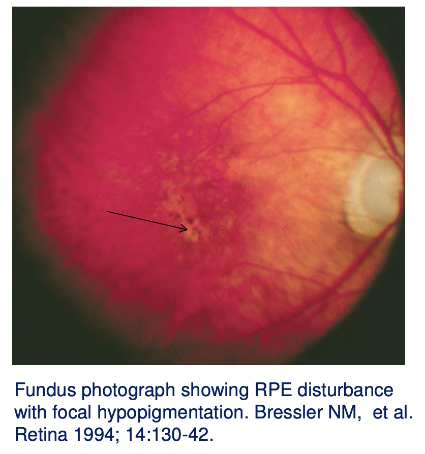
Where can pigment clumping be seen in geographic atrophy?
At the edge of the area of GA
Table that shows the risk of progression of early to advanced (wet / GA) AMD
1 risk factor for large drusen in each eye + 1 risk factor for pigmentary changes in each eye = total score 0 - 4

What is geographic atrophy (GA)?
Confluent areas >175 µm of RPE cell death

Why do photoreceptors die in geographic atrophy?
Photoreceptors are metabolically dependent on the RPE so RPE death leads to photoreceptor death and visual loss → begins parafoveally spares fovea until later
Why are underlying choroidal vessels visible in GA?
Loss of the overlying RPE allows the choroidal vessels to become visible
What can precede geographic atrophy on autofluorescence?
Areas of increased autofluorescence possibly associated with RPE lipofuscin
What events can precede development of GA?
Drusen regression
Flattening of a PED
Involution of CNV
How commonly is GA bilateral?
~50% of patients
What proportion of registered blindness due to AMD is attributed to GA?
~20%
Can GA and CNV occur together?
Yes → 2 - 4% incidence of CNV over 2 years in patients with bilateral GA
What is choroidal neovascularisation (CNV)?
Growth of new blood vessels from the choroid that proliferate beneath the RPE or into the subretinal space

How may CNV appear on fundus examination?
As a green-grey lesion
Why does CNV cause haemorrhage and leakage?
The newly formed vessels are fragile
bottom left = right arrow is SRF
top right = IRF

What retinal findings are commonly associated with CNV?
Subretinal or intraretinal haemorrhage
Hard exudates
Intraretinal fluid
Pigment epithelial detachment

In neovascular AMD what does repeated leakage of blood, serum and lipids stimulate?
Stimulates fibroglial organisation → formation of disciform scar (retina replaced by scar tissue so no visual sensitivity)

What is rapid vision loss in neovascular AMD due to?
Exudates and haemorrhage
Secondary cell death
Formation of disciform scar
How rapidly does untreated CNV affect VA?
~1 line logMAR acuity loss in 3 months and 3 lines by 1 year
What can stimulate CNV development?
Ischaemia
Oxidation
Inflammation
How do ischaemia oxidation and inflammation promote CNV?
They increase expression of proangiogenic growth factors such as VEGF-A
How can CNV cross Bruch’s membrane?
Sub-RPE and subretinal CNV can occur through breaks in Bruch’s membrane
Where does a pigment epithelial detachment (PED) occur?
Between the RPE basement membrane and the inner collagenous zone of Bruch’s membrane

What can happen to a PED over time?
Flatten
Tear
Usually leaves an area of atrophy or subretinal fibrosis
What characterises a drusenoid PED?
Irregular surface
Overlying pigmentary changes
Slow enlargement

What characterises a serous PED?
Smooth sharply demarcated dome-shaped PED
Rapid bright uniform filling on fluorescein angiography

How strongly is serous PED associated with CNV?
>80%
How may an avascular serous PED develop?
Thickening and increased hydrophobicity of Bruch’s membrane impair movement of fluid towards the choroid so fluid accumulates beneath the RPE
What is the relationship between serous PED and CNV?
CNV leakage may cause serous PED (alternatively existing avascular PED may promote CNV)
What characterises a fibrovascular PED?
Irregular appearance
Breaks in RPE exposing underlying CNV
Neovascular membrane elevates the RPE
Large adjacent areas of subretinal fluid

What structural changes occur in photoreceptors in AMD?
Cell loss initially mainly affects parafoveal rods; cones are lost later
What structural changes occur in the RPE in AMD?
Lipofuscin accumulation and RPE cell death
What structural changes occur in Bruch’s membrane in AMD?
Thickening and increased deposition of hydrophobic material beyond normal age-related changes
What is the consequence of Bruch’s membrane thickening in AMD?
Impaired transport of oxygen; fluid; growth factors; retinoids; waste products etc.
What structural choroidal changes occur in AMD?
Impaired choroidal blood flow and choriocapillaris dropout adjacent to areas of CNV and GA
What are the main mechanisms implicated in AMD pathogenesis?
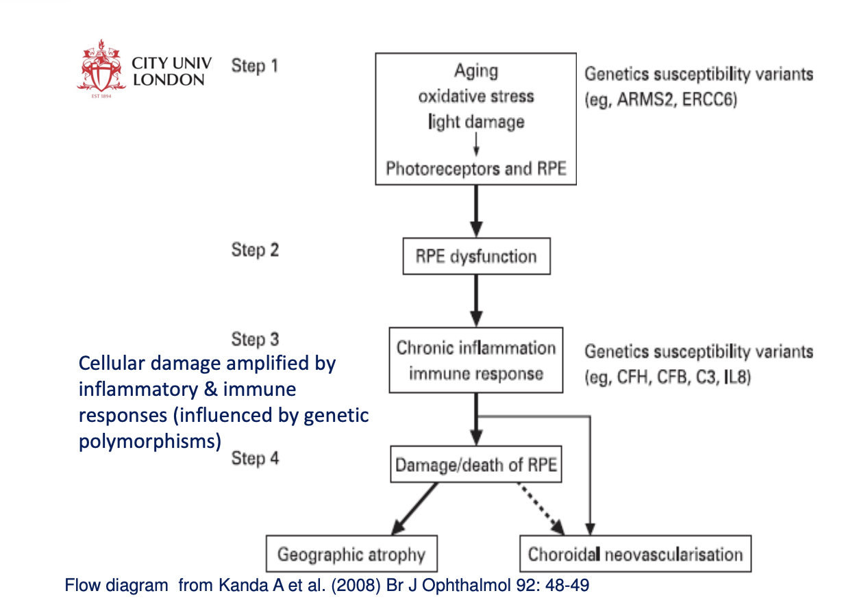
Oxidation
Immune response / inflammation
Ischaemia
Genetic predisposition
What is oxidation?
Removal of one or more electrons from an atom or molecule
Oxidising agent (B) accepts these electrons and so is reduced
Compound A (oxidised) needs to accept electrons to become oxidising agent

Why can oxidation cause cellular damage?
It changes the structure of cellular macromolecules and produces abnormal materials
What are reactive oxygen species (ROS)?
Free radicals
Hydrogen peroxide
Singlet oxygen
They are unstable and extract electrons from other molecules making those molecules unstable in turn
How are ROS normally produced?
As a side product of cellular metabolism
What factors increase ROS production?
Irradiation
Cigarette smoke
Ageing; inflammation
High partial pressure of oxygen
Why are photoreceptor outer segments prone to oxidative damage?
Their membranes contain high levels of polyunsaturated fatty acids which are susceptible to oxidation by ROS
Why does light exposure make the retina susceptible to oxidative damage?
The retina experiences high cumulative light irradiation (short-wavelength light is harmful)
How do retinal photosensitisers contribute to oxidative damage?
Chromophores such as rhodopsin and lipofuscin absorb light and trigger chemical reactions that generate ROS
What other features make the retina particularly susceptible to oxidative damage?
It has the highest O₂ consumption of any tissue in the body and RPE phagocytosis of photoreceptor outer segments generates ROS
What is lipofuscin?
An age-related pigment found in the RPE that progressively accumulates with age
How is lipofuscin formed?
From incomplete degradation by the RPE of abnormal oxidised material
Why is excess lipofuscin harmful?
It reduces functional cytoplasmic space
Short-wavelength excitation generates ROS
High levels are associated with RPE and photoreceptor degeneration
What is A2E and why is it important?
A component of lipofuscin that inhibits lysosomal function reducing the RPE's ability to process waste and inducing apoptosis
How can oxidative stress promote neovascular AMD?
Oxidative stress causes RPE upregulation of VEGF-A and activates neovasc
What are the effects of lipid oxidation in AMD?
Advanced lipoxidation end products accumulate in RPE and Bruch’s membrane → lipid peroxidation damages cell membranes and causes cell death
How does oxidation affect proteins and nucleic acids?
Oxidised proteins lose functional integrity
Oxidation of nucleic acids contributes to ageing and age-related disease
What evidence supports oxidative damage in AMD?
Higher antioxidant intake / serum levels inversely related to AMD prev
Macular pigment (antioxidant and blue light filter) may be protective
Sunlight exposure associated with AMD
Smoking associated with AMD
RPE lipofuscin correlates with AMD
Oxidative end products occur in AMD
ROS production peaks at the macula
RPE mitochondria are vulnerable to oxidative stress
Oxidative stress can promote CNV
Pathogenesis: immune response - what is the complement pathway?
~30 proteins forming the innate immune response
How can an unregulated complement response damage the retina?
It can directly damage host tissue and recruit immune cells to the area → Complement proteins stimulate RPE release of VEGF-A - C3a and C5a (CP) recruit proangiogenic leukocytes to the choroid
How are oxidative stress and complement activation linked in AMD?
Oxidative stress upregulates the complement pathway
What evidence links drusen with inflammation?
Drusen contain immune response proteins choroidal dendritic cells and antigen-presenting cells
Which complement components were demonstrated within drusen in the lecture?
Complement factor H and membrane attack complex C5b-9
What genetic evidence links the complement system to AMD?
Strong association with CFH polymorphisms; associations also occur with complement components C2 C3 and C7 and complement factors B and I
What is the relationship between CRP and AMD?
High plasma C-reactive protein which is a systemic marker of subclinical inflammation is significantly associated with AMD
Which inflammatory cells/processes contribute to AMD pathology?
Macrophages fibroblasts and leukocytes contribute to CNV RPE atrophy and breakdown of Bruch’s membrane
What evidence supports a genetic contribution to AMD?
High concordance in monozygotic twins
First-degree relatives → 6–12× higher risk
CFH
What sequence summarises the interaction of oxidative and inflammatory mechanisms in AMD?
How do drusen and thickened Bruch’s membrane contribute to hypoxia?
They further reduce oxygen availability and create a barrier to VEGF-A transmission to the choriod causing choroidal atrophy → further deposition in Bruch’s membrane → further hypoxia
How does hypoxia promote CNV?
Hypoxia → hypoxia-inducible factor (HIF) → VEGF production → neovascularisation

Where has HIF been identified in AMD?
CNV membranes
How might hypoxia contribute to geographic atrophy?
HIF can also cause apoptosis = GA