Histology Exam 3 Flashcards
 Call Kai
 Learn
 Practice Test
 Spaced Repetition
 Match
 Flashcards
 Knowt Play
1/52

Earn XP
Description and Tags
Old exam questions/practice questions
53 Terms
which lymphoid organ normally becomes less functional with age?
thymus

Identify the structure in this photomicrograph
lymph node
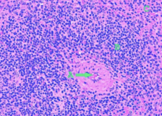
Identify the letter that best corresponds to the area that is on or pointing two the periarteriolar sheath in the white pulp
B
The immune system organ that exhibits both an afferent and an efferent lymph vessel is the…
lymph node
What type of lymphoid tissue is NOT found in the dog
hemal nodes
open and closed circulation are present in the _____ and swollen RBCs are removed by the _____ of this organ during open circulation
spleen, macrophages
What is the function of the thymus
T-lymphocyte selection/differentiation
Which of the following statements about lymph nodes is INCORRECT
blood vessels enter at the periphery and exit at the medulla along with the lymph vessels
What cell type is primarily associated with the humoral immune response?
B-lymphocytes
A ‘primary lymph organ’ is referring to a lymphatic organ where:
lymphocytes are created, lymphocytes mature

The darker staining cells that surround the individual lymph nodules are…
mantle

The cells that can be predominantly be found in the later of dark cells surrounding the lymph nodule that the white arrow is pointing to have what FUNCTION
they are primarily inactive B-lymphocytes
Identify the CORRECT statement regarding tonsils
tonsils have no afferent vessels, only efferent vessels

What specific cell type do you find here taking up the black stain in the marginal zone of this lymphatic organ?
macrophages
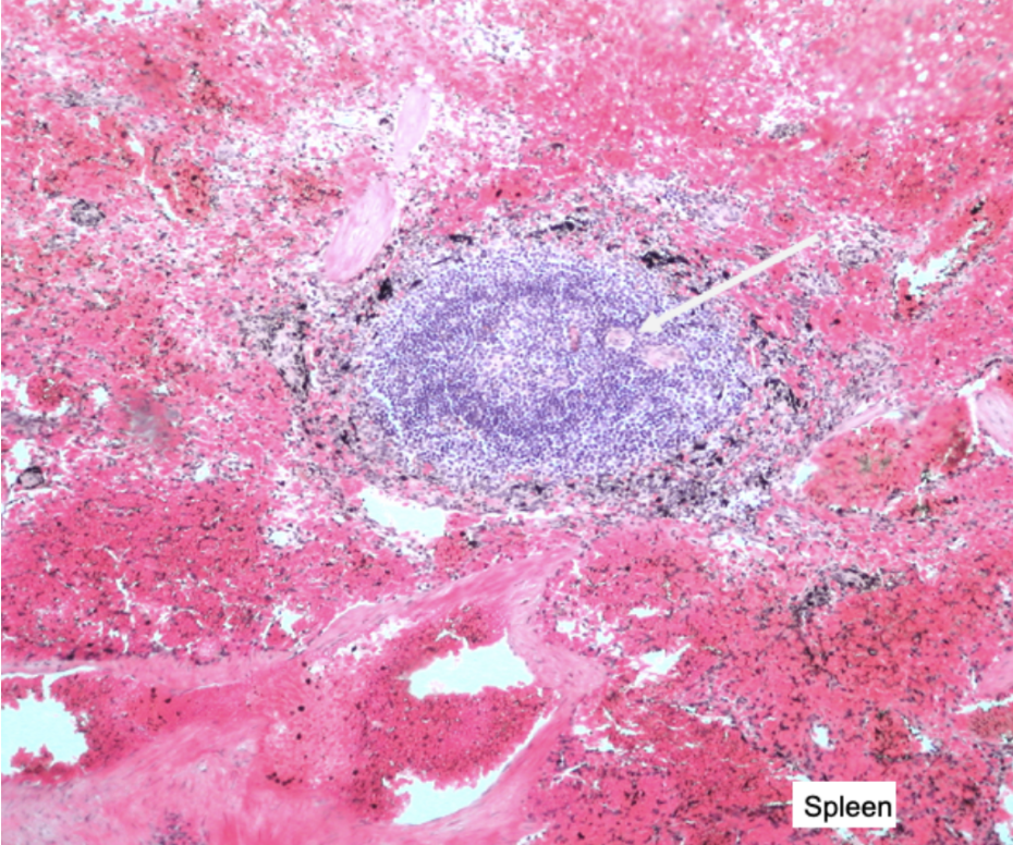
What acidophilic staining feature at the tip of the white arrow
artery of the white pulp
Which of the following is NOT a function of the spleen
All of these are answers
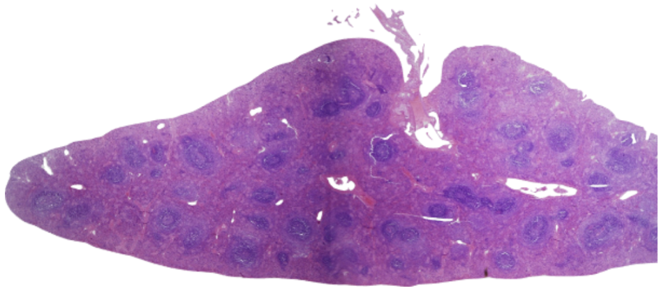
The general term for the basophilic areas of tissue on this organ is the ____ pulp
white

In which location would you expect to find the distal convoluted tubules
A

Which location would you expect to find the papillary duct
C
where can you typically visualize the macula densa cells
near the vascular pole of the glomerulus
Which of the following conditions would NOT be expected to increase the amount of filtrate produced at a glomerulus?
vasoconstriction of the afferent arteriole
at what region of the nephron will you find the most water being re-absorbed
at the descending loop of Henle

The tubules shown here on both longitudinal and cross section are best described as…
collecting ducts

Which of these histological sections is an example of the loop of Henle where the descending loop curves back to become the ascending loop
B
the proximal convoluted tubules and proximal thick tubules of the nephron have specializations to increase surface area for re-absorption. What is the specialization?
they have a brush border
what is one unique feature of the ureters of the equine that most other species lack?
the equine glands in the wall of the ureters that make urine is cloudy and foamy
Identify the correct statement about the urinary bladder
the detrusor muscle is smooth muscle
what type of epithelium does the urinary bladder have
transitional

Identify the structure indicated by the yellow arrows
diffuse lymphoid tissue

identify the structure indicated by the double arrows
primary lymphoid nodule

identify the structure labeled by the double arrows
primary lymphoid nodule

identify the structure indicated by the double arrows
secondary lymphoid nodule

Identify the structure indicated by the double arrow
germinal center
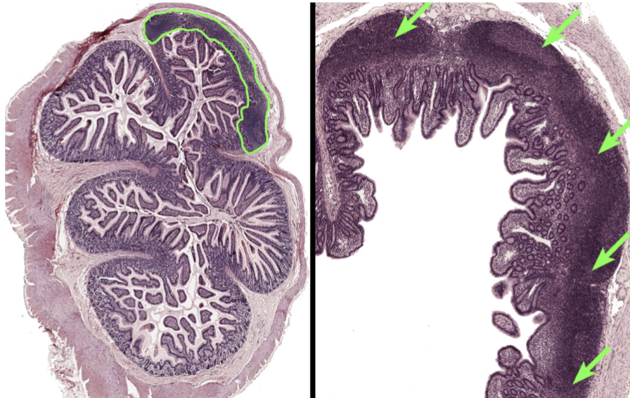
identify the indicated histological structure/tissue
Peyer’s patches

identify the indicated histological structure/tissue
lymphoid nodules

identify the indicated histological structure/tissue
diffuse lymphoid tissue
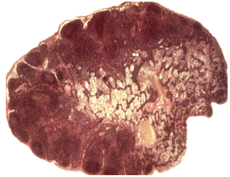
Identify the organ
lymph node

Identify the structure indicated by the arrows
subscapular sinus

Identify the structure indicated by the arrows
trabecular sinuses

Identify the structure indicated by the arrows
medullary cords

identify the structure indicated by the arrows
medullary sinuses
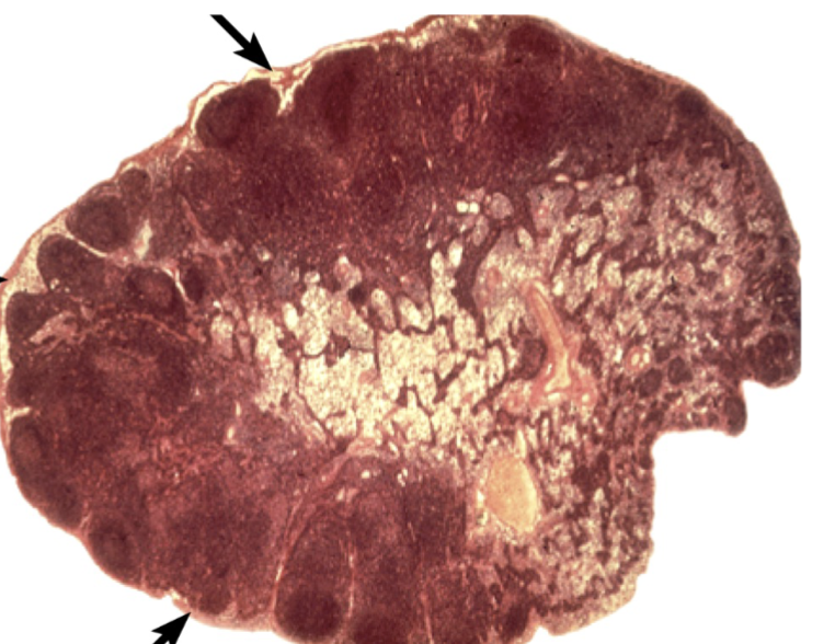
identify the structure indicated by the arrows
capsule

identify the histological structure/tissue
lymphoid nodules
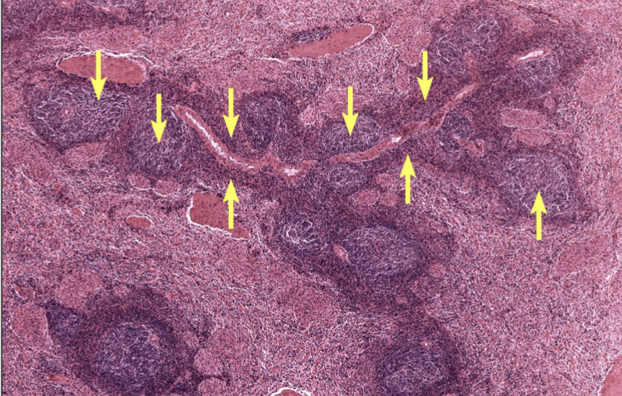
identify the indicated histological structure/tissue
white pulp

17 identify the indicated histological structure/tissue
red pulp

identify the indicated histological structure/tissue
PALS

identify the indicated histological structure/tissue
PALS
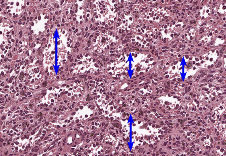
identify the indicated histological structure/tissue
splenic sinuses

identify the indicated histological structure/tissue
splenic cords (cords of Billroth)

identify this organ
thymus tissue

identify the structure indicated by the double arrow
Hassall’s corpuscles

identify the aggregate of lymphoid tissue
tonsil

identify the regions outlined in this image of a lymph node
paracortex