Upper Respiratory Tract Conditions in Small Animals: Nasal Discharge
 Call Kai
 Learn
 Practice Test
 Spaced Repetition
 Match
 Flashcards
 Knowt Play
1/31

There's no tags or description
Looks like no tags are added yet.
32 Terms
Describe the approach to nasal examination and discharge?
Note the location: unilateral / bilateral
What type of discharge?

What is the nature of serous discharge?
• Normal
• Viral infection (Cats > Dogs)
• Non-infectious inflammatory disease
• Early sign of aetiology of mucopurulent discharge
What is the nature of micropurluent discharge?
• Colour comes from neutrophil enzymes released during inflammation
Purulent does not equal infection
• Purulent is nonspecific - any cause of inflammation
Infectious, foreign bodies, neoplasia, polyps, extension of disease from oral cavity
Epitaxious - is blood
Sanguineous - mucus containing blood
What are common differentials for epistaxis?

What are food containing causes?

What are important history-taking considerations for upper respiratory associated diseases?
• Clinical signs are not pathognomic
• Location and character can help to narrow differential diagnoses
• Duration and progression of signs
Acute nasal discharge: < 3weeks
Chronic nasal discharge: > 3weeks
• Acute: Foreign body, cat flu, bacterial rhinitis
• Chronic: Fungal disease, neoplasia, chronic idiopathic rhinitis
Other Considerations:
• Sneezing, reverse sneezing or coughing?
• Strertor? (snoring)
• Head shyness or reluctance to eat?
• History of dental disease, trauma?
• Infectious aetiology (e.g., cat flu); check vaccination, travel history

What is paroxysmal respiration or “Reverse Sneezing”?
• Involuntary reflex, rapidly suck air into the nose instead of blowing it out (opposite of a sneeze)
• Spasm like episode
• Loud snort, honk, or choking noise
• Can last 30 - 60 seconds
• Generally harmless (owners can get alarmed)
• If frequent may require investigations
What are important clinical exam considerations for upper respiratory disease?
• Full general physical examination
Thoracic auscultation, mandibular lymph nodes
• Assess head for:
Facial asymmetry or focal swelling
Facial pain
Exophthalmos, 3rd eyelid protrusion, inability to retropulse the eye
Ulceration, crusting or depigmentation of the rhinarium-
Intra-oral exam: dental disease, holes, masses, halitosis
Ocular ulceration, oral ulceration (cat flu)
**Assess the character and side of the nasal discharge
**Assess the nasal airflow through both nostils (Consider blockage or obstruction), can use a slide to look for condensation
**Look for other signs of bleeding that may flag a coagulopathy
What are some further diagnostics or investigation indicated in upper respiratory diseases?
• Radiography
Widely available
Superimposition of nasal structures, poor definition of soft tissue changes, inability to distinguish between soft tissue and fluid
• CT/MRI
More sensitive and accurate
More expensive, less readily available
CT is the preferred imaging modality
What are the most important views and positioning for radiography use to diagnose upper airway disease?
• Requires anesthesia and good positioning
• Basic views:
• Dorsoventral and/or ventrodorsal
• DV intra-oral*
• Right and left lateral, oblique views reduce superimposition
• +/- Additional views:
• Skyline
• Open mouth VD
What pathologies might radiography reveal for upper airway diseases?
• Loss of turbinate pattern
Increased soft tissue opacity
Increased lucency
• Bony destruction
• Vomer bone destruction
• Extra-nasal signs, e.g., frontal sinus opacity or destruction, soft tissue swelling, dental changes
• Radio-opaque foreign bodies
Although most are plant based and not visible

What pathologies might be visible on a CT scan in regard to upper airway disease?
• Gold standard: provides greater diagnostic information
Pathology:
• Radiolucent foreign material (e.g. plant)
• Asymmetry
• Turbinate detail
• Bone lysis
What does rhinoscopy allow you to appreciate?
• Evaluation of nasal passages
• Retroflexed view - nasopharynx/choanae
• Visualisation of foreign material, fungal plaques, stenosis, mass lesions
• Scope guided biopsies
• Therapeutic - Aspergillosis soaks
What does nasal biopsy allow you to appreciate?
• Histopathology, cytology impression smears, culture
• Biopsy under direct visualisation using rhinoscope or image guided or
• Blind biopsy
Avoid penetration of cribriform plate (Separation of nasal cavity from the brain)
Measure distance from nose to medial canthus
• If epistaxis must evaluate coagulation disorders first:
Platelet count, coagulation screen, buccal mucosal bleeding time
Especially for patients with underlying coagulopathies, might want to keep overnight
What does nasal flush allow you to appreciate?
• Endotracheal tube cuffed (dog) and place throat packs
• Retrograde hydropuplusion
Dislodge foreign body
• Soft foley catheter positioned caudal nasopharynx
Samples for cytology
Dislodge foreign body
What does nasal swabs allow you to appreciate?
• Cytology and culture
• Can be done conscious
• Care with interpretation - nose has diverse flora of normal commensals
Primary bacterial rhinitis rare
Many normal bacteria which live there, primary infection of bacteria is rare (Mycoplasma is a concern however)
Often what is cultured is a secondary growth from a primary underlying issue
• Mycoplasma requires specialist media
• Viral swabs in cats
Describe the main features of Feline Upper Respiratory Infections.
• Typically, acute onset
• Bilateral nasal discharge
• Serious to mucopurulent
• Sneezing, +/- ocular discharge, +/- oral ulceration
• Refer to cat flu notes and lecture content
Describe the main features of bacterial rhinitis.

Describe the main features of nasal foriegn bodies.
• Often plant material
• Acute onset signs
Unilateral discharge
Sneezing, pawing at face
+/- Epistaxis +/- Facial pain
• Chronic signs
As above
+/- halitosis
+/- regional lymph nodes enlarged
Hx of temporary improvement to antibiotics
• Treatment and Evaluation
General anesthetic for evaluation
• Examine both nasal cavities and nasopharynx
Do imaging before
• Rhinoscopy retrieval
Try otoscope in practice
Flushing nasal cavity
• May need to refer for rhinoscopy +/- surgery
What are the main features of Canine Fungal Rhinosinusitis.
Imaging
Diagnosis
Treatment
Aspergillus - is a normal commensal, culture not always useful
Fungus caused bone destruction - seen on X-ray and can damage other bony structures
Sino-Nasal Aspergillosis
• Chronic, progressive signs
• Unilateral or bilateral discharge
• Mucopurulent to epistaxis
• Sneezing, reverse sneezing
• Head shyness
• +/- rhinarial depigmentation
Imaging
See radiolucency
CT - gives greater idea of the extent
Scoping
• Rhinoscopy:
Turbinate destruction
Visualise plaques
Biopsy plaques - culture
Biopsy turbinates - fungal hyphae on histopathology
• +/- Sinoscopy
25% of Aspergillosis just in sinuses
Diagnosis
• No single test diagnostic
Combination of clinical signs, destructive rhinitis, visualisation of fungal plaques, fungal hyphae on cytology/histopathology
Culture plaque
• Culture of nasal discharge of no diagnostic value: Aspergillus is a normal commensal organism
• Aspergillus serology:
• Not sensitive so not useful (e.g. a negative result doesn't rule out disease)
Treatment
• Refer for diagnosis and management
• Debride fungus via rhinoscope
• Anti-fungal agents
Topically: nasal instillation +/- trephination of sinuses - best use
Systemically? poor efficacy, systemic side-effects, ££
• Prognosis fair to good:
Treatment expensive and multiple often required
Relapses can occur

What are the main features of Feline Aspergillosis?
• Rare
• Sino-nasal and sino-orbital form
• Sneezing, serous to mucopurulent discharge
• +/- epistaxis, stertor, granuloma, soft tissue mass protruding from nares, exophthalmia, bone lysis
What are the main features of Cryptococcus?
• Uncommon opportunistic systemic fungal infection
Cats > dogs; check FIV/FeLV status
• Nasal cavities, paranasal tissues or lungs
Disseminates skin, eyes, CNS
• Chronic nasal discharge, sneezing, facial swelling, wounds
• Cytology - Nasal discharge, aspirate of mass
• Latex cryptococcal antigen agglutination test
• Oral antifungals, +/-surgery
What are the main features of Blastomycosis?

What are the main features of Coccidioidiomycosis?

What are the most common nasal neoplasias seen in cats and dogs?

What are common clinical signs of nasal neoplasia?
**Many are asymptomatic

Describe the diagnostics used for nasal neoplasia?
• Imaging
• +/- Rhinoscopy
• Nasal biopsy
Describe the treatment of nasal neoplasia.

Describe the main features of chronic rhinitis/rhinosinusitis.
Chronic nasal discharge, no other clinical signs, results reveal chronic inflammation, but that has a wide variety of causes
Suspect immune mediation
Still need to rule out viral diseases, dental diseases

What treatment options exist for chronic rhinitis?
• Good communication with owners essential
• Frustrating disease - unlikely to resolve
• Prognosis for resolution of signs poor
• Prognosis for survival is good

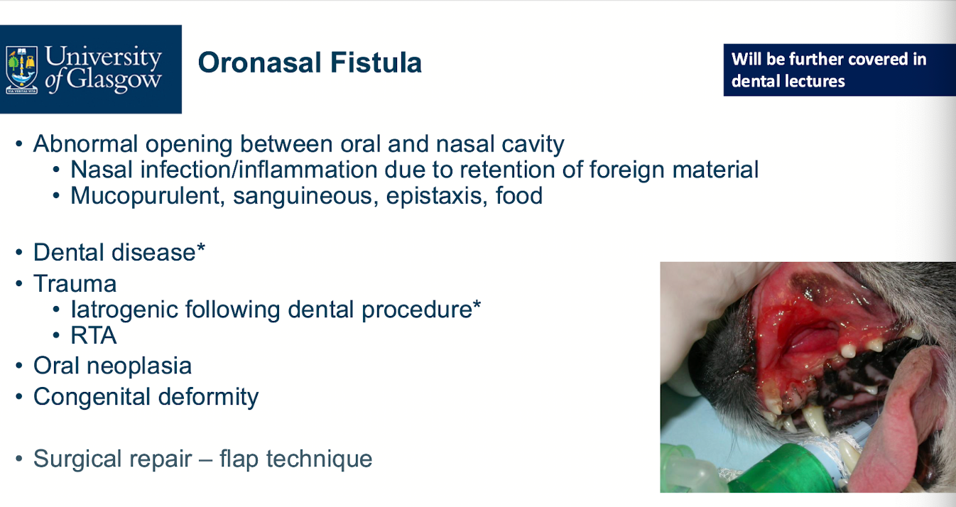
Describe the main features of oronasal fistulas.
Describe the main features of dentigerous cysts.
