internal topography
 Call Kai
 Learn
 Practice Test
 Spaced Repetition
 Match
 Flashcards
 Knowt Play
1/58

There's no tags or description
Looks like no tags are added yet.
59 Terms
what are ventricles lined with
ependymal cells
T/F ventricles have CSF
T(true)
where are ventricles found
cerebrum, brainstem, and spinal cord
there are two lateral ventricles, what does each one contain
anterior horn, body, posterior horn, inferior horn
where are the third and fourth ventricles
midline
what are basil ganglia
masses of grey matter in each cerebral hemisphere
what are the 5 parts of the basil ganglia
cuadate nucleus, putamen, globus pallidus, subthalamic nucleus, substantia nigra
what parts of the basil ganglia make up the lenticular nucleus
putamen and globus pallidus
what parts of the basil ganglia make up the striatum
caudate nucleus, putamen, globus pallidus
why do we pay attention to the basil ganglia
heavily involved in the motor movement system
what commissural fibers make up the corpus callosum
rostrum, genu, body, splenium
what does the commissural fiber, corpus callosum do
transfer sensory, motor, and cognitive information between left and right
what does the anterior commissure do
shares visual, auditory, and memory signals between left and right
what does the posterior commissure do
oculomotor functions (pupillary light reflex)
Association fibers, ____________ connect adjacent regions and ______________ connect distant reguins
short fibers, long fibers
what are the three large association fibers
superior longitudinal fasciculus, unicate fasciculus, inferior longitudinal fasciculus
what does the superior longitudinal fasciculus do
connect frontal to occipital and temporal lobes
where is the arcuate fasciculus and what does it do
in superior longitudinal fasciculus and connects brocas to wernickes
what does the unicate fasciculus do
connects frontal and temporal lobes
what does the inferior longitudinal fasciculus do
connects occipital lobe to temporal lobe
what is the internal capsule made up of
anterior limb, genu, posterior limb
what is the functions of the anterior limb in the internal capsule
connects frontal lobe and prefrontal cortex to subcortical areas, emotion, mood, executive functions, and motor planning
what is the function of genu in the internal capsule
connects motor cortex to brainstem, controls muscles of facial expression, mastication, speech, and swallowing
what is the function of the posterior limb in the interior capsule
connects motor cortex to descending motor pathways, connects thalamus to sensory cortex (somatosensory signals)
what are the 3 borders of the third ventricle
thalamus, epithalamus, hypothalamus
what is the function of the thalamus
relay and processing of sensory signals, sensory integration, attention and arousal from connections with reticular formation in brainstem, regulate behavior and mood with limbic system
what does the hypothalamus connect
infundibulum to the pituitary gland
what is the function of the hypothalamus
endocrine function via pituitary gland, homeostasis, mamillary bodies help with spatial naviftion and episodic memory
what roles of homeostasis does the hypothalamus effect
thermoregulation, energy metabolism, feeding and satiety, circadian rhythms
what is the function of the epithalamus
pineal gland secretes melatonin, regulats circadian rhythms, regulates motor pathways (limbic system with basil ganglia)`
what transverses the cerebral peduncles
CST, CBT, corticopontine fibers from all lobes

what is this a picture of
lower medulla
where do the CST cross
lower medulla

what is this a picture of and what is formed at this level
middle medulla and medial leminiscus

what is this a picture of
upper medulla

what is this a picture of
pons (middle section of brainstem)

what is this a picture of
inferior colliculus

what is this a picture of
superior colliculus

what is this a picture of
lower medulla; motor decussation

label 1-6
central canal, fasciculus gracilis, fasciculus cuneatus, anterolateral system, pyramids (CST), pyramidal decussation

what is this a picture of
middle medulla; sensory decussation

label 1-10
medullary reticular formation, internal arcuate fibers, central canal, nucleus gracilis, fasciculus gracilis, fasciculus coneatus, nucleus cuneatus, anterolateral system, medial lamniscus, pyramids

what is this a picture of
upper medulla

label 1-7
medullary reticular formation, 4th ventricle, anterolateral system, medial lemniscus, pyramids, olive, inferior olivary nucleus

what is this a picture of
lower pons

label 1-7
cerebellum, 4th ventricle, medial lemniscus, anterolateral system, descending motor tracts, pontine reticular formation, middle/inferior cerebellar peduncles
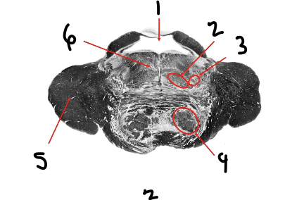
what is this a picture of
middle pons

label 1-6
4th ventricle, medial lemniscus, anterolateral system, descending motor tracts, middle cerebellar peduncle, pontine reticular formation

what is the a picture of
superior pons

label 1-4
cerebral aquaduct, anterolateral system, medial lemniscus, descending motor tracts

what is this a picture of
inferior midbrain

label 1-6
cerebral aquaduct, inferior colliculi, anterolateral system, medial lemniscus, cerebral peduncle, substantia nigra

what is this a picture of
superior midbrain

label 1-7
cerebral aquaduct, superior colliculi, anterolateral system, medial lemniscus, cerebral peduncle, red nucleus, substantia nigra

identify what level these pictures are
cervical, thoracic, lumbar, sacral

what section is this picture taken in and label 1-6
transverse, lateral ventricle (AH), caudate nucleus, internal capsule, lentiform nucleus, thalamus, lateral ventricle (PH)

what section is this a picture of, is it in the front of the back, label 1-3
coronal, back, occipital lobe, lateral ventricle (PH), cerebrellum

what section is this, is it front or back, and label 1-4
coronal section, front, superior frontal gyrus, middle frontal gyrus, inferior frontal gyrus, corpus callosum