Physiology Exam 2
 Call Kai
 Learn
Learn Practice Test
 Spaced Repetition
 Match
Match Flashcards
 Knowt Play
1/143

There's no tags or description
Looks like no tags are added yet.
Name | Mastery | Learn | Test | Matching | Spaced | Call with Kai | Chat |
|---|
No analytics yet
Send a link to your students to track their progress
144 Terms

describe the different parts of a neuron
A = dendrites
B = cell body
C = Axon Hillock
D = Axon
F = axon terminal
nervous system organization
Central nervous system — brain + spinal cord (integration center)
Peripherial nervous system — crainal & spinal nerves
define afferent neuron, efferent neuron, & interneuron
afferent = SENSORY
conduct impulses INTO CNS →
efferent = MOTOR (2 types)
conduct impulses ← OUT CNS
1. somatic — voluntary / reflex
2. autonomic — involuntary
interneuron = CNS neuron in between afferent & efferent
the flow of information in a neuron
sensory receptor detects something is happening
sends a information up the AFFERENT neuron
signal reaches CNS thru an interneuron
CNS sends signal down the EFFERENT neuron
reaches effector organ to carry out the movement
what are the effects of myelination on a neuron
can send signal much faster
allows it to skip thru axon in nodes of ranvier
different types of neurons & functions
afferent
efferent
somatic
autonomic
interneurons
different types of neuroglial cells & functions
supporting cells
shwann cells — PNS & MYELINATE PNS
oligodendrocytes — CNS & MYELINATE CNS
astrocytes
microglia — phagocytic
ependymal cells — line the CNS
difference in the PNS & CNS for neural regeneration
CNS — doesnt regenerate
PNS — can regenerate from schwann cells
how is an AP produced
excitable tissue
compare & contrast action potentials & graded potentials
graded: doesnt reach threshold
A.P.: able to reach threshold and continue w/ stimulus
characteristics of graded potentials
can be either depolarizing or hyperpolarizing
never reaches threshold
decremental (decreases)
how are action potentials conducted & characteristics of them
they reach threshold (-50 @ axon hillock)
not decremental (always the same magnitude)
all or none
describe the refractory period
absolute: cannot make more AP
either when Na+ channels are open / inactivated
prevents overlap of signals
relative: can send another AP
but you need a STRONGER stimulus & to get this you need higher frequencies of AP
hyperpolarized, more difficult to get it to threshold

define depolarization
less negative (than -70)
Na+ channels opening → Na+ rushes IN
define repolarization
returning back to RMP (-70)
Na+/K+ pumps maintain this process
define overshoot
when the membrane potential goes above 0 mV & becomes POSITIVE
define hyperpolarization
more negative (than -70)
Na+ channels inactivated & K+ channels activate → K+ rushes out
permeability of K+ & Na+ during AP & how does this change affect membrane potential
depolarization = Na+ most permeable & K+ least permeable
repolarization = Na+ least permeable & K+ most permeable
Na+ v. K+ gating channels
Na+ VOLTAGE gated channels
open: during depolarization
inactivated: during peak of AP
closed: at rest
K+ Voltage gated channels
what does “all or none” principle for AP
its either at threshold or it is not
what is saltatory conduction
when action potentials jump thru rodes of Rav.
dont happen in myleine
electrical v. chemical synapses
Electrical synapse
Cells are directly connected
Signal passes straight from cell to cell
Very fast
Uses gap junctions
Electrical = direct + fast
Chemical synapse
Cells are NOT directly connected
One cell releases a neurotransmitter
Neurotransmitter crosses the small gap to the next cell
Slower than electrical
what does propagation mean
moving down the axon
what is the sequence of events in neural communication across a synapse
Action potential reaches terminal end of the presynaptic neuron
triggers Ca2+ to rush IN thru Ca2+ voltage gated channels
Ca2+ triggers neurotransmitters to get exocytosed out synaptic vesicles
neurotransmitters bind to ligand gated channels on the POSTsynaptic neuron
induces synaptic potential & causes Na+ to rush in
can cause the next neuron to send or inhibit AP
excitatory v. inhibitory postsynaptic potentials
EPSP — less negative / depolarize (Na+ rushes IN)
can be strong enough to reach threshold & AP forms on postsynaptic cell
IPSP — more negative / hyperpolarizes (Cl- rushses IN)
more difficult to send AP and reach threshold
GABA opens ligand gated channels
differentiate: temporal v. spatial summation
WILL ALWAYS BE EXCITATORY
temporal: if 1 presynaptic input sends multiple outputs to reaches threshold
spatial: if +2 send a signal at SAME TIME, they get added together to reach threshold
what is a ligand gated channel
where neurotransmitter binds to
compare: actions of an excitatory neurotransmitter & an inhibitory neurotransmitter on the POST synaptic neuron
mechanically gated channel:
excitatory — opens Na+ channels & rushes in → depolarize & can reach AP
inhibitatory — opens Cl- channgels & rushes in → hyperpolarize & cannot reach AP
action of acetylcholinesterase
breaks down acetyl choline & found in synaptic cleft
contains neurotransmitter reuptake receptor
how do G protein coupled signal pathways produce synaptic potentials
polar molecule binds to receptor
G proteins released
G protein binds to channel
opens channel
describe protection of the CNS
bone is the thickest layer & outermost layer
meninges: 3 layers
dura mater by bone = solid matter
arachnoid matter = middle
subarachnoid space
pia mater = directly on the brain
what is the function of cerebral spinal fluid (CSF)
buoyancy
protection
maintenance of chemical environment
what produces CSF
epidenmal cells
what connects the right & left hemisphere
corpus callosum
what are the 5 lobes of the cerebral cortex
what are the function of all of them
occipital — VISUAL
perception and interpretation of visual images
temporal — AUDITORY
perception & interpretation of auditory info
parietal — SENSORY
somatosensory — receives info
perception of senses
frontal — MOTOR
voluntary movements
executive reasonings
insula — VISCERAL RESPONSES
deepest layer / on inside
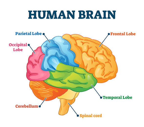
what divides the frontal and parietal lobe
precentral gryus — frontal
postcentral gryus — parietal
functions of the right hemisphere v. functions of the left hemisphere
which one is dominant
Right — depth perception (visuospatial) & patterns/reading maps
Left — language & analytical ability
THE MOST DOMINANT
what is the difference between broca’s area v. wernicke’s area
brocas area: frontal lobe
fine motor functions involved in speech
wenickes area: in between pariteal/occipital/temporal region
hearing & understanding language
formulating words
what is aphasia?
broca’s aphasia
wernicke’s aphasia
aphasia: a brain-related language disorder that makes it hard to speak/understand speech/read/write
Broccas Aphasia: cannot talk/difficult responding BUT can understand what is spoken
Wernickes Aphasia: cannot understand spoken/written language BUT can speak using made up words
what are the different types of memory
Memory:
short term: <30 sec
long term: >30 sec
non-decalarative: memory of simple skills (ex: tying shoes)
declarative: memory of facts & events (ex: where were you…?)
what are the different types of memory consolidation
temporal lobes (amygdala & hippocampus) involved in converting short term → long term
sleep is needed for this
STRESS IMPAIRS THIS
locations & functions of the thalamus & hypothalamus
location: diencephalon
functions:
body temp
thirst & urine
food intake
pituitary hormone
circadian rhythm
control ANS
what are the structures & functions of the midbrain
motor movements of the eye
auditory processing
what are the 2 proteins that impact alzheimers disease
beta amyoild plaques
tau
these both inhibit & alter movement of ions
what are the structures & functions of the brainstem
critical link between between spinal cord & higher brain regions
vegetative funcitons = involuntary functions that keep you alive
made up of:
midbrain (top) — eye movement
pons — relay information station
medulla (bottom) — basal survival functions (breathing, heartbeat, dilation of blood)
function of the cerebellum?
planning, initiating, & timing motor movements
learning skilled motor tasks
ataxia
damage to cerebellum
UNCOORDINATED MOVEMENT
function of the limbic system
hippocampus + amygdala
emotions
aggression
fear
smell
goal directed behavior
what are the structures & functions of the reticular activating system
Structure: midbrain, pons, medulla
Fire to keep you awake & brain awake
inhibited: brain isnt awake
phases of sleep?
REM — rapid eye movement // eyes moving very fast
brain is buidling memories
Stages:
go into sleep
longer & deeper stage & body temp drops
GOLDEN STAGE = want to be here& body is repairing
cycling back to REM
what is the difference between: ascending v. descending tracts of the spinal cord
ascending: dorsal root
carry sensory information UP to brain (afferent)
ascending: ventral root
carry motor information DOWN to effector (efferent)
structures of the spinal nerve v. neural pathways of the reflex arc
PNS
all are mixed nerves: sensory & motor
dorsal root = sensory (afferent pathway)
ventral root = motor (efferent pathway)
Pathway:
stimulus is detected thru receptor neuron →
receptor sends singal thru dorsal root
signal enters intregating center (interneuron)
response gets sent thru ventral root
signal gets to effector & a response happens
what are the components of the withdrawal reflex
nociceptor detects pain
AP goest thru afferent pathway
hits integration center
AP sent by integration center thru efferent pathway
stimulates motor control
stimulate flexors
inhibits extensors
classification of cranial nerves
part of the PNS
12 pairs
what are the divisions of the efferent division of the PNS
somatic nervous system — voluntary
pre&postsynaptic
autonomic nervous system — involuntary
pre&postganglionic
organization of the AUTOnomic motor neurons
both are on, but which one is innervating MORE
sympathetic: stress
parasympathetic: rest
what is the structure of the sympathetic nervous system
ganglia
preganglionic v. post ganglionic
pathway
thoracolumbar region
SHORT pre & LONG post
preganglionic release acetyl choline & bind to nicotinic receptors on post
Postganglionic release norepinenorepinephrinephrine into adrenergic receptors on effector
what is the relationship between the sympathetic nervous system & adrenal medulla
modified part of the sympathetic system
preganglionic fibers secrete hormones into BLOOD
more epinephrine > compared to norepinephrine
structure & innervation pathways of the parasympathetic division of the ANS
craniosacral region
LONG pre & SHORT post
preganglionic release acetyl choline & bind to nicotinic receptors on post
Postganglionic release acetyle choline into muscarinic receptors on effector
cholinergic receptors v. adrenergic receptors
cholinergic receptors = nicotinic & muscrainic
parasympathetic
adrenergic receptors = nicotinic & adrenergic
sympathetic
what are the effects of adrenergic stimulation on different organs & what are the different types of receptors involved
Alpha & beta receptors on SYMPATHETIC
release NE & epinephrine
how does albuterol work in ANS
b2 agonist = BETA = sympathetic
inhaling N.E.
how does atropine work in ANS
blocks muscrainic receptors = parasympathetic
dilate pupils
how does beta-blockers work in ANS
block beta-adrenergic receptors
sympathetic
antagonistic & cooperative actions of the sympathetic & parasympathetic divisions of the ANS
parasympathetic & sympathetic work in OPPOSITION = ANTAGONISTIC
EX: parasympathetic = increase heart rate & sympathetic = decrease heart rate
cooperative effects: saliva & urination
what innervates an organ without dual innervation
ONLY SYMPATHETIC (increase or decrease)
EX: sweat glands
explain the term sensory transduction
converting a stimulus into an electrical signal (AP) so the brain can understand it
phasic v. tonic receptors
tonic: CNS is continually getting info
dont adapt
or adapt slowly
you know & understand the stimulus is there
phasic: you forget the stimulus is there once applied, but remember its there when its gone
constantly adapt
touch receptors in the skin
describe the nature & significance of the receptor potential
electrical change that happens in a sensory receptor when it senses a stimulus
can lead to an AP
influx of Na+ can produce receptor potentials
differentiate between sensation v. perception
sensation: collection of info (RECEPTORS)
perception: interpretation of what is sensed (where AP are sent)
4 steps of perception
stimulus
transduction: converting stimulus → AP
conduction: axons; conducting AP/electrical signals
perception: integrating center
define sensory acuity & explain factors that affect acuity
sensory: ability to tell two things apart when touching you
high: able to tell very specifics
low: able to tell general area
affect:
field size
# of receptors
area of somatosensory
what are the different types of sensory receptors
photoreceptors - light
mechanoreceptors - mechanical energy
thermoreceptors - temp regulation
osmoreceptors - osmolarity
chemoreceptors - specific chemicals
nociceptors - pain
proprioceptors - position of your body
externoreceptors - outside your body
interoreceptors - inside your body
what are cutaneous sensations
temp, touch, pressure, pain
contralateral - terminates here
what are the modalities of taste
chemoreceptors detect taste — housed in taste buds
tastant: taste chemical that dissolves thru salvia
salty — caused by chemical salts (NaCl)
sour — caused by acids
sweet — caused by glucose
bitter — caused by chemically diverse group of tastants
umami — savory taste
how is taste produce + conveyed in the brain
produce: binding of tastant w/ receptor cell & produces receptor potential
brain: insula detects gustatory & somatosensory cortex moves tongue

how do odorant molecules stimulate their receptors (polarity?) & how the information is conveyed to the brain
odorants — chemcial molecules that can be smelled
POLAR = water soluble solvents that dissolve into mucus layer & stimulate the olfactory bulb by binding to MEMBRANE bound receptors
smell reaches the limbic system & thats how you form memories w/ smells

describe hertz
number of cycles / second
what are the structures of the vestibular apparatus & how do they function to produce a sense of:
balance
equilibrium
position of the head
semicircular canals - ROTATIONAL MOVEMENT
anterior canals: head moves forward / back (nodding)
posterior canals: head is tilting left / right
lateral canals: spinning
otolithic organs - LINEAR MOVEMENT
utricle: left & right
saccule: up & down
what are the semicircular canals
bending of hair cells embedded in cupula = KINOCILIUM
what are the otolithic organs
utricle - horizontal (moving forward/backward) or left/right
saccule - vertical (up/down)
contain otoliths - crystals found in gelatinous substance that provides mass & helps move against gravity
neural pathways of the vestibular system
input: goes to vestibular nuceli in brain
cerebellum helps w/ balance
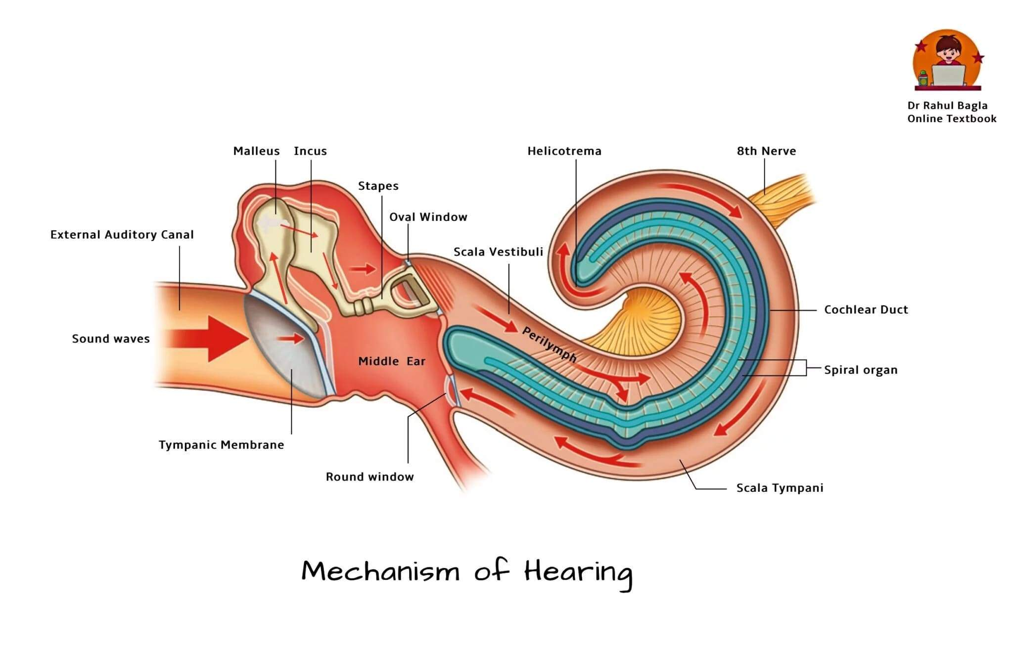
how do sound waves result in movements of the oval window & the basilar membrane
sound wave travels thru → hits the tympanic membrane → malleus → incus → stapes → hits oval window → oval window causes perilymph fluid to move in cochlea → fluid causes basilar membrane to deflect → the hairs on the basilar membrane move → sends AP
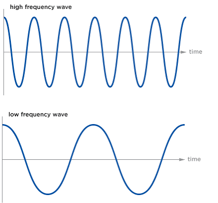
low pitch v. high pitch
higher frequency (#) = higher pitch
lower frequency (#) = lower pitch
soft sound v. loud sound
soft sound = smaller amplitude (height)
louder sound = larger amplitude (height)

how are loudness & pitch discrimminated
pitch:
low = distal (further) = helicotrema
high = against oval window (closer)
loudness
soft = hairs bent softer & closer
louder = hairs bent harder & further away = helicotrema
what are the structures of the eye
sclera - white portion of eye
cornea
iris - colored portion
pupil - black
vitreous humor - makes the shape & inside cell
aqueous humor - carries nutrients for cornea & lens
retina - rods & cones
how do the structures of the eye focus light onto the retina
cornea - where light first enters thru and bends
iris - controls the amount of light entering eye by changing pupil size
circular muscle
radial muscle
pupil - light passes thru here; doesnt move,, hte iris around it moves & adjusts the size
lens - changes shape helps to refract light on retina
flattens - near
rounder - far
retina - rods and cones
what is refraction
bending of light as it moves thru areas
cornea does this
what is accomodation
changing shape of lens for near/far away items
how to direct the bent light into eye
how is accommodation at different distances accomplished?
is this parasympathetic, sympathetic, or dual innervation?
REFRACTION OF LIGHT THRU ACCOMMODATION
near items: lens becomes rounder → parasympathetic INCREASES
ciliary muscles contract
suspensory ligaments slacken
far away items: lens becomes flatter → parasympathetic DECREASES
ciliary muscles relax
suspensory ligaments get pulled

how does the iris innervate & change
HOW MUCH LIGHT ENTERS
contains 2 muscles
circular muscle: CONSTRICT via parasympathetic nervous system
INNERMOST AREA
becomes smaller in bright light
radial muscle: DILATES via sympathetic nervous system
OUTERMOST AREA
becomes larger in dim light

common disorders of:
refraction
glaucoma
cataracts
cataracts: opacity of lens
glaucoma: increased pressure of built up aqueous humor in eye, causes damage to optic nerve
refraction:
myopia: nearsighted
unable to see far things
lens is too long, you need concave lens
hyeropia: farsighted
unable to see close up things
lens is too short, you need convex lens
presbyopia: stiffness of lens as you age
decrease in accomodation
what are the 3 layers of the retina
outermost layer = RODS & CONES
middle layer = bipolar cells
inner layer = ganglion cells
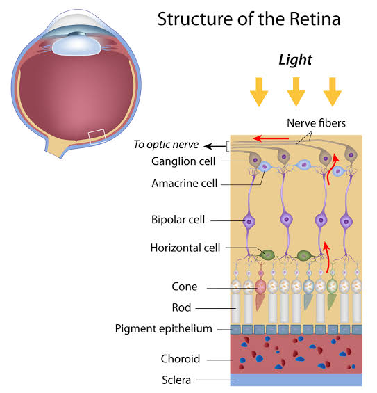
how does light affect rods & cones
outer segment detects light, inner segment = mitochondria, & synaptic terminal is where they release neurotransmitter
rods = night/gray colors
cones = day/color
describe dark & light adapatation
DARK:
AT PHOTORECEPTOR:
Na+ channels open
release neurotransmitter = GABA
GABA inhibits bipolar cell
No EPSP
No AP sent = cannot view anything in dark
LIGHT:
AT PHOTORECEPTOR
light closes Na+ channels
membrane becomes hyperpolarized & doesnt allow GABA to be sent
because GABA isnt sent, bipolar & ganglion cells can send messages
become EPSP →
there is AP in visual cortex & you are able to see light
compare the functions of rods & cones
(photopigments)
similarities:
consists of 2 components — opsin & retinal
opsin = protein that retinal binds to
retinal = absorbs light
cis or trans
Differences:
4 different types of photopigments
rods (1) = rhodopsin
absorbs all visible wavelengths
gray colors
cone (3) = red, green, blue
rods v. cones:
which has higher sensitivity & lower sensitivity
which has higher acuity & lower acuity
Rods: higher sensitivity because it has more receptors & low acuity
receptors CONVERGE into 1 pathway
larger number of receptors but they all converge
Cones: higher acuity because each cone has their own receptor / does not converge & low sensitivity
no convergence happening
receptors are their own individual one
smaller # of receptors
what is the importance of the fovea centralis
contains rods and cones
vision is best at fovea centralis
how is color perceived
depends on the amount of cones you have
red is the longest wavelength
green is middle
blue is shortest wavelength
the color you see is the light that is being reflected