Introduction to the Nervous System Lab (M10)
 Call Kai
 Learn
 Practice Test
 Spaced Repetition
 Match
 Flashcards
 Knowt Play
1/141

Earn XP
Description and Tags
142 Terms
Central Nervous System Functions:
Processes, interprets, stores sensory info; sends commands; generates consciousness
Peripheral Nervous System Functions:
Relays information to/from CNS; contributes to voluntary & involuntary actions
Sensory
Motor
Special Senses
Somatosensory
Viscerosensory
Somatomotor
autonomic motor
Parasympathetic
Sympathetic
Enteric
Central Nervous System
Brain and spinal cord, responsible for processing information and coordinating responses

a
nucleus of neuron
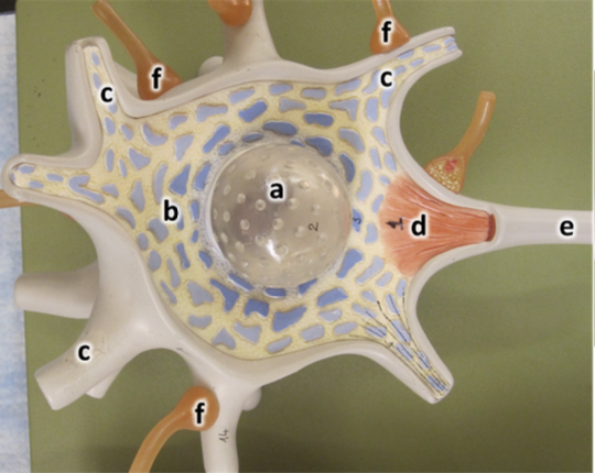
b
cytoplasm

c
dendrite
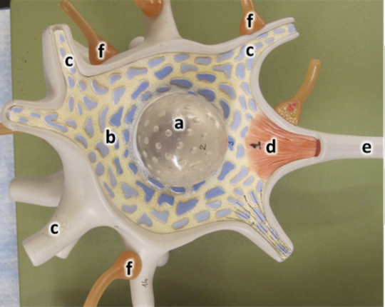
d
axon hillick

e
axon

f
axon terminal of neighboring cell

e
axon
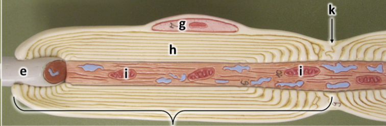
h
myelin

g
nucleus of schwann cell

k
node of ranvier

i
mitochondria
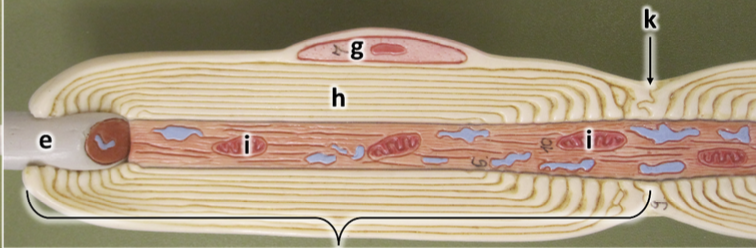
j
schwann cell
Dendrite:
receives signals from other neurons
Cytoplasm:
holds organelles and is where cell activities occur
Axon Hillick:
initiates nerve pulses
Nucleus of Neuron:
contains DNA and controls cell activities
Mitochondria:
produces energy (ATP)
Axon:
carries nerve impulses away from cell body
Schwann Cell:
produces myelin around the axon
Axon terminal of neighboring cell:
releases neurotransmitters to communicate w/ another neuron
Myelin:
insulates the axon and speeds up nerve impulses
Endoneurium:
connective tissue that surrounds and supports individual nerve fibers
Node of Ranvier:
gaps in myelin that help speed up nerve impulses
Nucleus of Schwann cell:
controls the activities of the schwann cell
larger axon =
faster traveling for nerve signals
If this axon dies, do the myelin and endonerium remain?
yes

M
endoneurim

G
Nucleus of Schwann cell
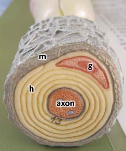
H
Myelin
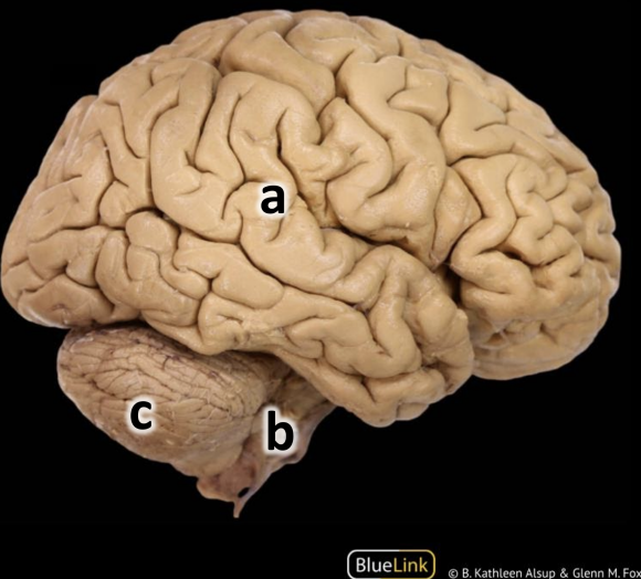
A
cerebrum
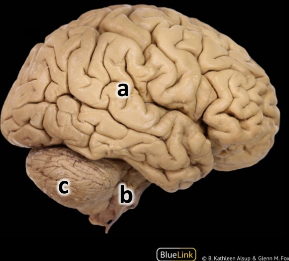
B
brain stem

C
cerebellum

D
dura mater

E
arachnoid mater

F
pia mater
Through what layer of meninges does the cerebrospinal fluid circulate?
Subarachnoid Space
Meninges:
Protective layers of connective tissue that anchor the brain & spinal cord to surrounding bones, & provide space for the CSF
Subarachnoid Space:
The space between the arachnoid mater and pia mater occupied by blood vessels, nerves, and cerebrospinal fluid
Blood Brain Barrier:
Tightly-sealed blood vessels that prevent pathogens and food-borne toxins (big proteins) from coming in contact with the CNS
Astrocytes:
Type of glial cell that exerts pressure on the lining of blood vessels in the CNS, forming a protective layer to inhibit transport of substances in/out
Oligodendrocytes:
Type of glial cell that creates myelin sheath around axons in the CNS
Ventricular System:
Spaces in the brain that do not contain cells, it is through this that cerebrospinal fluid flows
Gray Matter
The outer-most layer of the brain’s nervous tissue, but the inner-most layer of the spinal cord, these areas contain neuron cell bodies – so this is where action potentials originate
Myelin:
A lipid-rich structure that wraps around axons like insulation around an electrical wire, its function is to speed the transmission of an action potential down the length of and axon
Schwann Cell
Type of glial cell that creates myelin sheath around axons in the PNS and aids the establishment of new synapses between the axon and its effector
Ganglion
Cluster of neuron cell bodies in the PNS from which nerves emanate
Satellite Cell
Type of glial cell that creates a favorable habitat for neuron cell bodies in the PNS and aids the re-growth of severed axons
Nerve
Bundles of axons organized into fascicles and surrounded by protective layers of connective tissue
Perineurim:
The middle layer of a nerve’s connective tissue that contains collagen (so is structurally supportive) and creates a blood-nerve barrier to inhibit the movement of pathogens and food-borne toxins into axons
Where do neuron cell bodies live in the CNS?
brain and spinal cord
Where do neuron cell bodies live in the PNS?
ganglia
What protects the CNS?
Bones
What protects the PNS?
nerve coverings
Whats the source of Myelin for CNS?
oligodendrocytes
Whats the source of Myelin for PNS?
Schwann Cells
CNS injury response:
Damage usually permanent (e.g., paralysis), but plasticity possible (at younger ages, new dendrites and axon terminals can in-fill gaps left by injury)
PNS injury response:
Sometimes regenerates (e.g., severed axon re-grows, aided by Satellite cells) or show functional adaptation (e.g., new synapses form, aided by Schwann cells)

Cerebellum(a):
coordinates movement, balance, posture, and muscle coordination

Pre-central Gyrus(b):
controls voluntary skeletal muscle movements (primary motor cortex)

Pos-central Gyrus(c):
processes sensory info such as touch, pressure, pain and temperature

Central Sulcus(d):
separates frontal and parietal lobes; divides motor and sensory areas
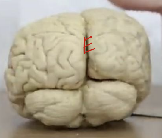
Superior Longitudinal Fissure(e):
separates right and left central hemispheres

Lateral sulcus(f):
separates temporal lobe from frontal and parietal lobes

Temporal Lobe(g):
Responsible for hearing, memory, and language comprehension

Frontal Lobe(h):
Involved in decision making, planning, personality, and voluntary movement
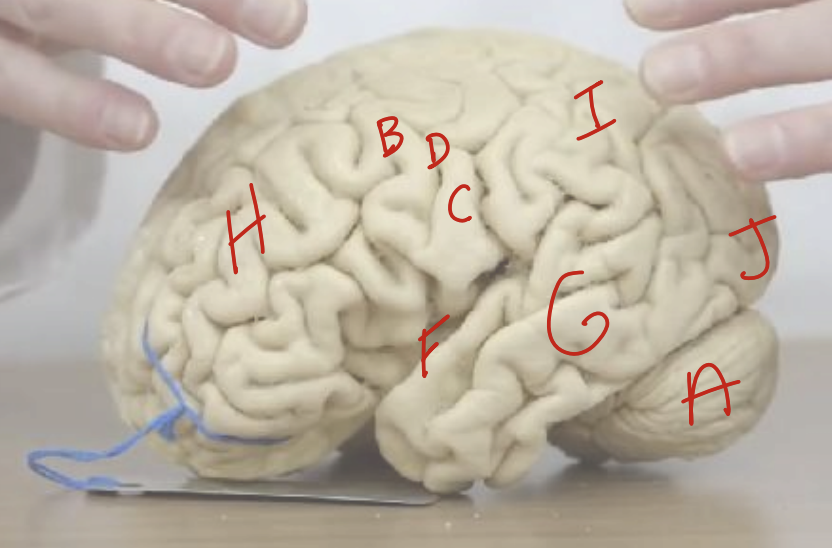
Parietal Lobe(I):
processes touch and other sensory information; helps w/spatial awareness
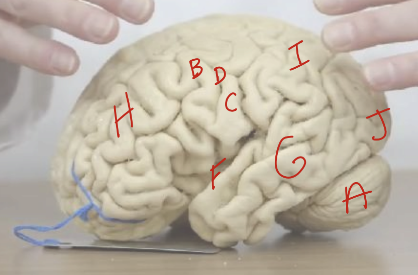
Occipital Lobe(J):
processes visual information
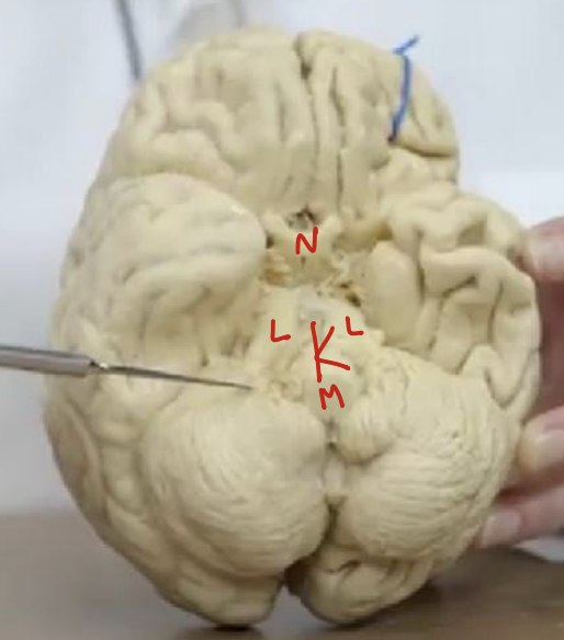
Brain Stem(k):
controls basic life functions & connects brain to spinal cord

Pons (of brain stem)(L):
Helps regulate breathing, sleep, and communication between brain regions

Medulla (of brain stem)(m):
controls heart rate, blood pressure, and breathing

Optic Chasm(n):
some optic nerve fibers cross, allowing visual info from both eyes to be processed

Insula Lobe(o):
involved in taste, emotions, and awareness of internal body sensations

Parieto-Occiptal Sulcus(p):
separates parieta and occipital lobes

Corpus callosum(q):
connects right and left cerebral hemispheres, allowing communication between them
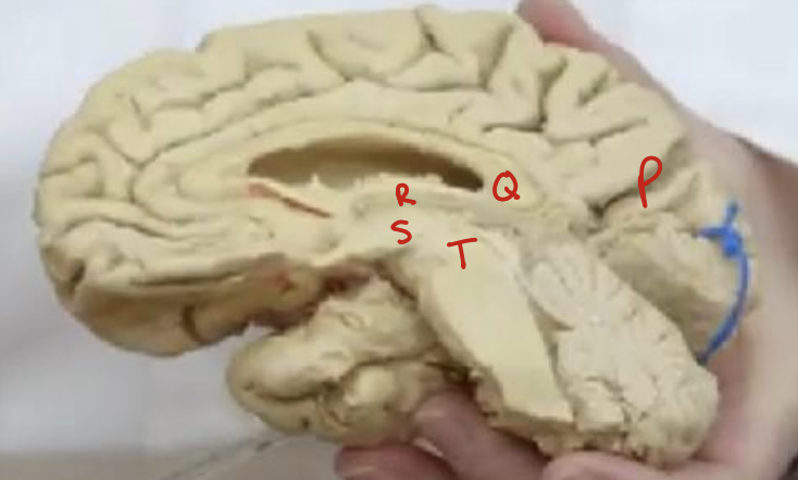
Thalamus (diencephalon)(r):
relays most sensory info to cerebral cortex

Hypothalamus(of diencephalon)(s):
regulates body temp, hunger, thirst, and homeostasis

Midbrain (of brain stem)(t):
eye movements, visual and auditory reflexes, and motor control
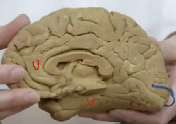
Cingulate Gyrus (of limbic lobe)(u):
emotions, memory, and behavior

Collateral Sulcus(v):
separates parahippocampal gyrus from nearby brain regions, including fusiform gyrus

A
Cerebellum

B
Precentral Gyrus

C
Poscentral Gyrus

D
Central Sulcus
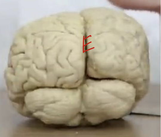
E
Superior Longitudinal Fissure

F
Lateral Sulcus
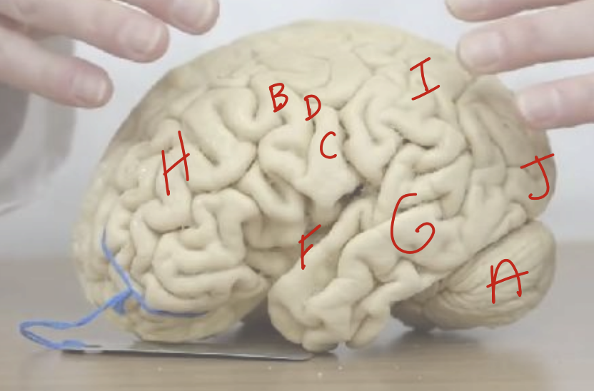
G
Temporal Lobe

H
Frontal Lobe