EKG Only PowerPoint 1 Cardio
 Call Kai
 Learn
 Practice Test
 Spaced Repetition
 Match
 Flashcards
 Knowt Play
1/11

Earn XP
Description and Tags
Cardiology Exam 1
12 Terms
Basic (3‐Lead) Electrocardiogram (ECG)
Four electrodes are placed on both of the patient’s arms and on the left leg.
.
The potential difference recorded between the electrodes on the person’s left arm (LA) and right arm (RA) is called lead I of the ECG.
.
The other limb leads are recorded between the right arm and left leg (lead II) and left arm and left leg (lead III).
Basic (3‐Lead) Electrocardiogram (ECG) (Photo)

Basic Electrocardiogram (ECG) Parts (8)
P wave
QRS complex
T wave
PQ segment
PR interval
ST segment
ST interval
QT interval
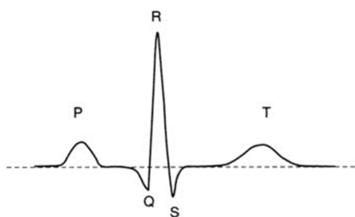
P wave
Depolarization of the atria
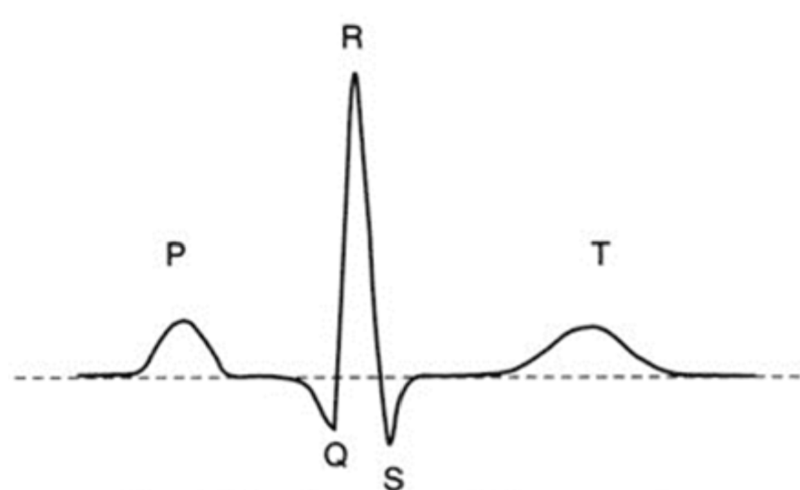
QRS complex
Depolarization of the ventricles

T wave
Repolarization of the ventricles

PQ segment
Time when the impulse is traveling through the AV node, bundle of His, and bundle branches

PR interval
Onset of P wave to middle of QRS complex

ST segment
End of QRS complex to onset of T wave

ST interval
End of QRS complex to end of T wave

QT interval
Onset of QRS complex to end of T wave

Overdrive Suppression
- The AV node and the Purkinje fibers are LATENT PACEMAKERS that may exhibit automaticity and override the SA node if it is suppressed.
.
The intrinsic rate of phase 4 depolarization (and heart rate) is FASTEST in the SA node and SLOWEST in the Purkinje fibers.
