Parathyroid Endocrinology & Calcium Regulation
 Call Kai
 Learn
 Practice Test
 Spaced Repetition
 Match
 Flashcards
 Knowt Play
1/36

There's no tags or description
Looks like no tags are added yet.
37 Terms
Calcium Homeostasis:
Essential for:
bones and teeth
all cells
nerve and muscle function
blood clotting
3 hormones responsible (a 3 chapter “story”):
Parathyroid hormone (PTH)
Calcitonin
1,25(OH)2Vitamin D3 (calcitriol or “Vitamin D”)
Distribution of Calcium
Total body calcium = 1 kg
99% bone + teeth
0.9% soft tissue cells
0.1% ECF
0.05% protein-bound or in inorganic complexes
0.05% free ionized (≈500 mg)
Only free ionized Ca2+ is bioactive
Calcium Regulation
Both plasma (ECF) concentration of ionized calcium and total body calcium are regulated:
Free ionized Ca (0.05%) in ECF by:
Exchange between bone and ECF
Renal excretion
Minute-to-minute regulation
Total body Ca by:
GI absorption of Ca
Renal excretion
Long-term regulation
Calcium Regulation Compartments
Small intestine (dietary calcium)
ECF
Bone (storage)
Kidney (excretion)
Cells (low levels of calcium)
Parathyroid Gland Histology
Chief cells and oxyphil cells:
Chief cells have a calcium ion sensing receptor!

Parathyroid Hormone (PTH)
A peptide hormone (84 aa)
Control by a negative feedback loop:
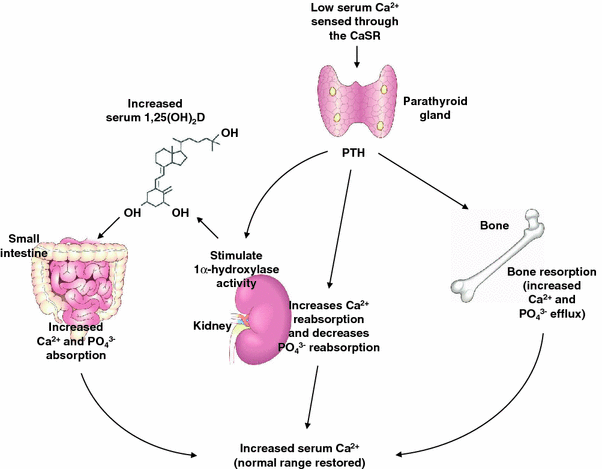
PTH synthesis & secretion stimulated by low plasma ionized Ca2+
PTH raises free ionized Ca2+ in ECF
PTH activity is essential for life
PTH acts on bone, kidneys, and - via Vit.D - GI tract
PTH Response to Changes in Plasma Calcium
PTH secretion goes down as plasma Ca goes up

PTH Effects on Bone
Bone remodelling (...ongoing throughout life; complete skeletal turnover every 10 years)
Osteoblasts create new bone by secreting organic matrix – osteoid – and then mineralizing it with calcium phosphate crystals
Osteocytes arise from entombed osteoblasts; involved in calcium regulation, but do not create bone
Osteoclasts dissolve bone (bone resorption) by solubilizing the crystals

Osteoblasts
Create bone throughout life → bone deposition
Dividing chondrocytes add length to bone
Chondrocytes produce cartilage
Old chondrocytes disintegrate
Osteoblasts lay down bone on top of cartilage
Epiphyseal plate is the site of bone growth

Osteoclasts
Dissolve bone → bone resorption
Secrete acid and enzymes → dissolve bones (apical membrane)

PTH Stimulates:
Movement of ionized calcium from bone fluid to ECF (fast response time)
Bone dissolving activity of osteoclasts (slower response time; coincident release of calcium and phosphate)
Tips bone remodelling in favour of resorption
In a fast exchange:
Calcium is moved from the labile pool in the bone fluid into the plasma by PTH-activated calcium pumps located in the osteocytic-osteoblastic bone membrane
In a slow exchange:
Calcium is moved from the stable pool in the mineralized bone into the plasma through PTH-induced dissolution of the bone by osteoclasts
Osteocytic-osteoblastic Bone Membrane
Formed by filmy cytoplasmic extensions of interconnected osteocytes and osteoblasts
RANK Ligand (RANKL) Binding:
Stimulates macrophages/osteoclasts
Differentiation of macrophages into osteoclasts + suppression of osteoclast apoptosis
Increased osteoclast action
Osteoclast action outpaces osteoblast action
Decreases bone mass
Osteoprotegerin (OPG)
When RANKL is bound to OPG, it is not available to bind with RANK
Blocks action of RANK
Decreases osteoclast action
Osteoblast action outpaces osteoclast action
Increases bone mass
PTH Effects on Kidneys
PTH increases renal reabsorption of calcium (decreases urinary calcium excretion)
and increases excretion of phosphate
PTH induces activation of Vitamin D
Calcitonin
Effects on bone remodelling opposite to PTH:
A peptide hormone (32 aa)
Decreases movement of ionized calcium from bone fluid to ECF
Inhibits activity of osteoclasts; therefore, decreases bone resorption
Increases renal calcium excretion
Inverse regulation of PTH and Calcitonin release:
Act through the same calcium-sensing receptor mechanism

When plasma calcium is low:
Parathyroid glands increase PTH and increase plasma calcium
When plasma calcium is high:
Thyroid C cells increase calcitonin and decrease plasma calcium
Vitamin D
Bioactive form is 1,25(OH)2 Vitamin D3
Calcitriol
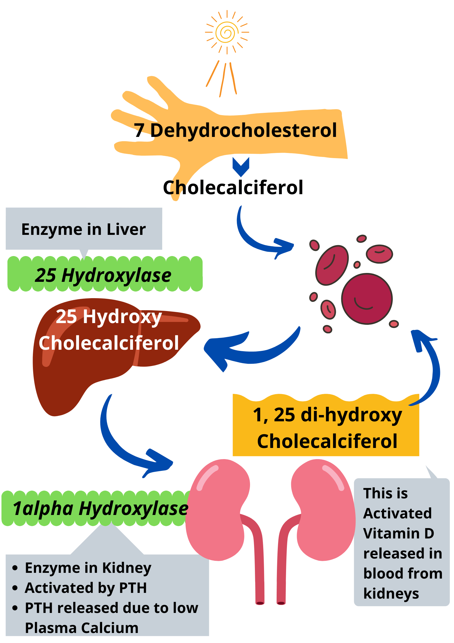
Derived from inactive vitamin D produced in skin by action of sunlight; present in some foods
The biologically inactive precursor is called 7-dehydrocholesterol
Vitamin D is a steroid hormone
Vitamin D activated by addition of 2 hydroxyl groups:
1st OH added in liver → 25-OH Vitamin D
2nd OH added in kidneys → 1,25(OH)2 Vitamin D3
Promotes intestinal absorption of calcium and phosphate
Kidney hydroxylation enzyme is stimulated by:
PTH
Low plasma ionized calcium
Low plasma ionized phosphate
Prolactin (during lactation)
Final product: VitD3= (1,25 (OH)2 D3) = Calcitriol
When plasma ionized calcium is high, renal hydroxylation:
Produces an inactive form: 24, 25(OH)2 Vit D3
Effects of Activated Vitamin D3:
Increases total body calcium by increasing GI absorption of calcium
Dose-dependent response
Slow, long-lasting response
Adjusts Ca2+ intake from diet to match needs via changes in ratio of active/inactive Vit D
Increases ECF ionized calcium by enhancing response of bone to PTH (resorption)
Problems of Calcium Homeostasis
Hyperparathyroidism
Hypoparathyroidism
Vitamin D deficiency
Osteoporosis
Hypoparathyroidism
A rare condition usually caused by autoimmune disease or physical damage to the glands
Low PTH secretion results in low plasma ionized calcium level:
Causes increased neuromuscular excitability by increasing nerve membrane permeability to Na+
Paresthesia (e.g., “pins and needles” sensation)
Tetany (muscle spasms)
Potentially lethal due to failure of respiration
Hyperparathyroidism
Primary form usually due to hypersecreting parathyroid tumour
Hypercalcemia results from high PTH
Due to chronic renal disease or vitamin D deficiency causing hypocalcemia
Hypocalcemia causes high PTH production
High PTH level with elevated Ca2+ causes:
Depressed muscle and nerve excitability:
High ECF [Ca2+]
Increased risk of fractures
Demineralization of bone
Risk of kidney stones
High concentration of calcium in renal filtrate
Hyperparathyroidism with Vitamin D Deficiency
Impairs GI absorption of calcium
Resulting high PTH causes bone demineralization
In children, result is rickets
In adults, result is called osteomalacia
Vitamin D supplementation is a consideration for Northern residents

Skin Pigmentation Affects Vitamin D Production
2007 study of U. Toronto students:
Plasma Vit D levels measured in Jan/Feb
Results by ethnic origin:
European ancestry – 34% were deficient (<30ng/ml)
East Asian – 85% deficient
South Asian – 93% deficient
African – 100% deficient
Vitamin D Deficiency:
Elevated risk of cancer, cardiovascular disease, autoimmune disease, multiple sclerosis, osteoporosis…
Meta-analysis of 18 controlled experiments showed that Vit D supplements lower risk of premature death by 7%
1,25-(OH)2-Vitamin D3 Activates more than 1000 Different Genes in at Least a Dozen Cell Types Including:
Bone
Breast
Fat
Intestine
Immune cells
Kidneys
Liver
Neurons
Pancreas
Parathyroid gland
Prostate
Skin keratinocytes
Vitamin D3 receptor is expressed everywhere in the body!
Osteoporosis - Brittle Bones
Condition in which bone resorption is greater then bone formation
Reduced osteoblast activity, increased osteoclast activity
Plasma calcium, phosphate, PTH, and Vitamin D levels are usually normal
Results in reduction of bone mass

Osteoporosis occurs in both men and women:
Estrogen inhibits bone resorption by decreasing osteoclast activity and increasing osteoblast activity
Increased incidence in post-menopausal women when estrogen levels are reduced
Osteoporosis prevention includes:
Getting adequate calcium and Vitamin D throughout life
Doing weight-bearing exercise
Not smoking