BIO - Section 2
 Call Kai
 Learn
 Practice Test
 Spaced Repetition
 Match
 Flashcards
 Knowt Play
1/93

There's no tags or description
Looks like no tags are added yet.
94 Terms
mutation(s)
a base change in DNA
not all base changes impact gene expression, but lots of mutations might
promoter methylation
promoter - on DNA
methyl - a sequence that RNAp binds to
promoter methylation - methyl group added to the DNA of promoters
methyl groups
H3C: covalent bond to bases in the promoter
prevents RNAp from connecting to promoter and prevents transcription
histones
protein DNA wraps around to form tight chromosomes to fit inside cellsh
histone acetylation
acetyl groups added to histones
more groups = loosely wrapped DNA
increased acetylation → increased transcription
miRNA (micro RNAs)
short RNA strands that recognize mRNA produced, and send these mRNAs for degradation so they can’t translate
high amounts of mRNAs → decreased gene expression
oncogenes
genes that exist in all cells that promote growth and increase speed through the cell cycle
ex. Proto-oncogenes
tumor suppressors
genes that exist in all cells that slow down growth and decrease speed through the cell cycle
Example 1:
Would a histone acetylation that promotes gene expression of an oncogene promote or prevent cancer?
Promote
Why:
Promoting gene expression of a cancerous cell
Example 2:
Would an miRNA targeting mRNA of a tumor suppressor promote or prevent cancer?
Promote
Why:
miRNAs send mRNAs for degradation, which blocks translation. If you don’t have translation, you cannot create the necessary proteins
there are no ‘breaks to counter promotion-at-neutral gear’
Example 3:
When a promoter methylation is increased at a promoter of a gene that is a tumor suppressor, would this promote or prevent cancer?
Promote
Why:
no transcription is occurring
increased promoter methyl → decreased gene expression
preventing gene expression of a tumor suppressor → ‘removing breaks’
if you’re not increasing tumor suppressors, you’re increasing methylation
Precancerous
Not yet cancerous, but has a high likelihood of developing it
benign
noncancerous/precancerous
has not yet crossed the basement membrane
malignant
cancerous
has crossed the basement membrane
neoplasia
cells have crossed the basement membrane, and are invading neighboring tissues
this can become cancerous, and is now considered malignant
hypertrophy
cells enlarge
benign
atrophy
cells shrink
benign
hyperplasia
cells increase in number
benign
dysplasia
cells became disorganized, stacked on top of each other
benign
metaplasia
cells look like others found in the body
benign
initial tumor
primary tumor; where cells begin to cause issues
secondary tumor
the distant site in the body
metastisis
cancer cells travel from the initial tumor to a distance site in the body
cancer progression
from the development of a tumor, to metastasis to different sites
intravasation
cancer cells enter the blood or lymph vessels
extravasation
cancer cells exiting the blood/lymph vessels
microstatises
small secondary tumors
macrometastises
large secondary tumors
Requirements for metastasis to occur (7)
invasion of neighboring tissue
cancer cells make it to the blood stream or lymph vessels
intravasation
they travel in blood/lymph to other areas in the body
extravasation
form micrometastises
have macrometastises
What are the three most common sites for secondary tumors?
lungs
bones
brain
metabolism
process of breaking down nutrients for energy
a form of catabolism
catabolism
breaking down larger molecules into smaller ones
anabolism
building large molecules for smaller ones
What leads to changes in behavior between normal and cancerous cells?
it’s all due to differences in gene expression
differential gene expression
comparing expression of genes in a tumor cell to expression of genes in a normal cell so we can identify the alterations
cancer cells begin to increase rate through the cell cycle
also begin to metabolize differently
ATP
energy currency
focusing on how sugar (glucose) is used for energy (ATP)
Steps of Cell Metabolism (3)
Glucose passes through the plasma membrane through glut receptors
glycolysis - splits apart a C6 molecule of glucose → 2 C3 pyruvate molecules
during glycolysis, ATP and NADH+ are produced
this happens in the cytoplasm
pyruvate enters into the mitochondria matrix of the mitochondria. Pyruvate then enters…
Kreb’s Cycle
ATP, NADH, FADH2, CO2 are produced (CO2 is what we breathe out)
NADH and FADH2 go to the inner mitochondrial membrane
they then enter the electron transport chain
this process is called oxidative phosphorylation
NADH and FADH2 donate electrons (e-) to proteins in the inner mitochondrial membrane
these protons (H, H+) then build up in the intermembrane space
H+ moves across ATP synthase like a windmill
when protons pass by, ATP is generated
At the end, this entire process creates 32-36 ATP for 1 cell. The range is not always the same and varies based on the amount of oxygen present

lactic acid
build up of glucose in the muscles/bloodstream instead of metabolizing properly when oxygen levels aren’t available as needed; lactic acid helps protect muscles from injury while being exerted
contributes to soreness
causes muscle cramps
lactic acid fermentation
pyruvate is converted to lactic acid instead of acetyl-CoA to produce NAD+ e- carriers for glycolysis
the only ATP produced now will be from glycolysis
result: less ATP
lactic acid fermentation is seen in human cells
anaerobic respiration
oxygen isn’t present in cells
this is a form of cell metabolism
as a result, they can’t do the electron transport chain (nothing is there to catch e- and p+)
instead: lactic acid fermentation
Steps of anaerobic respiration
Glucose enters cell through glut receptors
Glucose enters glycolysis, which occurs in the cell cytoplasm
ATP and NADH are produced
Since NADH can’t be converted into ATP using the electron transport chain, because there’s no oxygen, NADH isn’t useful in this process
The goal: make all ATP from glycolysis to keep the glycolysis process running
this would consume a lot of sugar
we also need NAD+ so it can be converted to NADH, which is needed to be made during glycolysis
Things to note about anaerobic respiration:
we want to get NAD+
we end with 2 pyruvate seen as 2 C3 molecules
1 glucose = 4 ATP → inefficient compared to when oxygen is present
aerobic glycolysis or “Warberg Effect”
even though oxygen is present, they undergo lactic acid fermentation
angiogenesis
cancer cells build blood vessels
when thinking about tumors, there is mass cell division, but still need nutrients, oxygen, etc that typically travel through the blood stream, so cancer cells need access to that
these new blood vessels are extremely hap-hazardous, leaky, and inefficient
Treatments for Cancer (3)
surgery - anatomical removal of all or part of a tumor/lesion/organ/tissue
typically high success rate
goal(s): to cure cancer, manage illness, palliative - keep patient comfortable/relieve pain
chemotherapy - kills rapidly dividing cells
cancer cells rapidly divide, but can kill healthy rapidly-dividing cells
ex. immune (white blood) cells, hair cells
cool caps - prevent chemotherapy from getting to hair cells because it will kill hair cells
decreases head temperature to decrease blood flow; chemo. is in bloodstream, so not as much will get to hair cells
targeted therapies - drugs targeted to specific proteins that are up-regulated in cancer
ex. tamoxifen
radiation - energy transmitted through particles/waves that target tumor cells to promote cancer death
multiple types an tends to be very successful
goal: target tour cells to promote cancer cell death
external source: patient go es to appointment and receives radiation
internal source: implant is inserted and can give radiation that way
Limitations: the organ affected has to be reachable and not under another organ
sometimes tumor radiation → other new tumors
radiation causes DNA damage; if radiation “hits” healthy tissue, could → mutations that cause new cancers, prevalent especially if it’s a young patient
high DNA damage → tumor; cells typically die off;
science of radiation: high DNA damage = cell death
immunotherapies - vaccines to treat cancer, using one’s immune cells to identify and eliminate cancer, or using antibodies to target certain drugs to an area to promote cancer elimination
Tamoxifen
drug for patients with high amounts of estrogen receptors
estrogen with a receptor → promotes cell growth
tamoxifen targets estrogen receptor, preventing estrogen from binding…prevents growth of cancer/tumor
Radiation
multiple types an tends to be very successful
goal: target tour cells to promote cancer cell death
external source: patient go es to appointment and receives radiation
internal source: implant is inserted and can give radiation that way
Limitations: the organ affected has to be reachable and not under another organ
sometimes tumor radiation → other new tumors
radiation causes DNA damage; if radiation “hits” healthy tissue, could → mutations that cause new cancers, prevalent especially if it’s a young patient
high DNA damage → tumor; cells typically die off;
science of radiation: high DNA damage = cell death
Mechanisms of cell death
Apoptosis - commonly triggered by DNA damage
Autophagy - when cell death is triggered → cells eat their own organelles → die
Circulatory system
transports gases and nutrients to different areas of the body
Arteries
vessels that carry blood away from the heart
Arterioles
smaller vessels that carry blood from the heart (‘carry old blood the heart produced’)
Capillaries
small vessels where nutrients are exchanged with cells and tissues
Venules
smaller vessels that carry blood back to the heart
where capillaries go to
then, venules dump their blood into veins
Veins
larger vessels that carry blood back to the heart
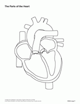
Heart
the major organ in the cardiovascular system
it is a pump that pumps blood around the body
humans have a 4-chambered heart
Blood Flow Through the Heart
when blood returns through the veins, it enters the vena cava - these veins take blood back to the heart
this blood is low in oxygen (deoxygenated blood), but high in CO2
deoxygenated blood enters → right atrium
→ deoxygenated blood → right ventricle
→ deoxygenated blood → pulmonary artery - takes deoxygenated blood → lungs
→ deoxygenated blood → enters capillaries of lungs; CO2 in blood now exits and
→ enters lungs; oxygen in lungs goes → blood →
→ blood is now oxygenated - high oxygen, and low in CO2 →
→ oxygenated blood returns → heart through pulmonary veins - brings blood back to heart, then →
→ enters left atrium, then → left ventricle →
→ now, oxygenated blood goes out the aorta - dumps blood into arteries
Explain the two (2) types of gas exchange that occur in the entire circulatory system
external respiration - capillaries of lungs: take CO2 from blood and go to lungs, then O2 from lungs to the blood
getting cases from outside the lung capillaries
internal respiration - capillaries by the cells deep in the body; oxygenated blood has left heart → arteries → arterioles → capillaries at cells deep in body; O2 leaves blood → cells; CO2 leaves cells → blood
no external interaction; all deep in body now (capillaries → lungs)
List the steps of blood circulation and where they take place (9)
Deoxygenated blood:
Enters vena cava (1)
goes to right atrium (2)
passes through tricuspid valve and enters right ventricle (3)
enters pulmonary artery (4) and exits into the lungs (5)
in the lungs: (O2 leaves and enters bloodstream; CO2 leaves bloodstream and enters lungs)
Oxygenated blood:
enters from bloodstream through the pulmonary veins (6) and enters the left atrium (7)
passes through bicuspid valve and into the left ventricle (8)
exits through the aorta
circulatory vs. lymphatic system
Circulatory - brings blood around the body
Lymphatic - drains fluid in the body (ex. decreases swelling)
liquid is called lymph: lymph vessels → passes through lymph nodes
is consistently draining; recirculating → immune system, or eventually the kidneys
Cells in the blood and what they do
Red blood cells:
anucleate (no nucleus)
replenish a lot
carrying oxygen → if they can’t, then → metabolic issues (eg. electron transport chain)
White blood cells:
immune system
lots of different kinds (B-, T-, macrophages, granulate…)
List and define the different blood cancers discussed (2)
lymphomas - cancers developed in lymphatic system
leukemias - cancer developed form abnormal cell division of white blood cells
List and define the types of lymphomas (2)
note: any type results from uncontrollable multiplication of lymphocytes at the nodes or other tissue (ie. tonsils)
Hodgkin’s - large white blood cell with 2 nuclei
less common; often I adolescents and young adults
Non-Hodgkin’s - also just called ‘lymphoma’
more common (often in adults 65+)
one nucleus
List and define the types of leukemia discussed
acute lymphoblastic (A.L.L.) - large # of lymphocyte cells
common in kids ages 3-6 and 65+
prognosis is good, usually → chemotherapy treatment
acute myeloid (A.M.L.) - most common in 65+
success/prognosis varies
chronic lymphatic (C.L.L.) - overproduction of B-cells
most common in North America
many don’t realize they have it for years
chronic myelogenous (C.M.L.) - overproduction of granulocytes
often in older adults
prognosis/survival rate 90%
(chromosomal) translocation b/w chromosomes. 9 (BCR) and 22 (ABL) → Philadelphia chromosome
increased cell division - proliferation
treatment: Gleevac
Gleevac
targeted therapy onto the BCRABL protein
cures over 90% of patients
inhibits protein from promoting cell division
Describe the kidney function
filters blood, specifically at nephrons - tells the body what to keep or get rid of
waste products and liquid sent out of nephron to ureter - makes up urine
bladder - stores urine
urine released from bladder out the urethra
What is the goal of the respiratory system?
to bring air (O2) into the body and remove ‘waste’ (CO2)
Inhalation vs. exhalation
inhalation - process of bringing air in
exhalation - process of forcing air out
Respiratory system process
Air enters nares (openings of nose) →
nasal cavity (warms air and hairs catch particles/bacteria/viruses and prevent their furthering into the body) →
pharynx (back of the mouth) →
glottis (opening to the respiratory tract), covered by the epiglottis (prevents food from entering glottis) →
larynx (vocal cords) →
trachea (windpipe; has cartilage rings (help ensure trachea doesn’t collapse); also has small openings along the back, capable of passing small amounts of food to digestive tract) →
bronchus/bronchi (cartalige-rung to prevent collapse) →
bronchioles (inside lung) →
alveoli (starchy circular organs; covered with capillaries; this is where gas exchange occurs)
at alveoli, O2 → capillaries, CO2 exits and → alveoli [external respiration]
How does CO2 leave capillaries to enter alveoli, and how does O2 leave alveoli to enter capillaries?
diffusion - matter will go where it’s less concentrated
CO2 is highly concentrated in capillaries → will move to less concentrated (alveoli)
O2 is very concentrated in alveoli → will want a less concentrated area (capillaries)
What brings air in and what pushes air out?
inhalation:
diaphragm ( sits below ribs, pulls downward)
when it pulls down, → makes more room in chest cavity
pull air into lungs
exhalation:
diaphragm relaxes upward
makes chest cavity smaller
as a result, this process forces air out
What contracts breathing rate?
at brain stem, CO2 levels are monitored
higher levels → brain stem signals body to breathe faster
ex. exercise: ↑ metabolism → ↑ CO2, → ↑ breathing
lower levels → brain stem signals body to breathe slower
ex. sleep: ↓ metabolism → ↓ CO2 in blood, → ↓ breathing rate
Types of Lung Cancer discussed in class
mesothelioma - cancer of lung lining and largely connected to asbestos exposure
asbestos - naturally occurring chemical compound; used as building insulation (illegal since 80s)
in walls, doesn’t cause harm (still safe there) unless disrupted
ex. tear-down; airborne if protocols aren’t followed [if airborne, this is what can cause mesothelioma]
SCLC - “oat cell” - begins in bronchi
very aggressive
classified under microscope
NSCLC - most common
have different subtypes
named after cell type it derives from
“lung carcinoma”
carcinogens - chemicals known to cause cancer
What carcinogens do
cause mutations in DNA
typically, you can’t isolate what is making the change occur
lots of heterogeneity - understand impact on cellular behavior
Differential gene expression
comparing cancer cell(s) to normal cell(s)
What tools are used to compare (in differential g.e.)
microarray - used to see what expression changes are caused
Microarray steps (5)
mRNA is isolated from tumor and normal cells
Synthesize: cDNA-complements DNA sequences; sourced from mRNA
Hybridize cDNA to existing DNA on chip
nucleotide sequence in probe connects → complementary
Analysis
how much color stuck
green = normal expression
red = cancerous expression
yellow = both have expression
black/clear = not expressed in either
Use of data
green → lower expression of this gene in tumor tissue compared to normal tissue
red → higher expression of this gene in tumor tissue compared to normal tissue
yellow → expression of this gene is the same in tumor and normal tissues
Sputum Cytology
study of cells from a sample
thorascopy
removing a sample of a tumor
adenocarcinoma
cancer from lung’s epithelial tissue
three common areas of the body affected by metastasis
brain, bones, lungs
wedge resection
surgical removal of a lung tumor and surrounding tissue
cisplatin function
binds to DNA, preventing replication
pacitaxol function
halts cells in M phase; prevents mitosis
carboplatin
derivative of cisplatin
“non-cell cycle specific drug”
damage cells at different stages
Taxol (paclitaxol)
used to treat cancer of ovaries, breast, NSCLC
what system the skin is considered to have
integumentary system
layers of the skin from surface → inward
epidermis - surface layer of skin
dead keratinocytes
melanocytes
living keratinocytes
basal layer - layer between epidermis and dermis
dermis - secondary layer of skin
nerve endings
blood vessels
hair follicles
sweat ducts and glands
sub-Q (fatty layer)
ends with a barrier before the blood
compositions of skin layers
dead keratinocytes - dead skin cells on the epidermis; shed over time
melanocytes - produce melanin
melanin - dark product made in the body; absorbs UV light
more UV exposure → increased gene expression of melanin
living keratinocytes - take the melanin produced by the melanocytes to protect themselves from UV
Types of UV light
A - reaches dermal and sub-Q → connected to premature aging and wrinkles; cancer-causal
B - reaches epidermis → less associated with aging; cancer-causal
C - stops at Earth’s ozone layer (layer is gradually shrinking)
what UV can do to the body (DNA)
causes DNA changes → creating thymine dimers
thymine dimers
caused by H2 bonds being broken down between double-stranded DNA
DNA “bubbles” → affects transcription and ultimate protein produced
though we have proteins (enzymes) to remove them, some people may have too many for the system to catch up or some may never be detected → possible cancer in long-term

Cause of a sunburn
UV rays causing too much damage
→ tissue damage: blood rushes to area (red appearance)
feeling hot with sunburn → inflammatory response
if bad enough: sunburnt cells/UV → triggers cell death → dead cells peel off
sun poisoning
extreme tissue damage → even stronger immune response
scrape-like patches where skin has died off
Types of Sunburns
First-degree - only impacts epidermal layer
Second-degree - impacts epidermal and dermal layer
Third-degree - all layers (epidermal, dermal, sub-Q)